Phospholipid Biosynthesis
Overview of Membrane-Associated Lipids:
The average lipid content in mammalian membranes is ~50%, but can be as low as 20% in the inner mitochondrial membrane, or as high as 80% in the myelin sheath.
The most common mammalian phospholipids are:
Phosphatidylcholine
Phosphatidylethanolamine
Phosphatidylserine
At physiological pH, it is negatively charged.
Sphingomyelin
Glycolipids are always present on the extracellular side of the membrane.
Galactocerebroside exists abundantly in myelin sheaths along with sphingomyelin.
Gangliosides are negatively charged due to presence of sialic acid.
GM1 acts as a cell surface receptor for Cholera.
An excessive accumulation of GM2 is observed in Tay-Sachs disease.
Glycerophospholipid Structure:
Also known as phosphoglycerides.
Composed of 2 fatty acyl chains esterified to glycerol-3-phosphate-X (i.e. alcohol).
Compose most of the lipid bilayer of membranes.
Fatty acyl chains are usually saturated (C1) and unsaturated (C2).
Glycerophospholipids and Head Groups:
Phosphatidic Acid → —H
Phosphatidylcholine → (CH3)3+NH2CH2C—
Phosphatidylethanolamine → H3+NH2CH2C—
Phosphatidylserine → -OOC(H3+N)HCH2C—
Phosphatidylinositol → O5H11C6—
Synthesis of Glycerophospholipids through a Phosphatide Intermediate:
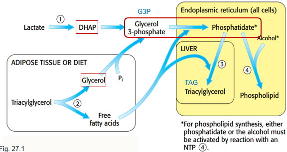
Occurs on the ER membrane.
Formation of (G3P): dihydroxyacetone phosphate (from gluconeogenesis or phosphorylation of glycerol).
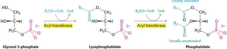
Successive esterification with acyl groups to form phosphatidic acid (PA.
Addition of two head groups via two routes*.
G3P to Phosphatide
Acyl CoA contributes to fatty acid additions that makes G3P into phosphatidate. The glycerophospholipid backbone is added with ethanol. This anabolic reaction requires an activated component in the form of phosphatidate or alcohol.
*Route 1: Phospholipid Synthesis via Activated Phosphatidate.
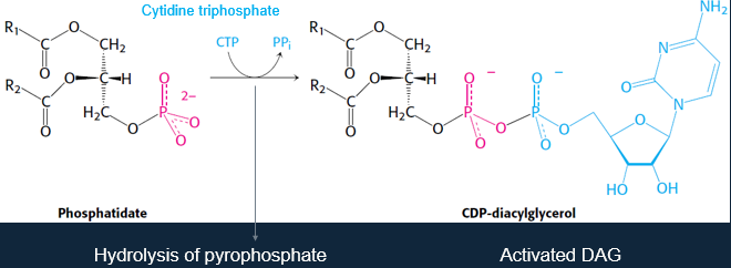
The phosphatidyl unit can then react with alcohol to form a phosphodiester linkage.
If the alcohol is inositol, then phosphatidylinositol and CMP form.
The phosphatidyl unit refers to cytidine diphosphate-diacylglycerol, an activated intermediate in phospholipid biosynthesis.
It consists of a diacylglycerol (DAG) backbone linked to cytidine monophosphate (CMP) via phosphate.
*Route 2: Synthesis via Alcohol Activation
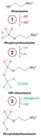
This involves the formation of a CDP-head group and transfer of phosphoryl head group to DAG.
Head Group + ATP → Phosphoryl-Head Group + ADP
Phosphoryl-Head Group + CTP → CDP-Head Group + PPi
CDP-Head Group + DAG → Phospholipid + CMP
— — — — — — —
Other Pathways - PC and PS:
Phosphatidylcholine (PC):
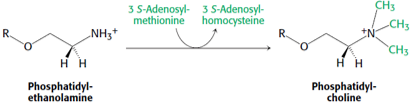
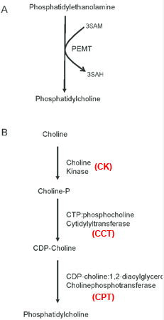
Can be synthesised in the liver by methylation of the head group of phosphatidylethanolamine (PE).
Phosphatidylserine (PS):
Synthesis by base-exchange reactions from PC or PE.
Phosphatidylcholine + Serine → Choline + Phosphatidylserine
Phosphatidylethanolamine + Serine → Ethanolamine + Phosphatidylserine
Membrane Phospholipids:
Phosphatidylcholine (PC):
Most common phospholipid in mammals, making up ~50% of membrane mass.
Precursor of DAD signalling molecule.
Synthesis from PE (A):
PEMT - PE methyl transferase in liver pathway when dietary choline is insufficient.
Kennedy Pathway (B): Synthesis from Dietary Choline:
CCT - amphitrophic enzyme; ligand regulator is the membrane itself.
CDP-Choline - the rate-limiting step.
— — — — — — — — — —
Phosphatidylethanolamine (PE):
Makes up 15-25% of total phospholipids in mammalian cells.
Cone-shaped, non-bilayer.
Synthesis pathway is similar to that of PC.
Diverse cellular functions:
Precursor for phosphatidylcholine.
Substrate for important post-translational modifications.
Influencing membrane topology.
Promotes cell and organelle membrane fusion, oxidative phosphorylation, mitochondrial biogenesis, and autophagy.
— — — — — — — — — —
Phosphatidylserine (PS):
Makes up 5-15% of total phospholipids in mammalian cells.
Higher distribution in the inner membrane leaflet.
Synthesised by base exchange.
Roles:
Activator of protein kinase C.
Exposure to cell surface contributes to platelet aggregation and elimination of apoptotic cells.
— — — — — — — — — —
Phosphatidylinositol (PL):
Less abundant than PS in mammalian cells.
PL that can be phosphorylated:
7 forms of phosphatidylinositol phosphates (PIPs).
Roles in lipid signalling, cell signalling and membrane trafficking.
Has an unusual nearly fixed fatty acid composition:
Stearic acid at C-1.
Arachidonic acid at C-2.
— — — — — — — — — —
Other Glycerophospholipids:
Alkyl-acyl-glycerophospholipids:
Alkyl chains either-linked to C-1 of glycerol.
Plasmalogens:
Oxidation of either of alkyl-acyl-glycerophospholipids.
Vinyl Ether
Sheath ethanolamine glycerophospholipids.
Other Types of Phospholipids:
Sphingolipids:
Not glycerolipids → contains sphingosine.
Simplest one is ceramide.
Sphinomyelin is an important constituent of myelin.
Contains gangliosides - sialic acid-containing ceramide oligosaccharides.
Contains cerebrosides - ceramide monosaccharides.
Found in the outer leaflet of the eukaryotic plasma membrane, and has the highest concentration in the CNS.
Concentrated in ‘lipid rafts’ and caveolae, together with cholesterol. They compartmentalise proteins in the membrane.
Phospholipids and Membrane Structure and Formation:
Phospholipids are synthesised on ER membranes.
Choline phospholipids are flipped into the outer leaflet by flippase enzymes.
Membrane Formation:
A consequence of the amphipathic nature of phospholipids.
Salts of fatty acids form micelles.
Most phospho- and glycolipids in aqueous media are bimolecular sheets because:
Bulkiness
Hydrophobic Interactions
Van der Waals of hydrocarbon tails prefer closeness.
Electrostatic and hydrogen-bonding attractions between polar head groups and water molecules.
— — — — — — — — — —
Implications of Lipid Bilayer:
Held together by many reinforcing, non-covalent interactions (predominantly hydrophobic), they are cooperative structures with fluidity.
Biological consequences:
Inherent tendency to be extensive.
Tend to close on themselves. This means there are no exposed edges of hydrocarbon chains, meaning they can form compartments.
Self-sealing because holes are energetically unfavourable.
Tay-Sachs Disease:
Sphingolipids degradation in lysosomes.
Defects in enzymes leads to sphingolipid accumulation and mental retardation.
Hexosaminidase A deficiency leads to ganglioside GM2 accumulation.