Anatomy and Physiology Segment 1
Module 1: Basics of Anatomy and Physiology
1.01 Understanding Anatomy and Physiology
Anatomy: study of the body’s makeup, Physiology: how the body works
It is important to study these fields to understand how the body works and to keep records for medical reasons. It is essential knowledge for those in sports and medicine.
Anatomy identifies the structures of the body while physiology studies the body’s functions and structures in the body’s relation to one another to sustain life.
Important Word Meanings:
Prefixes:
A- : without
Anti : against or stop
Append : to hang
Auto : self
Bi-/di- : two
Bio : life
Ecto : outside
Endo : inside
Epi : above
Hist : tissue
Hyper : above average
Hypo : below average
Leuko : white
Sub : below
Roots:
Artho: joint
Cardio: heart
Cranio: skull
Cyto: cell
Derma: skin
Entero: intestine
Gastro: stomach
Hema: blood
Hepato: liver
Nephro/Rena: Kidney
Ortho: straight
Osteo: bone
Pulmo: lungs
Therm: heat
Suffixes:
-ase: enzyme
-blast: growing
-clast: to break
-cyte: cell
-emia: blood
-ic: pertaining to characteristic of
-ism: condition
-itis: inflammation of
-logy: study of
-pathy: disease
-scopy: to view
-stasis: to stop
Body Systems:
[Brain, spinal cord, nerves] Nervous System: responsible for communication between the parts of the body
[Pituitary Gland, thymus, hypothalamus, thyroid, pancreas, adrenal gland, testes, ovaries] Endocrine System: responsible for hormones in the body
[Nasal passages, trachea, lungs] Respiratory System: responsible for the process of bringing oxygen in and carbon dioxide out of the body
[Skeletal muscles, organ muscles, tendons] Muscular System: responsible for the movement of the body
[Hair, skin, nails] Integumentary System: responsible for the protection of the outside body and most major organs
[Stomach, liver, gallbladder, large intestine, small intestine] Digestive System: responsible for absorption of nutrients and removal of waste
[Epididymis, testes, mammary glands, ovaries, uterus] Reproductive System: responsible for creation of a zygote and support of a gamete
[Bones, joints] Skeletal System: responsible for protection of organs, structural support, and movement of the body
[Kidneys, urinary bladder] Excretory System: responsible for the filtering and removal of waste from the body
[Heart, blood vessels] Cardiovascular System: responsible for movement of blood throughout the body
[Thymus, lymph nodes, spleen, lymphatic vessels] Lymphatic System: responsible for providing immunity from pathogens and maintaining proper water balance
Specialties within anatomy and physiology:
Anatomist:
Gross anatomy involves body structures visible to the naked eye
Microscopic anatomy involves microscopic views of tissues, cells, and molecules that make up the body
Regional anatomy looks at regional anatomical structures, like the head
Systemic anatomy involves identifying a system within the body
Physiologist:
Cell physiology is the study of chemical and molecular processes inside and between cells
Special physiology is the study of chemical processes and functions of specific organs such as the heart
Systemic physiology is the study of the cooperative functions of all the organs in a system
Pathological physiology is the study of the effects of diseases on organs and systems (pathogens)
1.02 Anatomy Basics
Axial means pertaining to the head, neck and trunk of the body. The prefix of the axis is the central line running down the middle of the body.
Appendicular means pertaining to the upper or lower limbs of the body. The prefix means to hang something, and is used because the limbs are attached (‘hanging’) to the trunk of the body.
Body positions are important to describe how the body can be viewed from different angles and are important to describe different body positions.
Anatomical position: the primary point of reference used to describe a location on the body. It means the body is facing forward, arms down by their side, palms facing forward and the thumbs point to the side
Supine: the body's laying face up, in anatomical position
Prone: the body is lying face down
Lateral: the body is lying on the side, where an arm is stretched outwards beyond the head.
Anatomical Terminology is always used in relation to the anatomical position. Anatomical term of position is referencing a position of the body from the patient’s viewpoint as they are standing in anatomical position
Anterior refers to a ventral location or towards the front. For example, the sternum is ‘anterior’ to the vertebral column, or ‘ventral’ to the vertebral column
Posterior refers to a dorsal location, or towards the back. For example, the vertebral column is posterior to the sternum, or the vertebral column is dorsal to the sternum.
Superior refers to something towards the top. The nose is superior to the mouth, or above the mouth.
Inferior refers to something towards the bottom. The mouth is inferior to the nose.
Cranial or cephalic means towards the top as well, but refers towards the head and cranial. The skull is cranial to the neck.
Caudal refers towards the bottom, but towards the tail and is used in regards to the head and vertebral column. The neck is caudal to the skull.
Medial refers towards the midline, so the nose is medial to the ears. This would be looking at the ‘axis’ of the body, or the center.
Lateral refers towards the side, so the ears are lateral to the nose.
Superficial or external refers to something situated on the surface. So the skin is superficial to the bone.
Deep and internal refers to something situated towards the inside. So the bones are deep to the skin.
Proximal refers to something situated closer to the origin of the body. So the elbow is proximal to the wrist as the point of origin is the shoulder. Otherwise, the legs would refer to the hip.
Distal refers to something situated farther from the origin of the body. So the wrist is distal to the elbow.
Body planes are like x, y, and z axis where the body can be observed based on these invisible rectangles that span the area of the body.
The Sagittal plane is a lengthwise cut that divides the body into right and left portions. The two planes do not need to be fully equal.
The Transverse plane is a horizontal cut that divides the body into superior (top) and inferior (bottom) portions. It runs across the torso, right above the hips.
The Frontal (coronal) plane is a vertical cut at a right angle to a medial plane, separating anterior (front) and posterior (back) sections.
Body cavities are any hollow spaces in the human body containing organs and important parts. They comprise primarily from the dorsal and ventral cavities, where they are then subdivided into smaller cavities.
The Dorsal cavity goes from the brain down to the pelvis, including the cranial and spinal cavities and is seen from a lateral position.
The Ventral cavity is divided into thoracic and abdominopelvic cavities and is seen from anatomical position.
The Thoracic cavity includes the pleural cavity (lungs) and the pericardial cavity (heart)
The abdominopelvic cavity includes the abdominal cavity (intestines, kidneys, and adrenal glands) and the pelvic cavity (reproductive organs, urinary organs, and rectum)
1.03 Physiology Basics
Physiology studies how systems of the body work together to sustain life, and life is based on 5 characteristics:
Contains cells, grows, reproduces, maintains homeostasis, and responds to stimuli
They involve multiple types of cells that grow and communicate, depending on one another to maintain bodily processes.
Stem cells are the first to develop after the egg is fertilized and contain DNA for the entire body. During development, these cells undergo differentiation (process of a stem cell becoming a specialized body cell) and develop specialized proteins.
Stem cells develop into muscle cells, intestinal cells, skin cells, nerve cells, bone cells, liver cells, alveolar (alveoli, lungs) cells, and blood cells.
Levels of organization in the human body:
Atom: all matter is composed of atoms, and it is the smallest whole particle.
Molecules: particles made up of two or more atoms bonded together, like H2O or CO2
Macromolecule: joined small molecules creating very large molecules consisting of smaller molecular units bonded together and form the structural features of cells and allow these cells to perform the functions needed to support life.
Organelle: a specialized subunit within the cell, akin to an organ in the human body. Macromolecules make up the proteins to form organelles and are used to perform processes.
Cell: the basic unit of life and function in all living organisms. Individual cells can vary in size, shape, and function depending on usage and species.
Tissue: composed of similar cells to form a larger group of intercellular material. For example, muscle is called muscle tissue, as it is made of similar muscle cells.
Organ: specific types of tissues join together to make up the organs in the body. In the stomach, there are five different tissues that join together.
Organ System: a system of organs that work together to complete a specific function. For example, the digestive system is a system that breaks down and uses the nutrients and food.
With the formation of an embryo, the stem cells divide to form three germ layers (three layers of cells formed in early embryo) and specialize in specific tasks for organs and systems.
Ectoderm: forms structures like neurons and skin cells.
Mesoderm: forms structures like blood cells and heart muscle cells.
Endoderm: forms structures like the liver and pancreatic cells.
All tissue can be categorized into four tissue groups:
Epithelial: it serves to protect, secrete, absorb, and excrete from glands. It covers the body’s surface and lines internal organs. However, they lack blood vessels (avascular) and they are tightly packed and divide quickly to replace themselves, often forming multiple layers.
Ex: skin, capillary walls, inner lining of lung, urinary tract
Muscle: primarily serves to help in movement, it is attached to the bones, walls of hollow internal organs, and in the heart. They contract in response to stimuli and muscle tissue has many blood cells (vascular).
Ex: heart, digestive tract, blood vessels, and attached to the bones
Nervous: conducts impulses for coordination, regulation, integration, and sensory reception. It is located in the brain, spinal cord, and the nerves throughout the body. The cells can communicate with each other and other body parts, but has no vascular tissue to transmit blood. Nervous tissue is made up of tissues and support cells.
Ex: neurons, glial cells, brain tissue, spinal cord, sensory organs
Connective: binds, supports, protects, fills in spaces, stores fat, and produces blood cells. They can be found throughout the whole body and is part of the support system for other cells. They can be liquid or solid and are found on the outside of other cells. The tissues themselves can be avascular or vascular, but avascular examples can include cartilage.
Ex: blood, bone, cartilage, fat, support cells for other tissues
Histology is the study of the microscopic structure of tissues, utilizing identification and functional knowledge. Under a microscope, each tissue has a specific structure.
Epithelial: most often appears as cells stacked next to or atop one another. To identify, it is easiest to look for squamous, cuboidal, and columnar cell shapes.
Squamous: flat and wide, they appear like layers of lasagna.
Cuboidal: cube shaped (more like half hexagons forming a C) and have a slight certain border of red circular shapes surrounding the cells.
Columnar: tall instead of wide, they stand like trees in a forest. They are crowded together to form tight but tall layers of cells.
Muscle tissue is divided into three types, skeletal, smooth, and cardiac. Under a microscope, each type looks like pink smears, but they have specific characteristics to each.
Skeletal muscle: long strands of cells with many nuclei, they resemble clothing fibres.
Smooth muscle: resemble eyes, they are what you first imagine when you think of muscle structure. They are fat in the middle and narrow out at the ends.
Cardiac muscle: has short lines between the cells, forming small openings between each tissue. These discs are intercalated (inserted between layers) and help with the consistent beating of the heart. Their shape looks like metal bridge supports, where they cross to support the weight.
Nervous tissue: is the most identifiable, as they have the axon terminal that forms long strands from the dendrite. It resembles almost like tentacles, where the myelin sheath coats the terminal.
Connective tissue: they come in multiple varieties. Blood is a liquid connective tissue, while bone is a solid and hard connective tissue. Loose connective tissue can look like muscle tissue, but look for characteristics of muscle, epithelial, and nervous tissue before identifying the observed tissue as connective.
Homeostasis is the state where everything remains in balance and functions properly. The body attempts to maintain homeostasis across temperature, blood pressure, and pH.
The body relies on metabolism to accomplish homeostasis. Metabolism itself involves all physiological events and chemical reactions involving energy. It provides the energy needed to break down body tissues and energy stores, but also building it up.
1.04 Integumentary System
The skin is also called the “integument” and is considered an organ due to its characteristic of consisting of all four tissue types. The skin and accessory organs (glands, hair, nails) make up the integumentary system, which acts as a barrier that separates our internal systems from the environment.
The skin consists of the epidermis which acts as the barrier from water and pathogens, the dermis which acts as a sensory receptor and temperature regulator, and the hypodermis which acts as a cushion.
The epidermis is the outermost, superficial region of the skin. It varies in thickness and has specialized cells acting as a barrier for the environment. It is also home to melanocytes, cells that produce a pigment called melanin. It is crucial for protecting the body from UV radiation.
Merkel cells are found in the basal layer of the epidermis and allow us to sense what is happening outside of our body. This is where the dermis resides, a thick layer of dense and irregular connective tissue. It helps maintain body temperature (thermoregulation) and ensures the body’s temperature remains at a healthy level through sweat. The dermis is where blood vessels, hair follicles, and sweat glands are found.
The hypodermis is underneath the dermis and is made of loose connective tissue and fat. While it contains blood vessels and nerves larger than those found in the dermis, it is used primarily for fat storage.
The skin’s layers are made of different and unique cells to form the integumentary organ system.
Keratinocytes: flat and round, keratinocytes are found in the epidermis and sit on basal cells. They serve to protect the body from foreign substances and prevent heat and moisture loss.
Langerhans Cells: rod or dendritic in shape, they can be found in all layers of the skin. It is the immune system’s first defense against pathogens entering through a cut in the skin and defend by engulfing pathogens.
Basal Cells: small and round, these cells are found in the epidermis and produce new skin cells.
Melanocytes: these cells have a round, globular appearance around its nucleus. It is found in the basal layer of the epidermis and produces the pigment called melanin, protecting cell nuclei’s DNA from ultraviolet radiation and mutation.
Merkel Cells: oval in shape, they are fewer in number and are also found in the basal layer of the epidermis. They are touch receptors and interact with sensory nerve endings, allowing us to sense changes outside our body.
The skin can typically recover from wounds, though they temporarily hinder their functions.
Burns can be caused by UV, chemicals, or electricity. Burns have a special classification system based on the damage to the layers of the skin.
First Degree Burn: only the epidermis is affected and will heal very quickly with little permanent damage.
Second Degree Burn: the epidermis and the dermis are affected, leaving behind red and swollen skin and blisters as the skin attempts to form protection from the outside as it repairs itself.
Third Degree Burns: the epidermis and dermis are often destroyed, leaving behind dark colorations and destroying nerve endings. Sometimes, it will reach the hypodermis, depending on severity. Anything beyond this will affect the muscle and bone underneath.
Cuts are categorized based on the level of impact to the tissue and cells. Most can be treated with bandages, ointment, or stitches. Due to cuts, it can range from surface-level blemishes to a complete structural change. Depending on the damage, the epithelial cells and blood vessels will respond differently.
Shallow cuts typically only cause the epithelial cells to be stimulated to divide rapidly to fill in the cut. When the injury extends to the dermis, blood vessels break and the blood is released to clot the wound. After new cells are formed and tissue is replaced, the scab will fall off.
1.05 Biotechnology Honors
Disease occurs when something is changed and no longer functions as it should. This can be linked either to environmental or genetic factors like errors in DNA.
Genetic factors begin in fertilization, where the egg and the sperm come together to form an embryo. As half the genes come from the mother and other half from the father, certain diseases can be linked to the possession of recessive or dominant genes.
Cystic fibrosis is a genetic disease that causes thick mucus to build up in the lungs and pancreas, making it difficult to breathe and digest food.
Huntington’s disease is a disease affecting the nervous system, causing the nerve cells to break down and die over time. This causes physical movements beyond the person’s control.
Sickle cell anemia affects red blood cells, causing cells to misshapen and die early, leading to pain, infections, and vision problems.
Environmental factors like diet and exercise can also affect how a disease appears.
Allergies affect the respiratory system, caused by an overly sensitive immune system.
Asthma also affects the respiratory system. During an attack, the airways narrow, leading to wheezing, shortness of breath, and coughing, but can also be brought on due to environmental factors.
Biotechnology refers to the application of biological techniques, tools, and processes to enhance the understanding, study, and manipulation of living organisms at the molecular and cellular levels. It encompasses a wide range of applications and utilizes biological systems to develop solutions and technologies.
Molecular Diagnostics: techniques used to analyze and detect specific genes, proteins, or biomarkers.
Pharmacogenomic: studies analyze genetic variations to determine the most effective and safest drug therapies for patients. Personalized medicine improves treatment and reduces adverse drug reactions.
Bioinformatics: a combination of biology and computer science, allows for large datasets related to genomics to be quickly analyzed.
Bioengineering: techniques used to design and create artificial organs, prosthetics, and medical devices such as synthetic tissues.
Biotechnology harnesses biological systems and organisms, and as the basis of all living things are proteins, it is useful to understand how proteins are built synthetically.
They also use stem cells in their advancements. As stem cells can develop into different body cells, it provides a useful suggestion as to rebuilding or healing damaged organs. However, it provides an ethical dilemma related to its usage.
The umbilical cord is where donated cells can come from (with others), and bone marrow transplants have also provided evidence that stem cells can produce undamaged cells.
Biotechnology improves the quality of healthcare, and can help increase the quality of life for individuals suffering from injury and disease.
Tissue regeneration offers life-saving solutions for individuals suffering from organ failure.
Therapy can be used to expedite the healing of damaged cells, reduce pain, restore function, and minimize complications.
Targeted therapy can make treatment and detection more efficient, increasing survival rates and minimizing side effects in crucial illnesses like cancer.
Module 2: Support and Movement
2.01 Understanding Body Motion
The way bones and muscles are connected allows for specific ranges of motion (ROM) to occur.
Abduction: movement of a limb away from the midline
Adduction: movement of a limb towards the midline of the body
Supination: describes the movement of a hand, arm, or foot in faceup position.
Pronation: describes the movement of a hand, arm, or foot in face-down position.
Flexion: decreases the angle between two limbs of a joint
Extension: increases the angle between two limbs of a joint
The axial skeleton contains the cartilage and bones that support and protect the organs of the head, neck, and trunk. It is composed of the skull, ribcage, and spinal column.
The skull is made up of the cranium (brain case) and facial bones
Hyoid bone is located between the larynx and lower jaw, supporting the tongue and other muscles that move the tongue. It is not attached to any other bones but held in position by muscles and ligaments
The vertebral column is also called the spinal column, forming the central axis of the skeleton. It is made of many vertebrae bones separated by discs consisting of fibrous cartilage and gelatin-like cores. The vertebrae and other facial bones are examples of irregular bones, or bones with a variety of shapes.
The sacrum is part of the pelvis and is the fusion of the five vertebrae, forming a small tailbone. The tailbone itself is called coccyx.
The rib cage, also referred to as the thoracic cage, is made up of twelve pairs of ribs connected posteriorly to the thoracic vertebrae. Most of them are connected to the sternum, and are flat bones.
Axial muscles are of any muscles of the trunk or head.
The trapezius is a large, superficial muscle that extends from the cranium to the lower thoracic vertebrae. It is one of the muscles involved in the movement of the scapula, and is used to shrug your shoulders. (The triangles on your shoulders connecting to your neck to the center of your back to the middle of the torso)
Rotates, retracts, elevation, depression and supports the arm
The pectoralis major is the fan-shaped muscle on the chest, making up the bulk of the human chest and found under the breast in women.
Flexes, extends, adducts, and rotates the arm at the shoulder.
Pectoralis minor is a thin, triangular muscle found deep underneath the pectoralis major.
Draws the scapula forward and downward.
Latissimus dorsi (lats) are large muscles posterior to the arm and partially covered by the trapezius. It is the broadest muscle in the back.
Adducts, medially rotates, and extends the arm at the shoulder.
The external oblique is a broad and thin muscle found on the lateral and anterior parts of the abdomen. It is the outermost of the three flat muscles of the lateral anterior abdomen. (sides of the body)
Flexes the vertebral column by drawing the thorax inward, rotates and laterally flexes the vertebral column and compresses the abdomen.
The rectus abdominis muscle (abs) is a paired muscle running vertically on the anterior wall of the human abdomen. These parallel muscles are separated by a midline band of connective tissue.
Flexes the vertebral column (and torso), compresses the abdomen, assists in breathing.
The appendicular skeleton consists of bones in the upper and lower limbs, as well as the bones that anchor those limbs to the axial skeleton. Includes lower limbs, pectoral girdle, upper limbs, and the pelvic girdle.
The lower limbs are made up of:
Femur: thigh bone, and is a long bone (elongated bone with expanded ends)
Tibia: shin bone
Fibula: slender leg bone next to the tibia
Patella: kneecap, covering the area where the femur and tibia connect
Tarsals: seven ankle bones
Metatarsals: five bones in the inner part of the foot
Phalanges: fourteen bones of toes
The pectoral girdle is made up of the scapula (shoulder blade) and clavicle (collarbone). They connect the bones of the upper limbs to the axial skeleton and aid in limb movement.
The upper limbs are composed of:
Humerus: upper arm bone
Radius and Ulna: two forearm bones, like the tibia and fibula
Carpals: eight wrist bones. They are short bones (cube shaped bones with roughly equal lengths and widths)
Metacarpals: five bones of the palm
Phalanges: fourteen finger bones
The pelvic girdle is composed of the left and right coxae. They are attached to the sacrum posteriorly and to each other anteriorly. The left and right os coxae connect the lower limbs to the axial skeleton. The os coxae, sacrum, and coccyx form the pelvis, protecting the lower abdomen and reproductive organs.
Appendicular muscles are any muscles of the upper and lower limbs. They control the movement of limbs and also stabilize and control the movements of pectoral and pelvic girdles.
The deltoid forms the rounded shape of the shoulder.
Abducts, flexion, and extension of the shoulder.
The triceps brachii muscle (triceps) is the largest muscle on the back of the arm. There are three bundles of muscles with different origins, joining together at the elbow.
Extends the forearm and straightens the elbow.
The biceps brachii (biceps) is a two headed muscle and lies on the upper arm between the shoulder and elbow.
Flexes the elbow and suppinates (rotates) the forearm.
The gluteus maximus is a narrow and thick muscle that makes up a large portion of the shape and appearance of the butt. With the gluteus medius and gluteus minimus, they form the glutes.
External rotation and extension of the hip joint.
The hamstrings are a large muscle group occupying the back of the thigh. It is made up of the semimembranosus, semitendinosus, and biceps femoris.
Flexes the knee joint, rotates the knee joint laterally, and extends the thigh.
The quadriceps femoris is a large muscle group that occupies the front and sides of the thigh. It is made up of four muscles, the rectus femoris, vastus lateralis, vastus medialis, vastus intermedius.
Knee extension and hip flexion.
The gastrocnemius is the muscle on the back of the leg that forms part of the calf. It is a powerful muscle, and along with the soleus muscle, forms the calf.
Plantar flexion of the foot, flexion of the knee.
Bones and their adjacent muscles allow for movement of the body.
Shoulder movement involves the deltoid, biceps brachii, triceps brachii, clavicle, and humerus.
Leg movement involves the quadriceps femoris, hamstring, gluteus maximus, femur, and patella.
Ankle movement involves the gastrocnemius, hamstring, soleus, fibula, and tibia.
Abdominal movement involves the external oblique, latissimus dorsi, rectus abdominis, ribs and pubic bone.
Joints are the locations that occur between major bones. Movement occurs where the joints meet, and the range of motion is impacted by the flexibility of joints.
Ball and socket is when the surface of one rounded bone fits into the depression of another bone. (hip and shoulder)
Gliding is when two bones with smooth surfaces slide over one another to produce restricted movement. (ankle, wrist, spine)
Hinge is when two bones are molded together to allow movement in one direction or plane. (knee and elbow)
Pivot is a cylinder shaped bone that rotates inside another bone. (neck and forearm)
Saddle is a joint with a saddle shaped surface that is in the convex in one direction and concave in the other (only in the thumb)
Certain joints work together with groups of bones to support movement.
The knee joint consists of the femur, patella, tibia, and fibula.
The shoulder joint consists of the clavicle, scapula, and humerus.
The hip joint consists of the hip bone, sacrum, and femur.
Joints are connected to tendons and ligaments and soft tissues of the body offer connection and support for joints and muscles.
Tendon is a connective tissue that attaches muscle to the bone.
Ligaments are connective tissue that attach one bone to another bone.
2.02 Skeletal System
The bones of the body come in different sizes and shapes depending on their function in the body.
Long bones support a person’s weight, help with large movements of the body, longer than they are wide, and mostly seen in the appendicular skeleton. They are primarily in the limbs, and include the humerus, radius, fibula, and the femur.
Short bones provide stability and help with minor movements of the body. They are found in the wrist and ankle joints, cube shaped, and wider than they are long. They include the patella, carpals (wrist), and tarsals (ankle).
Flat bones protect the internal organs like the brain, heart, and abdomen. They are flat and of various lengths and sizes. They include the cranial bones, sternum, ribs, and pelvic bones.
Irregular bones protect internal organs and do not have any specific shape. They include vertebrae, sacrum, mandible, and hyoid.
Bone tissue first develops as soft tissue called cartilage, which is found in sections between bones after full development. During development, bone tissue arises and replaces most cartilage to form a harder bony skeleton and this is called ossification. During ossification, two types of bone tissues are formed.
Compact bone is very dense and made up of calcium phosphate. Due to its structure being tightly packed and hardened, it does not have its own blood supply so the canals and canaliculi must deliver and remove blood, nutrients, and wastes from compact bone.
Spongy bone has a much softer structure, is highly vascular, and provides its own blood supply. It is found at the epiphysis, or the rounded end of a long bone.
The diaphysis is the shaft of the long bone, and the epiphyseal line separates the two types of bone tissue.
Bone tissue is active, living tissue with organic materials contributing to structure, flexibility, and strength. The inorganic components are tightly packed, needle-like mineral crystals of calcium phosphate, accounting for the bone’s hardness.
The osteon is the dense bone that appears as a series of concentric rings of bony materials.
Canaliculi are small, hollow channels that are part of the concentric rings that link all of the bone matrix together so nutrients and wastes can be transferred throughout the bone.
A Haversian canal is at the center of each osteon, and the canal contains small blood vessels that supply the needed nutrients to the bone matrix.
Volkmann’s canals (perforating) connect the various Haversian canals. They move blood from the periosteum of a bone to the Haversian canals in bony tissue.
The hollow part of the bone is called the medullary cavity that holds the bone marrow.
The periosteum is the first layer of the bone and contains two layers. The outer layer contains nerves and blood vessels while the second layer contains cells that grow bone tissue.
The endosteum is another layer of the bone that lines structures that help bring nutrients to the bone but does not have cells that grow bones.

The bone matrix is developed through osteogenesis (process of bone formation, ossification). It requires two types of cells, osteoblasts and osteogenic stem cells. Osteogenic stem cells are undifferentiated cells and can become any cell needed for bone formation. Osteoblasts are scattered throughout bones and make new bone cells called osteocytes. Osteoclasts break down old bone when new bone layers form.
Bones are composed of four kinds of cells: osteogenic, a type of stem cell; osteoblasts, bone-building cells; osteoclasts, bone-destroying cells; and osteocytes, cells involved in communication, transport, and maintenance of the bone matrix. When a bone breaks, blood vessels are damaged and blood leaks into the area, forming a hematoma, or pool of blood, that develops into a clot. Osteocytes, disturbed by the fracture, communicate changes in the bone to the osteoblasts, osteoclasts, and osteogenic cells. As blood vessels infiltrate the site, a soft fibrous granulation tissue is laid down, and a soft callus of fibrocartilage begins to form. Osteoblasts produce a hard callus that adheres to the dead bone at the injury site. They also form a temporary bridge of spongy bone to hold the ends together. Finally, osteoclasts act to remove any excess bone and callus. Within four to six months from the date of injury, repair is complete.
Bones have to go through with multiple steps during healing, beginning from hematoma (inflammatory), to soft callus (blood vessel formation), and finally hard callus (healed fracture).
Every week, we recycle five to seven percent of our bone mass and 0.5 grams of calcium leaves/enters the adult skeleton each day. Osteoclasts are present to break down old bone when new layers form.
There is a delicate balance between osteoclasts and osteoblasts, where the bone density and blood matrix are affected if hormones or disease throw off the balance. If there are too many osteoclasts present, the breaking down exceeds normal, healthy limits, increased calcium levels, or decrease in bone density. Osteoporosis is a bone disease where bone breakdown by osteoclasts causes a decrease in bone density.
Bone terminology and vital signs:
Periosteum - Made up of two layers to surround the bone and offer blood vessels, nerves, and bone cells
Medullary cavity - Hollow part of a bone that contains the bone marrow
Osteogenic cell - Stem cell responsible for bone formation
Osteoblast- Bone-forming cells
Osteocyte - Mature bone cell that maintains the bone matrix
Osteoclast - Large cell that resorbs and breaks down the bone matrix
Haversian canal - Small tubes that weave throughout bone tissue to bring oxygen and nutrients
Volkmann's canal - Small canals that are perpendicular to the Haversian canals and bring blood from the periosteum
Perforating canal - Small canals that are parallel to the Haversian canals and bring blood from the periosteum
Canaliculi - Microscopic canals running through the bone solid matrix that allows for diffusions of substances into the bone
2.03 Bone Markings
The bumps, grooves, and holes in bones create vital connections, facilitate movement, and protect veins and nerves. There are several types of bone markings in the body. The structure of a bone marking is related to its function.
Projections and processes are bumps or raised areas that grow out from a bone.
Depressions are cavities that indent into the bone.
Openings are holes that extend through the bone.
Some projections help form a joint, where two bones are attached for the purpose of permitting movement.
The head is a bony expansion on a narrow neck, providing stability to where it is inserted
The facet is a smooth, nearly flat articular surface. Joints limit excessive movement where an over-extension would result in injury.
The condyle is a rounded articular projection. They provide the surface necessary for the gliding movement of many joints.
The ramus is a smooth, arm-like bar of bone. The smooth shape provides a surface for muscle.
Some projections serve as a site for muscle or ligament attachment to the bone.
The tuberosity is a large rounded or roughened projection, giving a roughened surface for muscles or ligaments to attach at the end of long bones.
The tubercle is a small, rounded projection and is smaller than a tuberosity. Serve to provide areas for attachment and sometimes involved in the formation of a joint
A crest is a narrow, prominent ridge of bone and provide surface for muscle attachment.
The epicondyle is a raised area above the condyle. It provides a raised surface for connective tissue.
The spine has sharp, slender projections and serve as areas for attachment.
The trochanter is a large, blunt, irregular surface only found on the femur. IT is larger than a tuberosity or tubercle.
There is more words to describe bone markings than just that, as some explain shape or size and others explain the purpose. Depressions, canals, and other openings usually make way for other structures such as nerves or veins.
Sinus: air filled space in the bone, filled with mucus membranes and air. A cavity, where a human skull contains four pairs. They filter and humidify the air and protect our vital structures.
Foramen: a rounded opening through a bone, they allow blood vessels, ligaments, and nerves to pass.
Protuberance: rounded section off of another object
Crest: ridge on bone
Fossa: a shallow, basin-like depression that generally receive another bone with which a joint is formed.
Head: large rounded surface of bone
Tubercle: small rounded projection
Condyle: small rounded surface of bone
Trochanter: process at the upper part of the femur
Sulcus: a groove, or furrow, shallow, elongated depression. These depressions can serve as channels for blood vessels, nerves, or a tendon.
Fissure: a narrow, slit-like groove or hole. They serve as channels for blood vessels and nerves.
Suture: a type of joint that only occurs in the skull, where the skull bones are fused together and form an immovable joint. Only a tiny amount of movement is permitted at these sutures.
Health professionals use grooves and bumps on bones as fixed landmarks to find muscles, ligaments, or arteries or to check bone alignment.
2.04 Muscular System
Skeletal muscles are made up of tissues that contract. Through these contractions, posture and movement are possible.
Origin: a bone that the muscle is anchored to; it is usually above the muscle being used.
Insertion: a bone or tendon that the muscle is anchored to, usually smaller than the origin bone and below the muscle being used.
Range of Motion: how far and in what direction a person can move a joint or muscle
Skeletal muscles are the organs of the muscular system. They contain nervous tissue, blood, and other connective tissues. They vary considerably and contain long filaments called myofibrils that can pull together when the fiber receives a nerve signal.
Skeletal muscle is separated from nearby muscles and provides support and protection for fragile muscle cells. They allow them to withstand the force of contraption and allows the parts to move somewhat independently.
Inside every muscle tissue, there are bundles of connective tissue called fascicles, categorized into the Epimysium (entire), Perimysium (surrounds), and Endomysium (surrounds each tiny fiber).
Skeletal muscles have an abundant supply of blood vessels and nerves to help with muscle contraction. The muscle must receive an impulse to contract. Typically, one artery and at least one vein accompany each nerve.
Sarcolemma is the cell membrane of a muscle cell. It is made up of a plasma membrane and an outer layer of polysaccharide material that contains numerous thin collagen fibrils. It receives and conducts stimuli and fuses a tendon fiber to form muscle tendons that then connect to bones.
The Sarcoplasmic reticulum is a membranous network of channels that surround each myofibril. It mainly stores calcium ions that release when the muscle cell is stimulated.
Extensive blood vessels and nerves weave throughout muscles make them a prime location for injury disease.
Tear and strain injuries involve a stretch or tear in the muscle, tendon, or ligament.
A splint is caused by inflammation of the muscle due to using the muscle too much.
Tendonitis is caused when a tendon is inflamed repeatedly without being allowed to heal.
Muscular Dystrophy is a disorder that causes muscle cell death and is highly progressive. It breaks down muscle cells and does not reform them, leading to atrophy and the inability to perform basic functions.
Tendinosis is similar to tendonitis and involves the body breaking down the tendons and forming scar tissue when the tendon is repeatedly damaged.
Tetanus is a bacterial infection of muscle that can cause stiffness and paralysis and can be prevented through vaccination and early treatment.
2.05 Muscle Contractions
Muscle fibers are the building blocks of muscle tissue and have specific characteristics that contribute to their role in movement and bodily functions. They can be excited, contracted, or extended for movement. Muscles are very elastic and allow them to return to their original shape.
Excitability: respond to stimuli by transmitting electrical impulses along other membranes
Contractibility: enables generation of force as they shorten and produce movement
Extensibility: allows them to stretch within limits
Elasticity: the ability of an object to return to its normal shape after being stretched or compressed
Muscles can be structurally compared to a rope. The smaller ropes bundled together are made up of threads called muscle fascicles.
Myofibrils: cells of our skeletal muscles are made up of myofibrils
Myosin: a myofilament with gold-club shaped heads attached to myofilaments and located at the center of a sarcomere
Sarcomeres: sectional divisions of myofibrils are called sarcomeres.
Actin: myofilament made up of thinner proteins. They surround the thicker myosin strand in the sarcomere. Actual number of strands would depend on the specific muscle.
Sliding filament theory of muscle movement:
Step 1. Muscles at Rest: when muscles are at rest, there are two tiny proteins called tropomyosin and troponin wrapped around the actin. ADP molecules are stuck to each myosin head when the muscle is in a relaxed state. All three of these keep the myosin heads from extending and attaching to the protected actin strand.
Step 2. Waiting Muscles: during rest periods, muscles build up an abundance of calcium ions that will be used when it is time for muscle contraction. The cell gets calcium ions through pumps of the sarcoplasmic reticulum, which is wrapped around each sarcomere.
Step 3. Excitation: When it is time for muscle movement, neurons surrounding the muscle send signals to stimulate action, and when this signal reaches the SR, the calcium pumps open wide and the ions flow.
Step 4. Ready for Bonding: At this point, the calcium ions bind to the troponin. It causes the troponin to change shape and rotate around the actin, moving the tropomyosin out of the way. Then, binding sites on the actin molecule are open and ready to bind with myosin.
Step 5. Building a Cross-Bridge: The myosin heads extend and attach to the actin, and this is called a cross-bridge. The contact causes myosin heads to bend toward the center of the sarcomere, shortening the overall length and shortening a muscle contraction.
Step 6. The Slide: as sarcomeres shorten, the myosin releases ADP but remains attached to actin. This occurs throughout each contraction until ADP is released, leaving actin in a fixed state until another ATP binds to myosin, causing movement again. The steps can repeat as long as ATP and calcium are present.
Step 7. Muscle Relaxation: When there’s no longer a nerve stimulus, the calcium ions diffuse back inside the sarcoplasmic reticulum. The troponin and tropomyosin return to resting positions. ATP molecules attach to the heads of myosin and the energy from ATP fuels muscle relaxation. ATP is needed to break myosin-actin bonds and are required to pump calcium ions back into the SR.
A voluntary muscle is a muscle attached to bones and the brain can actively decide to move them. They can include biceps, triceps, lats, abdominals, glutes, quadriceps, and hamstrings.
The goal of skeletal muscle is to create torque and tension to move the bone. Torque is rotational force exerted by muscles to support joints and ligaments. Tension is the generation of force by a single muscle fiber by the interactions between actin and myosin.
When picking up an object, the brain sends signals to stimulate muscle fibers, causing them to contract. Contraction generates tension, pulling on the tendons attached to the bone. This tension then creates torque around the joints as a rotational force and causes the forearm to lift.
There are two types of skeletal muscle fibers: slow twitch and fast twitch. They differ in contraction and endurance.
Slow twitch fibers are red in color, contain a lot of mitochondria, have increased ATP, higher endurance, and are most active during endurance activities.
Fast twitch fibers are pale in color, have fewer mitochondria, produce less ATP at one time, contract quickly but stop after a short time, and are ideal for activities like short sprints, power lifting, or catch.
Involuntary muscles are found in the intestines, stomach, uterus, blood vessels, and heart. They can be broken down into smooth muscles and cardiac muscles. They’re different as the brain does not have to make a conscious effort to move them and they are constantly contracting as long as the needed molecules are available for the contraction to happen.
Smooth muscle contraction means that they consistently push fluids and other substances through them. It keeps blood and digestion flowing throughout the day, including sleeping.
Cardiac muscle uses strong, strategic contractions of the muscles in unison to propel blood throughout the heart’s chambers and the body’s blood vessels.
The difference between voluntary and involuntary muscles can be distinguished through the following traits:
Voluntary muscle (skeletal) have cylindrical, unbranched, and fairly long cells. Those cells have multiple nuclei, have high energy, get fatigued, involved in body movement, and can be seen in the diaphragm, pharynx, and muscles under the skin.
Involuntary muscle (smooth and cardiac) has spindle-shaped, fairly small cells. Those cells have one nucleus each, require mostly low energy, do not get fatigued, and are involved in internal organ movement. They include ducts of glands, urogenital tracts, and the respiratory tract.
2.06 Muscle Metabolism
Metabolism is the chemical reactions that happen inside the cell to convert food to cellular energy. The energy to run our bodies come from the food we eat and the oxygen we intake.
We take in carbohydrates, fats, and proteins to fuel our cells. ATP is formed when glucose is broken down, turning sugars into energy.
Muscle cells get their ATP from the mitochondria.
The mitochondria’s chemical reactions convert glucose from food into cellular energy. Cells that require extra energy contain more mitochondria to accommodate their needs, like skeletal muscles.
The process of cellular (aerobic) respiration breaks down glucose from food into adenosine triphosphate. Red blood cells are attached to hemoglobin, transporting oxygen to the body.
Reactants in cellular respiration are glucose and oxygen, while the products are carbon dioxide, water, and energy. While there is energy needed to break down an ATP’s triphosphate bond, the amount released is significantly more than what is consumed. Think of nuclear fission and how as the subatomic particles break apart, the tremendous amount of energy holding the particle together is released.
While cellular respiration is good for long, sustained exercise, if there is no oxygen, then it cannot proceed. When this occurs, anaerobic respiration takes over, utilizing the process of fermentation.
Fermentation also produces ATP molecules, though not at the same volume. Both must take a glucose molecule and turn it into a pyruvate (end product of glycolysis that is converted to energy) through a process of glycolysis (anaerobic breakdown of glucose by enzymes, releasing energy and pyruvic acid and the first stage of cellular respiration or fermentation).
Alcoholic fermentation is what yeast and other microorganisms undergo through, breaking down glucose into pyruvate molecules which are further broken down to produce ATP, ethyl alcohol, and carbon dioxide.
Lactic acid fermentation occurs in animals and other organisms when oxygen is not available. It also breaks down glucose into pyruvate molecules but breaks them down further to produce ATP and lactate (lactic acid). This can be used in yoghurt.
Muscle fatigue can occur when a muscle is exercised for an extended period of time. Certain molecules build up in the muscle tissue, causing the fibers to develop reduced contractile function. They can be stuck if the sarcomere cannot contract properly. They are primarily caused by lactic acid and inorganic phosphate.
Lactic acid causes lactic acidosis. They create an excess of Hydrogen ions and interferes with the function of ATPases (enzymes breaking down ATP) and reduce the rate at which calcium is reabsorbed. It causes muscle aches, rapid breathing, and nausea. It can be reversed quickly by increasing water consumption and reduction in exercise. It typically lacks severity, but can be dangerous if persistent.
Inorganic phosphate is a necessary nutrient that works with calcium to assist muscle contractions. When muscle works anaerobically, creatine phosphate is broken down, releasing higher amounts of inorganic phosphates (hence why creatine powder is sold for workout). The muscle cannot contract as well as a result and cause cramping and soreness, and long term effects show that they can weaken the bones.
Module 3: Coordination and Control
3.01 Understanding the Nervous System
The nervous system is comprised of various parts that take in input and turn it into action throughout the body.
Brain: processes sensory information
Spinal cord: allows signals to be transmitted between sensory organs, neurons, and the brain
Nerves: send and receive signals for the body
Neurons: nerve cells that make up the nervous system
Interneurons: nerve cells found only in the brain and spinal cord that move nerve signals in both directions
Motor Neurons: cells of the brain and spinal cord that send commands from the brain to muscles and organs to carry out functions
Sensory Neurons: neurons that send signals to and from the body to the brain and spinal cord
Glial Cells: cells that provide physical and chemical support to neurons
The spinal cord within the vertebral column is made of different types of matter that helps signals pass through.
The outer layers of tissue around the spinal cord and brain is called the meninges. The meninges produce cerebrospinal fluid that protects and cushions the spinal cord and brain.
White matter is the next layer and found in the spinal cord and brain. It communicates between grey matter and the body.
The inner material is called grey matter. It processes information and releases new information to the white matter to communicate that information to the body.
The nervous tissue is composed of neurons, and the neurons have a different structure from regular cells.
Dendrites: Fibrous roots that branch from the cell body. They receive and process signals from other neurons. The number and size vary based on function of particular nervous tissue.
Cell Body: This is also called the soma. The cell body contains all of the cell's organelles.
Nucleus: House the genetic material of the cell.
Myelin Sheath: Fatty substance that coats the axon to speed up signals.
Axon Terminals: Smaller fibrous roots branch from the axon's end. They send signals to other cells of the body.
Axon: Long structure that transmits signals through the nerve cell to and from other cells or tissues.
Synapse: The space between one neuron to another neuron or cell body. The nerve impulse travels through the synapse in the form of neurotransmitters.
The nervous system can be categorized into two parts: the central nervous system and the peripheral nervous system.
Central Nervous System: the brain and spinal cord.
Peripheral Nervous System: the nerves that extend out from the spinal cord
Autonomic: controls involuntary reactions of the body
Sympathetic: fight or flight
Parasympathetic: brings the body back to homeostasis
Somatic: controls voluntary movement and sensory input
The primary parts of the nervous system are the brain and spinal cord, protected by the skull, vertebrae, sacrum, and coccyx. They are further protected by the meninges, a three layered section of connective tissues, blood vessels.
Dura Mater: the layer closest to the bone, it is composed of tough connective tissues.
Arachnoid Mater: the middle layer, the membrane includes subarachnoid space that contains cerebrospinal fluid.
Cerebrospinal fluid circulates through the space between the meninges and spreads the force of impact. It also removes metabolic wastes.
Pia Mater: the layer attached to the surfaces of the CNS, it supports and nourishes the underlying layers of brain and spinal cord.
The cranial and spinal nerves of the peripheral nervous system lead in and out of the brain and spinal cord. Peripheral nerves extend into the extremities of the body and help manage functions of the body. They interpret and relay information from all senses of the body.
Nervous system disorders are often referred to as neurological disorders and can affect the brain, spine, or any nerves connecting the two. Damage can occur through external injury, genetic factors, and environmental factors.
In the case of radiation, it causes a disruption or breakdown in the body. In other cases, these disorders are created by inflammation in the body. As a result, strokes and migraines can happen due to this disruption.
Bell’s Palsy is a condition where swelling occurs around a facial nerve on one side. It leaves a distinct drop and weakness in the muscles as the impulse is unable to travel to the muscles.
Parkinson’s disease begins with the death of cells in the part of the brain that directs motor functions. Symptoms are characterized as uncontrolled movements.
Multiple Sclerosis is a disorder that causes uncontrolled body movements due to a breakdown of the myelin sheath that covers the axon of neurons. The immune system attacks then and leads to a reduced ability to control specific movements of the body like lifting their arms.
3.02 Central Nervous System
The brain is a part of the CNS and controls every part of one’s daily life. It is similar to a supercomputer, as signals travel to and from the brain at speeds up to 170 mph. The brain is capable of storing five times the size of the Encyclopedia Britannica.
The average brain weighs about three pounds and appears like a walnut. It can be divided down a mid-saggital plane into cerebral hemispheres, with each hemisphere further divided into four lobes.
The brain has sulci (fissures), which are the grooves we see on the plane. Gyri are the bumps that we can see on the surface, and by folding the sulci and gyri, it increases the amount of cerebral cortex that can fit into the skull.
Parts of the Brain:
Cerebrum: The cerebrum is the largest region of the human brain. It is responsible for voluntary, or conscious, body activities and emotions. It is also the site responsible for intelligence, learning, and judgment. A deep groove divides the cerebrum into left and right hemispheres.
Cerebellum: The cerebellum is located behind the hindbrain and is tucked partially beneath the cerebrum. It is responsible for the coordination of movement. It receives sensory information from nerves near joints and muscles, as well as information from visual and auditory systems. It uses this information to provide unconscious coordination of movement and balance. Hand-eye coordination is one example of the cerebellum's function.
Cerebral Cortex: The outer layer of the cerebrum is called the cerebral cortex. It is made up of densely packed nerve cell bodies often called gray matter. The cerebral cortex controls body movements and processes information from the sense organs, thoughts, plans, and learning abilities. Each hemisphere of the cerebral cortex deals mainly with the opposite side of the body: Impulses from the left side of the body are processed in the right hemisphere, and those from the right side of the body are processed in the left hemisphere. Both hemispheres of the cerebrum can be divided into four lobes, each associated with different functions.
Corpus Callosum: The corpus callosum is a bundle of fibers that connects the right and left hemispheres of the brain, allowing communication between the two.
Pons: The pons is located just above the medulla oblongata, and they work together to relay sensory information and regulate blood and oxygen flow. The pons has roles in your level of arousal or consciousness and sleep and is important to keep the body alive and functioning.
Medulla Oblongata: The medulla oblongata is found in the lowest part of the brain stem, just above the spinal cord. It is responsible for controlling several autonomic body functions that occur even without you thinking about them. These functions include breathing, heartbeats, blood vessel activity, swallowing, vomiting, and digestion.
Thalamus: The thalamus is a pair of egg-shaped masses lying beneath each cerebral hemisphere in the brain. This region of the brain is a major relay station for information on its way to the cerebrum from the spinal cord. It sorts sensory information by type and then sends it on to the appropriate area of the higher brain for further processing.
Hypothalamus: The hypothalamus is the central area on the underside of the brain. It is important for regulating homeostasis, hunger and eating, thirst and drinking, emotions, and many other functions of basic survival. This section of the brain is the major link between the nervous system and the endocrine system.
Pituitary Gland: The pituitary gland is a small oval gland at the base of the brain that is controlled by the hypothalamus. This gland secretes many of the hormones that regulate the endocrine system.
Brain Stem: The brain stem is located at the bottom of the brain, connecting the brain to the spinal cord. It includes three regions: the midbrain, pons, and medulla oblongata. Each of these regions helps regulate the flow of information between the brain and the rest of the body. This region of the brain controls some of the body's most important functions, such as blood pressure, heart rate, breathing, and swallowing.
Frontal Lobe: The frontal lobe is the front part of each hemisphere. This lobe processes information from all other areas of the brain. This area of the brain is our emotional control center and the area we use for problem solving, judgment, impulse control, and social and sexual behavior. The frontal lobe in a human does not fully develop until around 25 years of age.
Parietal Lobe: The parietal lobe is the middle region of each hemisphere, lying beneath the crown of the skull. It is the cognition part of the brain. The parietal lobe works by instinct as it receives signals of different sensations—such as pain, warmth, cold, pressure, and movement—from the skin.
Occipital Lobe: The occipital lobe is found at the back of each hemisphere. Although it is located farthest away from the eyes, this lobe of the brain deals with visual interpretation. It receives measurements taken from the eyes' lenses and processes those signals into information about what the eyes are seeing.
Temporal Lobe: The temporal lobe is located on the side of each hemisphere, behind the ears. It is associated with speech, hearing, and memory skills. It houses the hippocampus (the area of the brain associated with memory) and contains the auditory centers responsible for hearing.
The brain is divided into two hemispheres, each specialized for specific behaviors. The hemispheres communicate through the corpus callosum, which is comprised of a thick band of 200-250 million nerve fibers.
The right side controls muscles on the left side, and the left side controls muscles on the right side. Damage to one side or information from one side will affect the opposite side of the brain and body.
The right side is dominant for spatial abilities, face recognition, visual imagery, and music. The left brain is dominant in language, calculations, math, and logical abilities.
The regions of the brain:
The forebrain is composed of the cerebrum, thalamus, and hypothalamus. It is responsible for thought, personality, senses, and voluntary movement.
The midbrain, or mesencephalon, is the smallest region near the center of the brain associated with vision, hearing, motor control, alertness, and temperature regulation. The midbrain, pons, and medulla oblongata are referred to as the brainstem.
The hindbrain is also referred to as the “Reptilian brain” because it is the oldest part of the human brain and is the most primitive. It is composed of the cerebellum, pons, and medulla. It is responsible for our most basic functions, such as breathing, heart rate, sleep, and respiration.
The spinal cord begins at the base of the cranium at the occipital and runs through the vertebrae and ends between the first and second lumbar vertebrae.
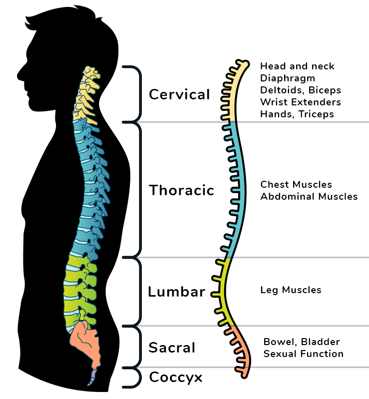
The spinal nerves are part of the peripheral nervous system divided into five regions:
Cranial: ear, eye, frontalis, lips
Cervical: biceps, diaphragm, heart, lungs, nose, tongue
Thoracic: armpit, lungs, rectus abdominis
Lumbar: big toe, knee, intestines
Sacral: bladder, gluteus maximus
Coccygeal:
The spinal nerves are attached to the spinal cord by two roots, the posterior dorsal roots and the anterior ventral roots.
The dorsal root comprises of sensory neurons that carry information towards the brain and spinal cord. A ventral root consists of motor neurons that carry information away from the brain and spinal cord.
The grey matter of the spinal cord is divided into two sections, the dorsal horns and the ventral horns.
The dorsal horn grey matter receives sensory information from the roots and spinal nerves, the grey matter of the ventral horn contains motor neurons that send signals for movement through the ventral horns.
The grey matter of the spinal cord consists of interneurons. They move signals in both directions and are only found in the central nervous system.
3.03 Peripheral Nervous System
The peripheral nervous system includes nerves throughout the body, which can be divided further into the autonomic and the somatic nervous systems.
Autonomic controls involuntary reactions of the body. It affects the cardiac muscle, smooth muscle, blood vessels, and glands. There are two neurons from the CNS to the effector. It excites or inhibits muscles with a nerve impulse.
Somatic controls voluntary movements and sensory input. It affects skeletal muscles and there is one neuron from the CNS to the effector. Its effective of nerve impulse on muscle is exciting them.
The somatic nervous system is made up of some cranial nerves and all spinal nerves. They include sensory and motor neurons and regulates conscious body movements and reflex.
Receptors in the skin, eyes, mouth, nose, ears, and muscles detect changes in the environment while the effectors are skeletal muscles. These receptors can be classified as either simple or complex.
Simple receptors are small and widely distributed throughout the body, governing sensory info.
Complex receptors govern the five senses of taste, smell, hearing, balance, and vision.
A reflex is an involuntary response of the nervous system to an internal or external stimulus. Does not include the brain. The entire process of the body acting involuntarily involves a reflex arc.
A reflex arc is a set of connected neurons that create a pathway for sensory input to be converted to a physical one. The brain is not used in a reflex arc.
Afferent neurons (sensory) carry signal from sensory organs into the body and to the spinal cord.
Efferent neurons (motor) carry the signal to muscles.
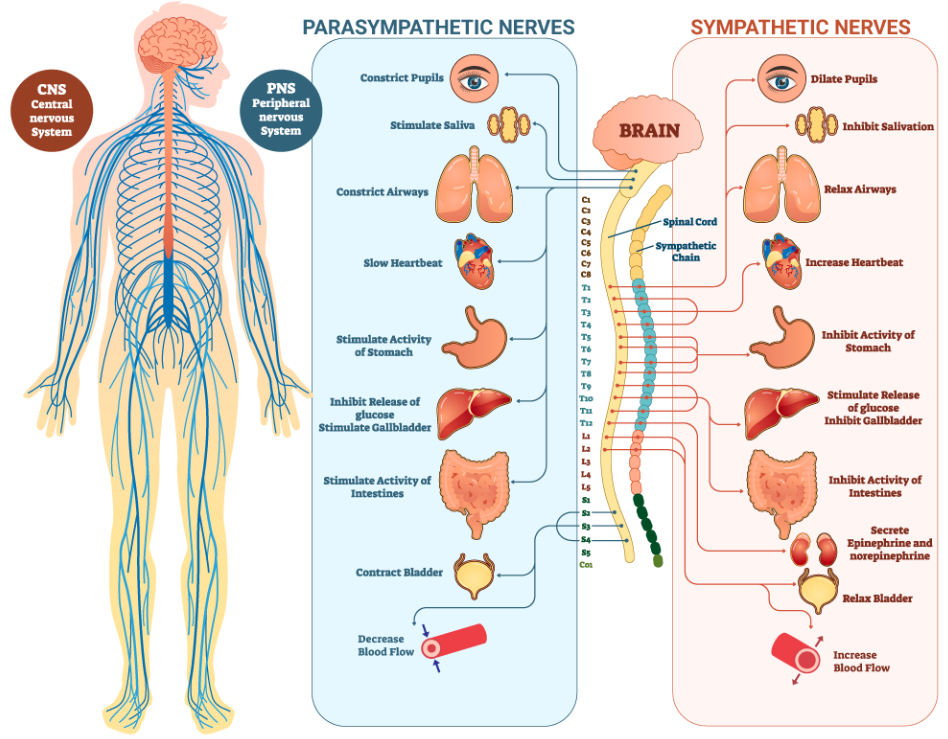
The autonomic nervous system controls voluntary actions of our skeletal muscles, while autonomic controls involuntary actions of cardiac muscles, smooth muscles, and glands. It is divided between sympathetic and parasympathetic.
The sympathetic nervous system dilates the pupil, stimulates sweat secretion, increases heart rate, constricts blood vessels to increase pressure, increases respiration, inhibits gastrointestinal tract, and stimulates breakdown of fat cells.
The parasympathetic nervous system constricts the pupil, decreases heart rate, dilates blood vessels to decrease blood pressure, decreases respiration, and stimulates movement in the gastrointestinal tract.
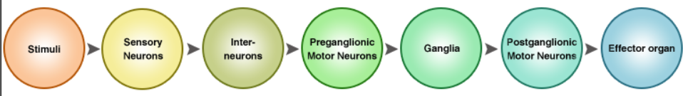
The nerve pathways of the parasympathetic and sympathetic nervous systems consist of one group of sensory neurons and two groups of motor neurons with a ganglia lying in between them. (Ganglia are clusters of cell bodies in the peripheral nervous system)
The first set of motor neurons sit in the CNS. They are preganglionic motor neurons because they come before the ganglia in the nerve pathway. Their nerve fibers extend into the ganglia of the PNS.
The second set are postganglionic motor neurons, extending from the ganglia to the body organs.
In the SNS, the ganglia are grouped in a long chain just outside the spinal cord. The ganglia receive the right emergency impulse from the spinal cord and then the entire body reacts.
In the PNS, the ganglia are located in groups along certain cranial nerves, with the most distinct cranial ganglion referred to as the vagus nerve (central). It transfers impulses to the heart, lungs, digestive tract, liver, and bladder. The PNS does not affect the whole body at once.
A neurotransmitter is a chemical that transfers the nerve impulse from the axon to the end of one neuron to the dendrites of another neuron.
In the SNS, the postganglionic motor neurons release the norepinephrine. The usage depends on the tissue, it can excite or inhibit organ functions.
Guillain-Barre Syndrome is an autoimmune condition where the PNS is attacked, provoked by illness or stress. In mild cases, the patient will have severe pain or limited use of a part of the body.
Diabetes is not a disorder of the PS, but it can cause nerve damage due to having too much glucose in the blood. The damage can cause numbness or paralysis, and the damage can require amputation as blood can no longer circulate.
3.04 Neuromuscular Junctions
Here are clean, organized, study-ready notes summarizing everything shown in the screenshots.
Neural Circuits – Notes
Types of Neurons
Sensory neurons – carry information to the CNS.
Interneurons – connect neurons within the CNS.
Motor neurons – carry signals from the CNS to muscles or glands.
Neural Pathways
Neurons send electrical impulses called action potentials.
These impulses travel down axons and jump between neurons.
Pathways work like communication roads—messages travel quickly from one area to another.
Nerve Conduction (How a Neuron Sends a Signal)
Resting Potential
Inside of neuron = slightly negative.
Outside of neuron = positive.
Maintained by sodium–potassium pumps:
Pump out 3 Na⁺ for every 2 K⁺ brought in.
This charge difference is the membrane potential.
Generating a Nerve Impulse
When stimulated, Na⁺ channels open → Na⁺ rushes in.
Inside becomes positive → depolarization.
After the spike, K⁺ channels open → K⁺ leaves cell → brings charge down again (repolarization).
A brief refractory period follows before neuron returns to rest.
Characteristics of Action Potentials
All-or-nothing.
Travel in one direction.
Extremely fast (up to ~100 m/s).
Steps of a Nerve Impulse (Theory in Action)
Step 1: Resting Potential
Neuron is negatively charged inside.
Stimulus opens Na⁺ channels → incoming sodium depolarizes the membrane.
Action Potential Graph Highlights
Threshold must be reached to trigger an action potential.
Depolarization: voltage spikes upward due to Na⁺ influx.
Repolarization: voltage drops due to K⁺ exiting.
Refractory period: neuron briefly hyperpolarized.
Returns to resting state.
Neurotransmission
Neurotransmitters
Chemicals that pass signals across synapses.
Examples: dopamine, norepinephrine, acetylcholine.
Synapse
Gap between axon terminal of one neuron and another cell (neuron or muscle).
How Signal Crosses a Synapse
Action potential reaches axon terminal.
Calcium channels open → Ca²⁺ enters.
Ca²⁺ causes synaptic vesicles to fuse with membrane.
Vesicles release neurotransmitters into synaptic cleft.
Neurotransmitters bind to receptors on next cell.
Neuromuscular Junction (NMJ)
Process of Muscle Activation
Action potential reaches motor neuron terminal.
Ca²⁺ flows into terminal.
Synaptic vesicles release acetylcholine (ACh).
ACh binds to motor end plate receptors on muscle membrane.
Ion channels open → creates end plate potential.
Muscle membrane generates its own action potential.
Muscle contracts.
Stopping the Signal
ACh is broken down by acetylcholinesterase to prevent continuous muscle contraction.
Nerves in Action – Effects of Substances
Caffeine increases synaptic transmission → alertness.
Drugs like LSD, mescaline, amphetamines can disrupt synaptic function → long-term health issues.
Key Structures and Definitions
Synaptic vesicles – store neurotransmitters.
Calcium channel – allows Ca²⁺ entry to trigger neurotransmitter release.
Axon terminal – releases neurotransmitters.
Synaptic cleft – gap between neurons or between neuron & muscle.
Neurotransmitter – chemical messenger.
Motor end plate receptor – on muscle fiber, binds ACh.
Slide Images (Microscopy Notes)
Neuromuscular Junction 10X & 40X
Shows where a nerve fiber meets muscle tissue.
Visible structures: axon, axon terminal, neuromuscular junction.
Here are clear, organized, and comprehensive notes from all the endocrine system screenshots you provided.
Endocrine System – Study Notes
Overview
The endocrine system works with the nervous system to maintain homeostasis.
It regulates:
metabolism
growth & development
reproduction
sleep cycles
heart rate
digestion
Uses hormones, which travel through the bloodstream to target cells.
Endocrine vs. Exocrine
Endocrine
Releases hormones into the bloodstream.
Controls long-term processes like growth, reproduction, metabolism.
Exocrine
Uses ducts to send secretions to external surfaces (ex: sweat, saliva, tears).
Does not release products into the blood.
Major Endocrine Glands & Their Functions
Hypothalamus
Location: near center of brain.
Function: links nervous & endocrine systems.
Releases hormones that control the pituitary.
Regulates:
hunger
thirst
temperature
sleep
mating & sexual behavior
water & salt balance
Pituitary Gland
“Master gland,” controlled by hypothalamus.
Has two parts: anterior & posterior.
Anterior pituitary hormones:
Growth hormone (GH): bone & muscle growth
Endorphins: reduce pain perception
Controls many other glands.
Pineal Gland
Releases melatonin.
Controls sleep cycles and daily rhythms.
Thyroid Gland
Releases:
Thyroid hormone (T3, T4) – metabolism, growth, development
Calcitonin – lowers blood calcium
Parathyroid Glands
Release parathyroid hormone (PTH).
Raises blood calcium levels.
Opposite function of calcitonin.
Thymus
Produces thymosin.
Develops/activates lymphocytes (immune function).
Pancreas
Produces:
Insulin – lowers blood glucose
Glucagon – raises blood glucose
Kidneys
Release:
Renin – regulates blood pressure
Erythropoietin – increases red blood cell production
Adrenal Glands
Two regions: cortex & medulla.
Cortex releases:
Cortisol – increases blood sugar during stress
Medulla releases:
Epinephrine & norepinephrine – fight-or-flight, boosts energy
Gonads
Ovaries produce estrogen & progesterone → female sex characteristics, reproduction.
Testes produce testosterone & androgens → male sex characteristics.
Summary Table (Condensed)
Gland
Hormone(s)
Function
Hypothalamus
releasing & inhibiting hormones
Controls pituitary
Pituitary
GH, endorphins
Growth, pain reduction
Pineal
melatonin
Sleep cycles
Thyroid
T3, T4, calcitonin
Metabolism; lowers blood calcium
Parathyroid
PTH
Raises blood calcium
Thymus
thymosin
Immune function
Pancreas
insulin, glucagon
Regulates blood glucose
Kidneys
renin, EPO
BP & RBC production
Adrenal
cortisol, epinephrine, norepinephrine
Stress responses
Gonads
estrogen, testosterone
Reproduction & sex traits
Endocrine vs. Nervous System
Nervous System
Uses electrical impulses.
Fast, immediate response.
Short duration.
Endocrine System
Uses chemical hormones.
Slower to act.
Longer-lasting effects.
Link Between the Two
The hypothalamus connects them.
Nervous system can trigger hormone release (ex: fight-or-flight).
Hormone Control Mechanisms
1. Neural Control
Nervous system directly stimulates glands.
Sympathetic system → adrenal medulla → releases epinephrine & norepinephrine.
Used for fight-or-flight or rest-and-digest adjustments.
2. Hormonal Control
One gland releases a hormone that controls another gland.
Example:
Hypothalamus → TRH
Pituitary → TSH
Thyroid → T3/T4
Uses negative feedback to maintain hormone balance.
3. Humoral Control
Glands respond to levels of substances in blood (ions, glucose).
Example:
High blood glucose → insulin released
Low blood glucose → glucagon released
Common Endocrine Disorders
Hyperthyroidism
Too much T3/T4.
Symptoms:
High metabolism
Rapid heartbeat
Anxiety
Weight loss
Eye problems
Hypothyroidism
Too little T3/T4.
Symptoms:
Low metabolism
Weight gain
Depression
Hair loss
Fatigue
Goiter may develop
Diabetes
Body doesn't make enough insulin or doesn't use it properly.
Results in high blood glucose.
Effects:
Kidney issues
Nerve & eye damage
Fatigue
Organ damage
Here are detailed, clear, and organized notes on all the information in the hormone-types screenshots.
Hormone Types – Study Notes
Hormones regulate:
growth
development
metabolism
homeostasis
reproduction
There are two major types of hormones:
Steroid hormones
Nonsteroid hormones
1. Steroid Hormones
Chemical Nature
Made from cholesterol (a lipid/fat).
Fat-soluble (hydrophobic).
How They Enter Cells
Easily pass through the lipid bilayer of cell membranes.
Bind to receptor proteins inside the cytoplasm or inside the nucleus.
How They Work
Hormone enters cell.
Binds to internal receptor.
The hormone–receptor complex moves to the nucleus.
It attaches to DNA and turns genes on or off.
A new protein is produced from mRNA.
Protein changes the cell’s activity.
Effect Profile
Slow-acting but long-lasting.
Strong effects because they change gene expression.
Examples
Testosterone
Estrogen / Estradiol
Progesterone
Cortisol
Aldosterone
Vitamin D (hormone form)
Glands That Produce Them
Ovaries
Testes
Adrenal cortex
Skin/kidneys (vitamin D processing)
2. Nonsteroid Hormones
Chemical Nature
Made from proteins, peptides, or amino acids.
Water-soluble (hydrophilic).
Cannot pass through the lipid bilayer.
How They Enter Cells
Bind to receptors on the outside surface of the cell membrane.
How They Work (Second-Messenger System)
Hormone (first messenger) binds to cell surface receptor.
Activates an enzyme on membrane.
Enzyme converts ATP to cAMP (second messenger).
cAMP activates proteins (protein kinase).
These proteins cause changes inside the cell.
Produces cellular response.
Effect Profile
Fast-acting but shorter-lasting than steroid hormones.
Do NOT change DNA directly.
Examples
Insulin
Growth hormone
TSH (thyroid-stimulating hormone)
Adrenaline/Epinephrine
T3 and T4 (technically amine hormones)
Glands That Produce Them
Pancreas
Pituitary gland
Thyroid gland
Adrenal medulla
Steroid vs. Nonsteroid – Summary Table
Feature
Steroid
Nonsteroid
Solubility
Fat-soluble
Water-soluble
Cross cell membrane?
Yes
No
Receptor location
Inside cell (cytoplasm/nucleus)
On cell membrane
Mechanism
Alters DNA → protein synthesis
Second-messenger system (cAMP)
Speed
Slow
Fast
Duration
Long
Short
Examples
Testosterone, estrogen, cortisol
Insulin, GH, adrenaline
How Hormones Find Target Cells
Hormones travel through blood until they randomly encounter the correct receptor.
Target cells have specific receptors (lock-and-key model).
Once bound, chemical reactions occur:
change pH
activate/deactivate genes
start metabolic pathways
trigger hunger, growth, etc.
Synthetic vs. Natural Hormones
Natural Hormones
Produced by the body.
Synthetic Hormones
Lab-made versions used for medical treatment.
Designed to mimic natural hormones.
Examples of Natural vs. Synthetic
Estrogen
Natural: Estradiol
Synthetic: Ethinylestradiol
Used in birth control and hormone therapy.
Growth Hormone
Natural: Somatotropin
Synthetic: Somatrem
Used for growth disorders or GH deficiency.
Why Synthetic Hormones Are Used
Replace missing hormones.
Treat hormone imbalance disorders.
Common uses:
Birth control
Menopause treatment
Thyroid replacement
Growth hormone deficiency
Risks and Side Effects of Synthetic Hormones
Some synthetic estrogens/testosterones may increase:
blood clotting risk
certain cancers
Overuse of synthetic GH can be harmful.
Must be used under medical supervision.