Histology - tissues I
Histological technique
LSMU’s laboratory uses formalin-fixed samples that are embedded into paraffin.
- Fixation
Chemical processes in which tissues are preserved from disintegration and prepared for further processing.
Fixed in 10% formalin solution.
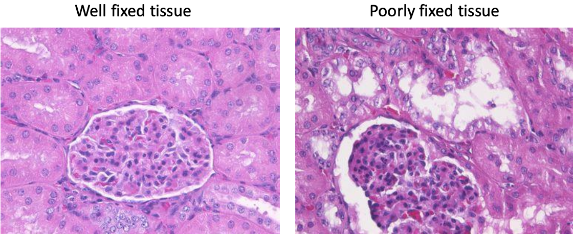
Factors determining fixation quality.
- Volume of fixation (10-20 times of the tissue volume)
- Sample size (fixative must be absorbed into all layers of the sample)
- Permeability of fixative (formaldehyde has fastest penetration 1mm/h)
- Start and duration of fixation time (immediately placed in fixative and stored 24h)
- Temperature (18-24 Celsius)
- Acidity (higher acidity 🡪 faster reaction, optimal pH 6-8).
- Removal of fixative material
- End of fixation, fixative is removed from tissue by washing in running water.
- Histological cartridges with date, name and number to avoid confusion.
- Dehydration
- Removal of free water because hydrophobic substances (paraffin/resin/waxes) will be used later.
- Ethyl alcohol, methanol, isopropyl alcohol, acetone
- Tissue dehydrated by increasing solution concentration 70% solution 🡪 80% 🡪 96%.
- Clearing
- Remove dehydration substance from tissue.
- Xylene, toluol or chloroform
- Refractive index of substances is close to protein index, which affects tissue making it translucent/transparent.
- To short clearing, may influence preparation: poorly impregnated, remain soft, fluffy and unsuitable for making thin incisions.
- To long clearing, the tissue solidifies, and protein desaturates.
- Impregnation
- With stiffening substances (paraffin), allowing the structures to be preserved ones cut thin.
- Preparation of paraffin sections
- Chilled paraffin block is cut by a microtome (a twisting handle) to 1-10 micrometers.
- Sections are placed in warm water and straightened, then placed on a glass and dried in a thermostat.
- Staining
- In laminar air flow chamber
- Different staining methods give rise to different highlighted structures.
- Golden standard: hematoxylin (pink-violet), eosin (blue)
- Xylene to remove paraffin.
- Rehydration with alcohol solution of decreasing concentration
- Stained 🡪 washed with running water 🡪 dehydrated 🡪 cleared with xylene.
- Histological glue on covering glass is applied.
Diversity of human cells
What to look for:
- Shape of cell
- Nucleus position
- Special features
- Location to other cells
Artifacts should be ignored.
- Folds
- Thick cuts
- Nucleus out of focus are not part of the focused cell.
- Different shape
- Reside between two cells.
Factors which determine morphology
- Environment
- Functions
- cytoskeleton
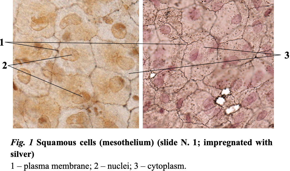
- Squamous cells/mesothelium/Epithelial tissue
- Flat cells
- Oval, central, middle nucleus
- Scale-like appearance
- No special features
- Reside side by side with other cells.
Avascular! Get their nutrition from connective tissue.
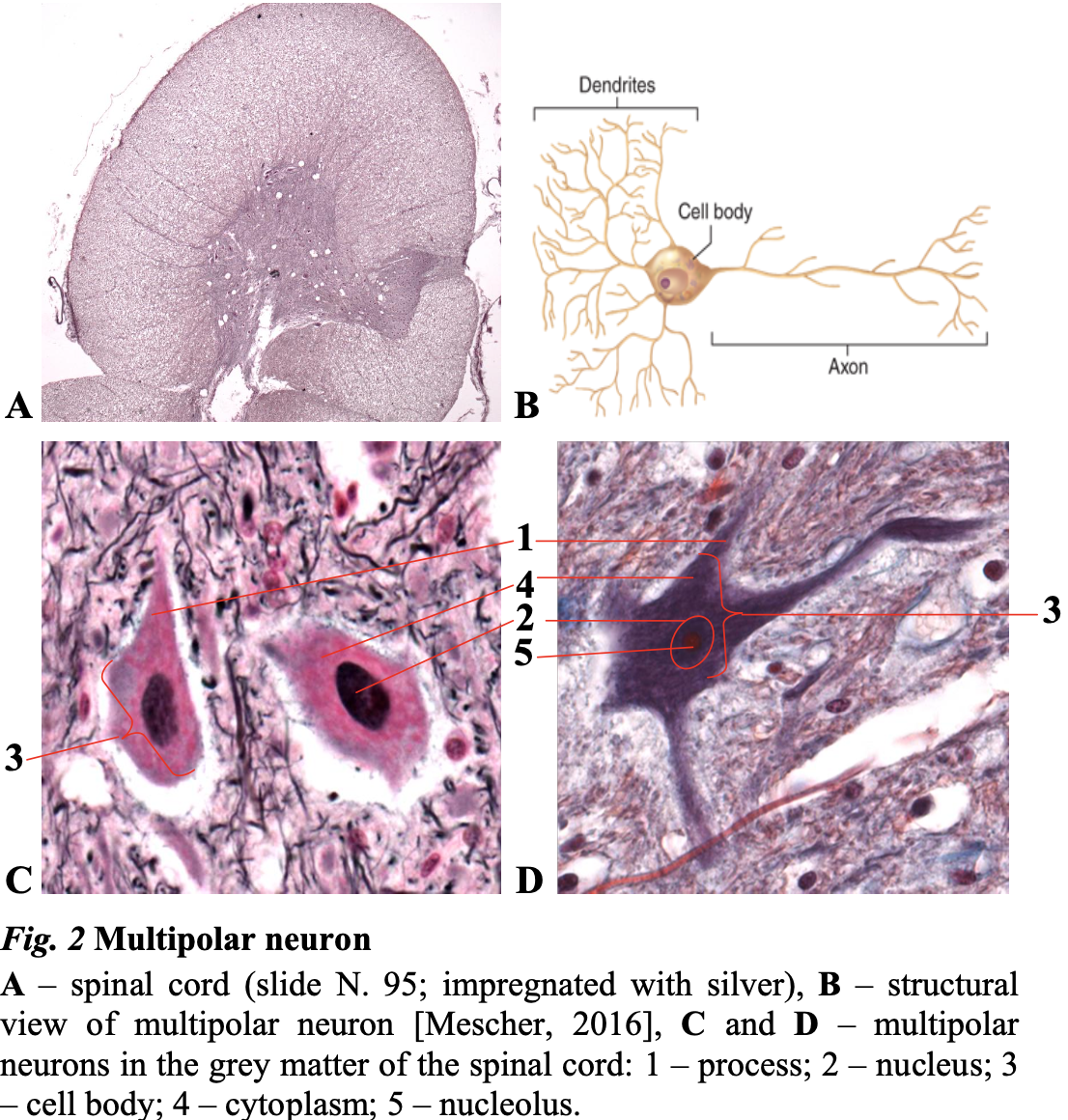
- Multipolar neurons
- Single axon extending from one end of the cell body.
- Several dendrites protruding from the other side of the cell body.
- Processes are either dendrites or axons.
- Posterior horn: Larger neurons
- Anterior horn: More in number
- Branched, oval cells.
- Large, oval, central nucleus with nucleolus
- Connections are not seen.
- Outer connective tissue is Neuropil with ganglia or other visible processes.
- Striated skeletal muscle fibers.
- Cylindrical shape
- Blunt ends
- Long
- Many nuclei peripheral
- Striations
- Vertically: y-band is lighter, A-band is darker
- Laterally: myofibrils
- Connective tissue
Cross-section
- Only one nucleus is seen.
- Myofibrils
- Cytoplasm is whiteish.
- Number of myofibrils determines blueness.
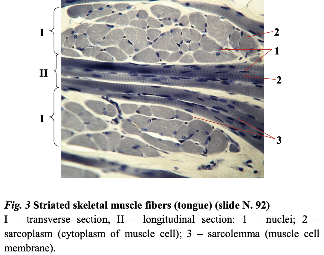
The tongue has striated skeletal muscle – epithelial tissue and connective tissue.
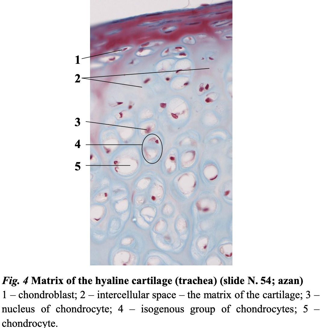
- Matrix of hyaline cartilage
- Fibers made up off type II collagen.
- Chondroblast/chondrocyte:
- Oval cells
- Oval peripheral nucleus
- Resides in clusters in even number: isogenous group.
- Territorial matrix in between cells in the clusters
- Interterritorial matrix is between cells.
- White cytoplasm because of lipids
- Connective tissue cells
- Maintain the formation of cartilage.
- Spherical cells
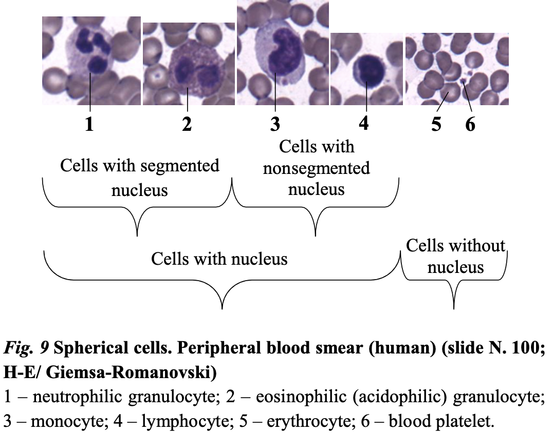
- Erythrocytes
- No nucleus
- Squamous center
- Biconcave
- 7,5 micrometers
- Most abundant
- Platelets
- Small
- Not cells
- Leukocytes
- Granulocytes:
- Basophils: bluer and granular
- Eosinophils: violet-pink-reddish, 2 segments
- Neutrophils: 2-5 segments, granulated, 15- 20 micrometers. Round
- Agranulocytes
- Monocytes: largest, round, horseshoe nucleus, 20-24 micrometers
- Lymphocytes: round, large nucleus, 8-11 micrometers
- Granulocytes:
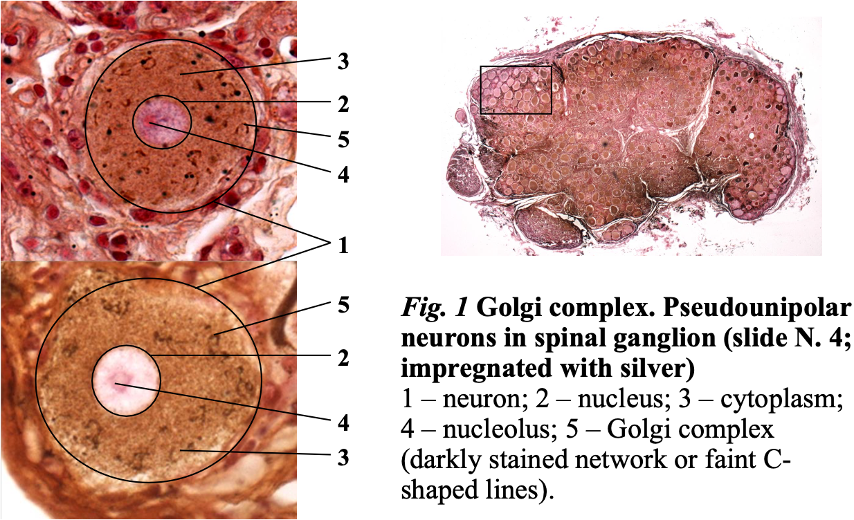
- Pseudounipolar neurons in spinal ganglion
Stained with silver salt 🡪 highlights membranous organelles, C-shaped lines are golgi
- On dorsal root ganglion
- Has one axon that projects a short period before splitting into two branches.
- Dark ganglion neurons: poor in neurofilaments
- Clear ganglion neurons: rich in neurofilaments
- Golgi complex is usually dark lines, C-shaped. This is seen because they produce neurotransmitters.
- Hepatocytes
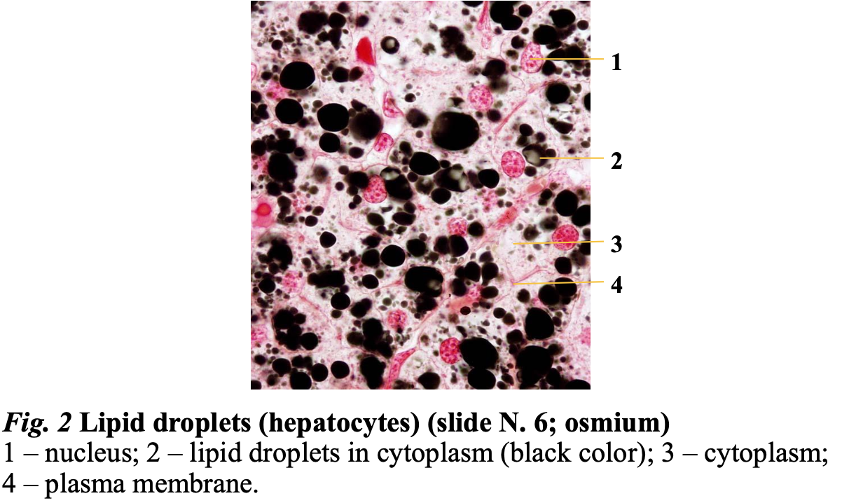
Freezes in cryostat and stained with osmium.
- Lipid droplets, various in size
- polygonal cells (20-30µm), usually diploid cells arranged in lines.
- abundant granular eosinophilic (pink) cytoplasm,
- centrally placed round to ovoid nuclei,
- prominent nucleoli
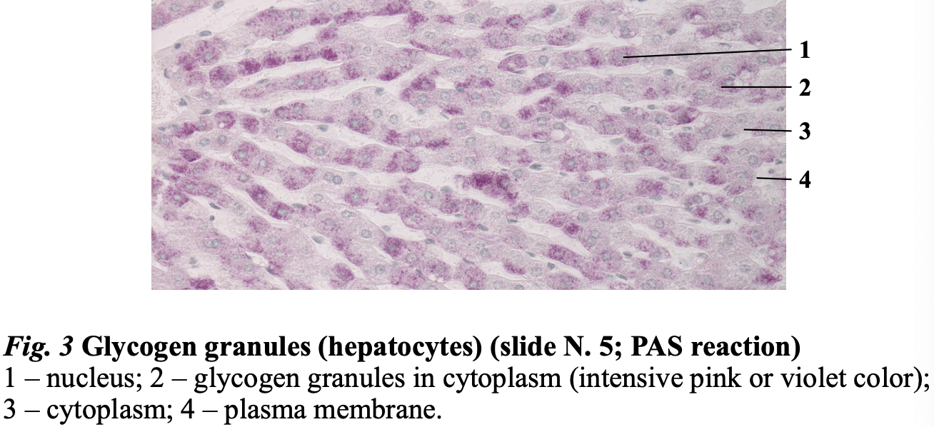
- Glycogen granules are the glucose storage. = nutritional inclusions
- Glycogen is shown with a PAS reaction.
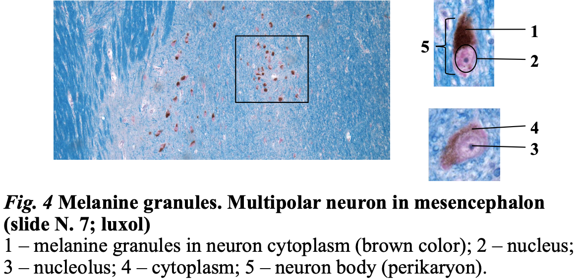
- Multipolar neuron in mesencephalon
- Melanin granules: dark brown pigment in dopaminergic neurons.
- Melanin granules, no staining needed.
- These neurons produce dopamine and then melanin is seen.
Organelles
Membranous:
- Nucleus
- ER
- Golgi
- Peroxisome
- Endosome
- Mitochondria
- Lysosome
Non-membranous:
- Ribosomes
- Centrioles
- Cytoskeleton
Cytoplasmic inclusion
- Nutritional inclusions
- Secretory inclusions
- Pigmental inclusions
- Endogenous (cells make melanin themselves)
- Exogenous (From the outside)
- Crystal inclusions, usually in cells that suffer a pathology, but Leydig cells have this.
Epithelial tissue
Tissue: an assemblage of similarly specialized cells united in performing a specific function
- Epithelial tissue
- Connective tissue
- Muscle tissue
- Nerve tissue
Lining of GI tract organs and other hollow organs and skin surface (epidermis)
Originates from all three germinal layers.
- Ectoderm: epidermis, sweat gland,
- Mesoderm: mesothelium
- Endoderm: Airways
Covering/lining group 🡪 simple or stratified
- Simple: mesothelium, endothelium
- Stratified: Epidermis
Glandular epithelium 🡪 exocrine glands or endocrine glands
- Exocrine: salivary glands
- Endocrine: thyroid or hypophysis
Pigmented,
spermatogenic,
sensory epithelium.
Properties
- Strictly polarized: Apical and basal poles and lateral surface
- Apical pole: covered in specialized structures, microvilli, stereocilia, cilia.
- Cells are always separated from other cells by basement membrane.
- Abundant junctions with other epithelial cells and with basement membrane
- Is avascular!
- Good regeneration, except sensory epithelium
Basement membrane
- Supports epithelial cells.
- Forms barrier, between epithelium and other tissues.
- Works as filter, not all molecules can pass.
- Composed of basal and reticular laminas.
- Basal: closer to the epithelium made of:
- Lucida: type IV, XVII collagen, integrins, laminins (Closest to epithelial cells)
- Densa: heparin, IV collagen, entactin
- Reticular: I, III, V collagen, proteoglycans, (closer to connective tissue: made of connective tissue cells) (closest to fibroblast)
- Basal: closer to the epithelium made of:
- Also composed of water!!
- No cells in basement membrane!!!
Intercellular junctions
- Hemidesmosomes
- cell to basement membrane with integrin and cytoskeleton (keratin filaments)
- Gap junctions
- not connected to cytoskeleton but have transmembrane proteins and allows transfer of small molecules between cells.
- Desmosomes:
- transmembrane proteins and have cytoskeleton (Keratin filaments), couple adjacent cells and strengthens tissue.
- Adherent junctions:
- transmembrane proteins and cytoskeleton filaments (actin filaments) and link cytoskeleton of cells and strengthen tight junctions. Not so strong as desmosomes
- Tight junctions: not all have this. Transmembrane proteins and actin filaments, seal adjacent cells and control molecule passage. Large molecules, bacteria can´t travel between cell membranes.
Specialized structures of apical pole
- Microvilli
- An extension of cytoplasm with cytoskeleton in them
- Increase surface area.
- Stereocilia
- huge microvilli,
- increase surface area.
- Cilia
- Motor proteins which can move this apical structure.
- Moves something.
General Functions
- Protect deeper tissue.
- Transports molecules and ions
- Secrete enzymes, mucus, hormones.
- Absorbs substances.
- Receives information from environment.
- Expels secretion of glands
Naming
Number of layers (simple or stratified) 🡪 cell shape (which is dominating in simple, and which is in the apical part in stratified?) 🡪 cell properties (has cilia or keratinized?) 🡪 Epithelium
Exception:
- Transitional epithelium
- Pseudostratified epithelium
- Simple squamous epithelium (the name does not clarify the function)
- One layer
- Squamous
- Irregular shape
- Alveoli
- Inner surface of cornea
- Hearth chambers
- Endothelium
- Mesothelium: peritoneum, pleura, pericardium
- Simple cuboidal epithelium
- One layer
- Basement membrane
- Bronchi
- Ovaries surface
- Excretory salivary gland ducts
- Kidney tubules
- Simple columnar epithelium
- One layer
- Basement membrane
- Can have microvilli, cilia or stereocilia.
- Mucosa of stomach and intestine
- Mucosa of gallbladder
- Mucosa of uterine tube and uterus
- Pseudostratified columnar ciliated epithelium
- Basement membrane and 4 types of cells
- All cells touch the basement membrane so one layer.
- Basal cells (basement membrane)
- Elongated cells
- Columnar cells (cilia which removes the debris into the mouth so we can cough or swallow)
- Goblet cells (secret mucus in the airways)
- Mucosa of airways
- Part of urethra
- Ductus deferens
Stratified epithelium
Only basal cells are attached to the basement membrane and the superior cells are attached by adherent junctions or desmosomes to other cells.
- Stratified squamous nonkeratinized epithelium (in all moist places)
- Basal layer (cuboidal or columnar)
- Intermediate layer (oval)
- Surface (squamous)
- Oral cavity
- Vagina
- Esophagus
- Cornea
- Anus
Lactobacteria in vagina converts glycogen (produced by epithelial cells) to lactic acid which lower pH to protect from other bacteria. Moistness and sugar attract bacteria.
- Stratified squamous keratinized epithelium (epidermis) Found on all the skin.
- Basement membrane and 5 layers (thick skin. hairless)
- Or basement membrane 4 layers (thin skin, hairy skin)
- Stratum basale
- keratin filaments
- Stratum spinosum
- polygonal cells
- Stratum granulosum
- squamous cells, few layers, start to lose organelles.
- Stratum lucidum
- not if there only are 4 layers.
- no organelles but keep the junctions.
- Stratum corneum
- thickest layer, with dead cells: keratinocytes.
- Start to lose their junctions and filled with proteins. The cells become a squam.
- They are impermeable and prevents water loss, protects from mechanical things and sun. No visible nucleus.
- Transitional epithelium
- Composed of basement membrane and basal and surface layers.
- Surface layer with specialized cells called umbrella cells.
- Prevent other cells from all harmful substances in the urine.
- When the organ extends 🡪 the cell layers will decrease because the epithelium also extends.
- Calyces of kidneys
- Ureters
- Bladder
Sensory epithelium
- Inner ear, tongue, olfactory epithelium
- Receive, convert and pass along information.
Spermatogenic epithelium (stratified)
- Testes – seminiferous tubules
Pigmented epithelium
- In retina to prevent light to affect deeper tissue, and bounce light to the receptor.
- Simple cuboidal epithelium which are pigmented
Glandular epithelium
- Endocrine (secrete hormones into the blood stream)
- Exocrine (have excretory ducts that go to the surface of something)
- Unicellular
- Goblet cell:
- In airway and intestine
- Merocrine/apocrine
- Secretes mucus.
- Microvilli
- Basal pole resides organelles and nucleus
- Secretory granules on the apical pole
- Goblet cell:
- Multicellular
- Simple glands (one duct + secretary portion)
- Compound glands (duct with branches + secretory portion)
- Secretory portion can be tubular, acinar, tubuloacinar
- Unicellular
- Merocrine: secretes but does not affect the cell
- Apocrine: secretes and loses part of the cytoplasm and plasma membrane
- Holocrine: disintegrates upon secretion
Connective tissue
- Fat and other soft padding tissue
- Bone
- Tendon
- Blood
- Cartilage
Classification
All connective tissue is derived from the embryo mesenchyme.
- Adult
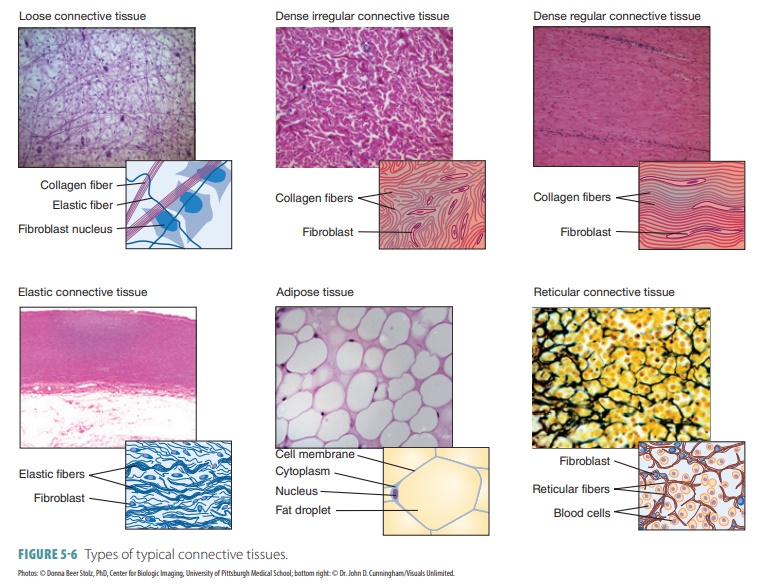
- Fibrous (proper): in between the cells and structures without functions, “the one we imagen”.
- Collagen
- Loose
- Dense
- Regular
- Irregular
- Elastic
- Collagen
- Specialized
- Blood & lymph
- Adipose
- White
- Brown
- Pigmented
- Reticular
- Skeletal
- Cartilage
- Hyaline
- Elastic
- Fibrous
- Bone
- Lamellar
- Woven
- Cartilage
- Fibrous (proper): in between the cells and structures without functions, “the one we imagen”.
- Embryo
- Mesenchyme
- Mucous
Connective tissue contains extracellular matrix and cells.
Extracellular matrix
- Ground substance (watery/gelatinous thing)
- Glycosaminoglycans (GAGs)
- Long chains of disaccharides longest is hyaluronan.
- Cells synthesize them directly and secrete them.
- Proteoglycans
- Chains of amino acids
- Synthesized in ER and get connected to GAGs in Golgi complex.
- Glycoproteins
- Strengthen and stabilize tissue and help cell migration thanks to areas which can join to other molecules.
- Glycosaminoglycans (GAGs)
- Fibers:
- collagen
- elastic
- reticular
Classification of fibers
- Fibrillar: type I, II, III collagen 90% of the human body
- I:
- bone tissue
- Fibrocartilage
- Pigmented CT
- Fibrous CT
- II:
- Cartilage
- III:
- Fibrous CT
- Glands
- Immune system
- Bone marrow
- I:
- Network/sheet forming: IV collagen.
- Linking/anchoring: Type VII collagen
Collagen (protein) is synthesized by cells (fibroblasts)
Fibers form only in ECM.
Reticular fibers
- III collagen
- Synthesized by reticular cells and fibroblast.
- Found in immune system and hematopoietic.
- Seen with silver salt staining.
Elastic fibers
- Composed of elastin and fibrillin (fibroblasts)
- Gives elasticity.
- Found in organs that change volume (lungs).
- Can form elastic membranes.
Cells in connective tissue proper
- Resident (true cells)
- Fibroblast, young cell 🡪 fibrocytes
- Actively synthesizes ECM components (build)
- Responsible for growth factors
- Oval euchromatic nucleus
- Developed ER and Golgi
- Fibrocytes
- Does not actively synthesize (does not build)
- Nucleus is tapered, heterochromatic.
- Can reverse to active form (fibroblast) when tissue is injured.
- Reticular cells
- In reticular connective tissue only synthesize type III collagen
- Partially cover fibers with their cytoplasmatic processes
- Pericytes
- Star shaped cells and can contract.
- Next to capillaries
- Mast cells
- Irregular shape
- Central, oval nucleus, not visible
- Basophilic granules
- Synthesize bioactive substances for immune response, inflammation and healing.
- Close to blood vessels and mucosa of organs
- Adipocytes
- In majority of organs
- Accumulate and storage lipids.
- Large groups are called adipose tissue.
- Pigmented cells (melanocytes)
- Fibroblast, young cell 🡪 fibrocytes
- Transient (traveling cells) All derived from blood.
- Granulocytes do not differentiate.
- Lymphocytes 🡪 plasma cells (look like monocytes)
- Derived from activated B lymphocytes by antigens.
- Produce immunoglobulins.
- 10 – 20 days life
- Cell is oval and heterochromatin clumps are visible in nucleus.
- Monocytes 🡪 macrophages
- Can phagocytose.
- Shape depends on activity.
- Mononuclear phagocytes system
Embryonic connective tissue
- Mesenchyme
- Mesoderm and partially from ectoderm
- Origin of all adult connective tissue
- Mesenchyme cells
- Mucous
- Found in umbilical cord.
- Does not found in adult because placenta is gone.
- Protects blood vessels of umbilical cord from strangulation due to movements.
- Mucocytes
- Hyaluronic acid in the ground substance attracts water.
Proper
Elastic connective tissue
- Elastic fibers prevail.
- Gaps can be filled with collagen, fibroblasts and fibrocytes.
- Ground substance is scarce.
- In ligaments and large arteries
- Fibrils are arranged in order.
Loose connective tissue
- Plenty of ground substance
- In submucosa of hollow organs and found almost everywhere, blood vessels.
- Fibroblasts prevails.
- Fibrils have no order.
Dense connective tissue
- Tons of densely packed tissue
- Ground substance and cells are scarce.
- Fibrocytes predominates.
- Irregular (no orderly arrangement)
- In dermis
- Regular (orderly arranged fibers)
- In tendons
- Irregular (no orderly arrangement)
Specialized connective tissue
- Reticular tissue
- Reticular cells
- Ground substance
- Reticular fibers (III collagen)
- Found in immune system and hematopoietic organs.
- Adipose tissue
- White
- Unilocular adipocytes and reticular fibers network
- Solitary cells are spherical.
- Nucleus and organelles are in the periphery.
- Secretes hormones (thermo-insulation) and pads, palms and soles.
- In adults
- Brown
- Small cells, polygonal, oval nucleus is central.
- Has lipid droplets in cytoplasm (multilocular)
- Have many mitochondria.
- Found in newborns vital organs.
- Well vascularized
- Production of heat which keeps the newborns temperature.
- White
- Pigmented tissue
- Variety of loose connective tissue (I, III collagen fibers, fibrocytes and fibroblasts + melanocytes)
- Irregular shaped cells with many branches and dark color, found in the choroid, iris and genital skin after puberty.
- Blood
- Composed of cells and extracellular matrix (plasma)
- Erythrocytes + leukocytes
- Granulocytes and agranulocytes
- Platelets (cytoplasmic processes of other cells, not cells)
- Red blood cells and platelets are never in the connective tissue but stay in the blood vessels.
- Neutrophils, secrete granules, segmented 2-5,
- Eosinophils have two segments, participate in allergic reactions and parasitic infections.
- Basophils have two segments but have high number of granules and that is why segments are not see. Allergy
- Lymphocytes
- T 🡪cytotoxins against cancers
- B 🡪 secrete immunoglobulins
- Monocytes travel to tissue and becomes macrophages which phagocytose.
Blood
Belongs to specialized connective tissue.
- 7-8% of body mass
- Erythrocytes 44%
- Leukocytes + Blood platelets <1%.
- Leukogram
- Neutrophils 50-70%
- Lymphocytes 20-40%
- Monocytes 2-8%
- Eosinophils 1-4%
- Basophils 0.5-1%
- Leukogram
- Plasma 55%
- Water 92%
- Proteins 7%
- Other solutes 1%
Hematocrit is percentage of red blood cells volume (40% women, 45% men)
Bright red is oxygenated.
Dark red is unoxygenated.
Denser than water
pH 7.35-7.45
Typical volume 4-6L
Functions:
- Distribution and transport of different materials
- O2 lungs 🡪 cells
- CO2 from cells 🡪 lungs
- Nutrients from GI
- Hormones
- Wastes from cells to excretory system.
- Regulation (maintenance of homeostasis)
- pH by albumin + bicarbonate
- Temperature
- Protection
- Platelets in coagulation seal vessel damage
- White blood cells
Plasma
- Water
- Albumins, globulins
- Clotting proteins (fibrinogen, prothrombin)
- Enzymes, hormones
- Nutrients
- Electrolytes
Serum
- Fluid that remains after blood clotting, lacks coagulation factors.
- Proteins not used in clotting, electrolytes, antibodies, antigens, hormones, exogenous substances.
Blood smear:
Erythrocytes
- Biconcave disc, thin middle, thick in periphery
- Spectrin allows shape shifting (elastic protein)
- No nuclei
- Few organelles: mainly hemoglobin carrier 33% of body.
- Anaerobic respiration
- 7,5 micrometer diameter
- Count: 4,1 – 6 x 10^12 per liter of blood (m), 3,9 – 5,5 x 10^12 per liter of blood (f)
- Live 120 days, phagocytosed in spleen, bone marrow and liver
Hemoglobin (Hb)
- Oxygen binding capacity
- Iron containing metalloprotein which transport oxygen.
- Bonds O2 to form oxyhemoglobin.
- Carries CO2 (30% carbaminohemoglobin), 10% is dissolved in blood plasma.
- 60 % CO2 is carried as bicarbonate buffer
- Carbon monoxide reacts with hemoglobin 🡪 carbon monoxyhemoglobin which displaces O2 on hemoglobin, reducing the oxygen carrying capacity of blood.
Reticulocytes are young erythrocytes with remain of the chromatin.
Anemia
- Deficit of erythrocytes or hemoglobin in periphery blood
- Determine blood count, morphology, Hb concentration.
- Sickle cell anemia: damaged synthesis of hemoglobin and changes shape of red blood cells (autosomal recessive disease), attaches easier to blood vessel wall and can block blood passage.
Leukocytes
- 1% of blood volume
- Count 4-8 x 10^9 per liter
- Granulocytes: Cannot divide and differentiate 20 days life span
- Basophil (dark granules) 0,5 – 1%
- 2-3 lobes nucleus
- 12-15 micrometers
- 6 hours circulation
- Few days lifespan
- Histamine (increase permeability of blood vessels and causes edema), heparin (decreases blood clotting) granules.
- Acts on allergens and produce intense adverse systemic response (anaphylaxis)
- Eosinophil (red/orange granules) 2-4%
- 8-12 days lifespan
- 6-8 hours in blood
- 2 nucleus lobes
- Phagocytosis of antigen - antibody complex and enzymes destroy parasites.
- Eosinophilia: Increase during allergic reaction. Accumulate in tissues and may cause injuries of the cells, chronic inflammation, parasitic infections.
- Neutrophil (47-70%)
- Circulate 8-12 h in blood.
- Diapedesis: Exit to tissue and do not return
- 2-5 nucleus lobes
- Young stab cells increase during inflammation (2-3%)
- Phagocytose bacteria and remains of dead cells.
- When activated they restore tissue microenvironment by releasing chemokines which attract other leukocytes and cytokines.
- Release glycogen to use glucose for energy.
- Infected wounds: Pus is apoptotic neutrophilic granulocytes, degrading bacteria, tissue fluid.
- Neutropenia is an abnormal decrease in number (AIDS, drugs, genetic factors induce)
- 10-12 micrometers
- 1-4 days lifespan in connective tissue
- Female granulocytes show a Barr body (appendix with sex chromatin)
- Primary granules: major role in degrading and killing microorganisms.
- Secondary granules: smaller and less dense
- Basophil (dark granules) 0,5 – 1%
- Agranulocyte
- Lymphocyte 19-37%
- Normally in resting condition
- Rapid response to antigen 🡪 proliferates and differentiate to functional active state.
- Migrate to tissue.
- B-lymphocytes: humoral immune response
- Develop in bone marrow🡪
- Secondary lymph organs (spleen, lymph nodes, tissue)
- Differentiate into:
- B-memory cells (circulate many years and remember antigens)
- Plasma cells (effector cells participate in humoral response) increase in cytoplasm producing immunoglobulins. Peripheral nucleus and more cytoplasm.
- T-lymphocytes: cellular immune response
- Bone marrow
- Mature in thymus.
- Outspread in secondary lymphoid organs.
- Differentiate into:
- Helpers
- B-lymphocytes: humoral immune response
Activate other cells.
- Memory
Live long and remember antigens.
- Cytotoxic (killers)
Primary effector cells, destroy foreign cells.
- Suppressors
Suppress immune response when antigen is removed.
- Assist B-lymphocytes in performing humoral immune response.
- Natural killer cells
- Reside in spleen.
- Circulate in blood.
- Differentiate into:
- Cytotoxic
- Destroy other cells wall infected with virus, cancer.
- Release citokins and modulate immune response.
- Reside in resting state in lymphoid organs and circulation.
- When antigen is present, they will migrate to the particular area and start a immune response.
- Monocyte 3-8%
- Bean shape nucleus
- 12-20 micrometers
- Mitochondria, Golgi, RER granules with enzymes
- 5 days in blood
- In tissue 🡪 macrophages
- 60-90 days
- Accumulate in place of infection.
- Phagocytose
- Release cytokins that help to accumulate forces of the organism to fight infection.
- Interleukins attract lymphocytes, neutrophils.
- Antigen presenting cells to T-lymphocytes.
- Red bone marrow, spleen, lymph nodes, connective tissue: macrophages,
- Langerhans cells: epidermis,
- Lungs: macrophages,
- microglia in nerve tissue,
- Macrophages in synovial joints.
Functions:
- Protection: microbes, toxines, parasites, cancer cells.
- Diapedesis: can slip between capillary wall, reside short time in plasma after they are released from blood marrow.
- Ameboid motion
- Chemotaxis: moving in direction of a chemical
- Leukocytosis: increase number in response to infection
MHC major histocompatibility complex binds to peptide fragments from antigens for recognition by the appropriate T-cells and activate a response.
Differences
- B-cells recognize cognate antigen in native form, recognize free antigen in blood or lymph using membrane bound immunoglobulins.
- T-cells recognize cognate antigen in a processed form, peptide fragment presented by an antigen- presenting cells MHC molecule to the T-cell receptor.
Antigens: genetically foreign material that generate immune response (protein, virus, foreign cells, tumor cells)
Antibodies: proteins, plasma globulins which recognize antigens
- IgA, IgD, IgE, IgG, IgM
Blood platelets
- Originate from giant polyploid cells in bone marrow (megakaryocytes)
- 150 – 400 10^9 per liter
- 10 days life span
- 2-4 micrometers
- Central dark part (granulomere) initiates thromb process
- Hyalomere is brighter and helps degranulation.
Function
- Coagulation by attaching to blood vessel wall collagen in basal laminas.
- Release glycoprotein which induces further platelets to attach.
- Fibrinogen from plasma
- Von Willebrand factor from endothelium
- Platelet factor 4 from platelet granules
- Forms fibrin polymer network
Thrombocytopenia: reduced number in platelets
- Damaged megakaryopoiesis
- Side effect of chemotreatment
- Autoimmune disease
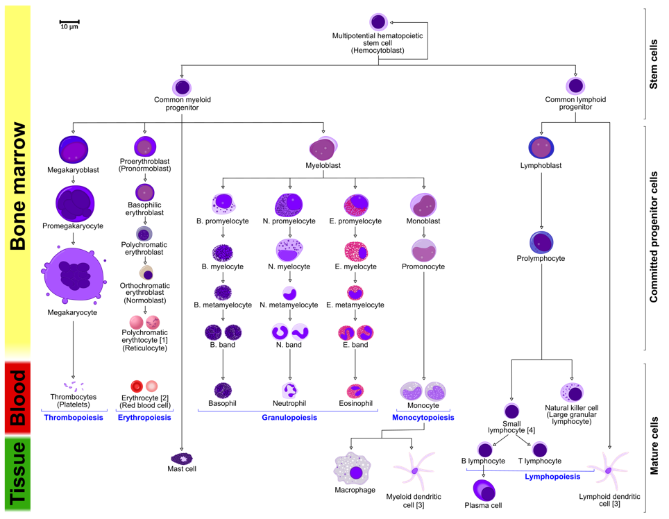
Hemopoiesis
- Mature blood cells have short life span and must be replaced.
- Yolk sac 🡪 spleen + liver 🡪 Red Marrow of specific bones in epiphysis: vertebra, sternum, rib, femur, tibia
Stem cells
- Unspecialized cell which has the ability to differentiate
- Asymmetric division
Properties:
- Proliferation – cell renewal
- Differentiation – develop to specialized cells
- Plasticity: ability of stem cells from one type of tissue to transform into stem cells of other tissue
- Lineage of stem cells: that retain ability to divide in vitro for long period without differentiation into specialized cell types.
In bone marrow are pluripotential hematopoietic stem cells
- Cell renewal and differentiation
Stem cells differentiate into:
- Lymphocytic stem cells
- Multipotent myeloid stem cells
- Give rise to major groups of blood cells (except lymphocytes)
- Burst-forming unit: erythrocytes
- Colony-forming unit: megakaryocytes
- Give rise to major groups of blood cells (except lymphocytes)
Bone marrow:
- Stroma: reticular connective tissue
- Modified fibroblasts 🡪 reticular cells
- Osteoblast
- Reticular fibers
- Parenchyma: hematopoietic cells arranged in irregular cellular cords or islands separated by thin-walled nervous sinusoids
- Red: active hematopoietic tissue
- Yellow (white):
Autologous transplantation from same patient
Allogenic transplantation from another patient
Erytrocytopoiesis is the development of erythrocyte from
- proerythroblast 🡪 erythroblast 🡪 reticuloblast 🡪 erythrocyte.
- Nucleus decrease in size and finally disappear.
- Stimulated by erythropoietin from kidney.
- Anemia and hypoxia stimulate the release of erythropoietin.
- Granulocyte
Myeloblast 🡪 promyelocytes 🡪 myelocytes 🡪 metamyelocytes (C-shaped nucleus)
- Megakaryoblast 25-50 micrometers (stimulated by thrombopoietin) 🡪 megakaryocyte 150 micrometers 🡪 Platelets.
- Monoblast 🡪 promonocyte 🡪 monocyte
Progenitors develop into B-cells in bone marrow, other progenitors travel to lymphatic organ systems and differentiate into B-cells, B-cells develop into plasma cells and release Ig.
Progenitor cells travel to thymus and maturate into T-lymphocytes.
Aplastic anemia
- Failure in bone marrow
- Bleeding (low platelets), fatigue (low erythrocytes), infection (low leucocytes)
- Pancytopenia is low count of erythroid and myeloid series.
- Caused by reduced hematopoietic stem cells.
Lymph
- Lymph plasm resembles blood plasma.
- Lymphocytes join lymph when it passes lymph nodes.
- Lymph capillaries are covered with endothelium.