Feces Elimination
The Gastrointestinal Tract
The digestive system consists of the liver, pancreas, gallbladder, and a series of hollow organs that originate at the mouth and terminate at the anus. These organs, which include the mouth, esophagus, stomach, small and large intestines, and anus, make up the gastrointestinal (GI) tract. The GI tract works with the organs of the digestive system in a coordinated fashion to digest the food and beverages a person eats so that the body can utilize them and properly function.
Food and liquid enter the body at the mouth. Once chewing begins, food is broken down and pushed into the throat and swallowed. A small flap of cartilage, the epiglottis, prevents food and liquid from entering the airway, but allows them to continue into the esophagus. In the esophagus, a process called peristalsis moves food toward the stomach. Once in the stomach, food and liquid are mixed with digestive secretions before slowly being emptied into the small intestine.
peristalsis- Contractions that occur throughout the digestive system that move food along a pathway to be digested.
In the small intestine, food and liquid continue to mix with digestive secretions from the pancreas, liver, and small intestine as peristalsis continues to aid in the transport of food to the large intestine. Digested nutrients are absorbed by the intestinal walls and enter the bloodstream, which transports them to other tissues for use by the body.
Digested products that are not utilized by the small intestine continue onward into the large intestine. Once in the large intestine, liquid is absorbed from the waste byproduct, and stool begins to form. Peristalsis continues to move the forming stool through the large intestine until it reaches the rectum and is pushed out of the body through the anus during a bowel movement.
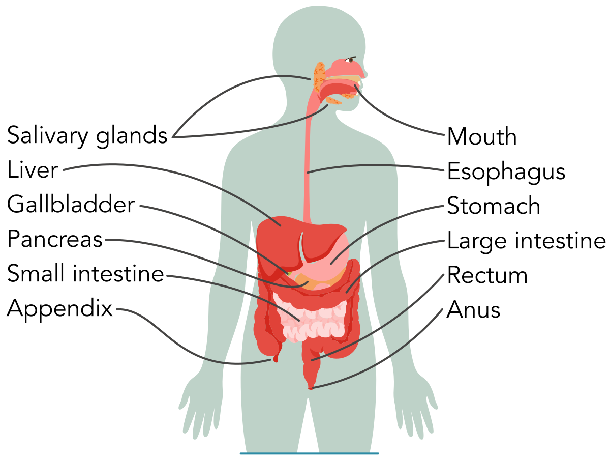
Digestive system- The digestive system consists of the liver, pancreas, gallbladder, and GI tract, which originates at the mouth and terminates at the anus.
Production of Feces
From the moment food and liquid enter the mouth, the production of feces begins. Food is broken down into smaller, easy-to-digest parts by chewing and by the action of digestive enzymes. Once food reaches the stomach, further digestion occurs through the mixing of stomach acid and other enzymes, aided by the stomach muscles, which help mix the contents. Once the stomach’s contents are emptied into the small intestine, the pancreas, liver, and gallbladder secrete more digestive enzymes into the small intestine to further assist with the breakdown of carbohydrates, fats, proteins, and vitamins. Bacteria within the small intestine also secrete their own digestive enzymes during this process. As food and liquid are transported through the small intestine, nutrients are absorbed that support the body by providing for energy, growth, and cellular repair. Once in the large intestine, stool forms from unused food, liquid, and cells from the GI tract. Bacteria in the large intestine continue to break down any remaining nutrients and produce vitamin K, a nutrient important for blood clotting and strong bones. At the terminal end of the large intestine, the rectum stores stool until it is expressed from the anus in the form of a bowel movement.
Like urine production, the amount of stool or feces produced depends on the amount of food and liquid consumed. The frequency of bowel movements varies significantly from person to person. Whereas some individuals may have daily bowel movements, others may evacuate stool one to three times a day, every other day, or only three times a week. Although the amount and frequency may vary, passing stool should not be difficult, and stool should be soft, not hard or liquid. The Bristol Stool Chart is a tool used to describe stool consistencies: it places stool on a continuum from constipation (types 1 and 2) to expected or ideal stools (types 3 and 4) to diarrhea (types 5 to 7).
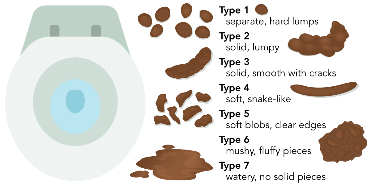
bristol stool chart- This tool is used to describe stool consistencies.
Dietary Considerations That Affect Stool Production
Diet can influence stool production. For example, fecal incontinence, otherwise known as accidental bowel leakage or the accidental passing of stool, can be affected by diet. Fecal incontinence can be caused by diarrhea, so avoiding dietary factors that increase diarrhea is an important preventive step. These factors include the following foods and beverages, which are known to increase the risk of diarrhea.
Alcohol
Caffeinated beverages and foods
Dairy (milk, cheese, cream, and ice cream)
Foods that are high in fat or are highly greasy
Beverages that contain fructose
Spicy foods
Apples, peaches, and pears
Products that contain sweeteners, such as sorbitol, mannitol, xylitol, and maltitol
Age-Related Considerations
As is true of many other organ systems, aging influences the function of the digestive system. With age, esophageal contractions decrease, but the passage of food or liquid from the mouth to the stomach does not change. Once food or liquid enters the stomach, age-related changes to the stomach lining may increase the risk of peptic ulcer disease, especially in clients who take aspirin or other nonsteroidal anti-inflammatory (NSAID) drugs. Stomach elasticity also diminishes with age, so the amount of food the stomach can hold changes, as does the rate of emptying. Movement through the small intestine is not significantly affected by age, but nutrient and vitamin absorption may be impaired by bacterial overgrowth. Lactose intolerance may develop as lactase levels decrease.
The pancreas, liver, and gallbladder may undergo cellular changes and size reductions in older adults, but their overall function may be preserved. During aging, minor changes in the large intestine and enlargement of the rectum may also occur. As the body ages, instances of constipation may increase, requiring greater straining to evacuate the bowel, a decrease in expected frequency, or stool classified as hard (types 1 and 2) per the Bristol Stool Chart. Specific changes in the digestive system related to aging can account for this change in stool characteristics. These physiological changes include decreased peristalsis and bowel muscle tone. Finally, changes in lifestyle often linked to aging can affect bowel movements, including inactivity, decreased fluid and fiber intake, and ingestion of medications, such as some antihypertensives and antacids.
Expected and Unexpected Findings Related to Elimination
Expected Elimination Characteristics
Elimination patterns can vary for many reasons and may not be a cause of concern. On average, urine should be clear, light yellow in color, and odorless. The amount and frequency will depend on the amount of fluids ingested, activity level, and medications taken, such as diuretics. Expected stool patterns can also vary. Some clients may have a daily bowel movement, whereas others may have fewer or more frequent bowel movements. Healthy bowel elimination should result in soft but formed stool that is easy to pass without straining.
Altered Bowel Elimination
Constipation
Constipation occurs when a client has infrequent bowel movements, often defined as three or fewer bowel movements per week. Additionally, these stools are hard, lumpy, and difficult to pass. Constipation can affect all age groups but is more common in older adults. The risk of constipation increases with low fiber intake, inadequate fluid intake, sedentary lifestyle, and certain medications (opioids, iron). Constipation is more common in clients who have neurological disorders.
constipation- Difficulty moving the bowels due to hardened stool.
Manifestations that require medical attention include abdominal pain, rectal bleeding, vomiting, low back pain, unexplained weight loss, inability to pass flatus, and abdominal mass. If constipation is chronic, fecal impaction can occur and lead to complications (bowel obstruction, perforation). Hardened stool clumps together, preventing evacuation of the bowel and potentially leading to intestinal obstruction or rectal injury. This extreme form of constipation happens more often in clients who are immobile or have a nervous system injury.
Interventions to treat constipation include eating a high-fiber diet, staying well hydrated, engaging in regular exercise, scheduled toileting/bowel training, and, when needed, using stool-softening medications to draw water into the bowel to help stool move or stimulant medications to help the bowels contract. If the constipation has advanced to an impaction, treatment options include manual removal (disimpaction) when stool is palpable in rectum, rectal enemas/suppositories for impaction in the lower bowel, and oral bowel-cleansing solutions for stool higher in the colon once mechanical obstruction has been excluded. Findings that may indicate the need for urgent medical intervention include severe abdominal pain and cramping with the inability to pass stool or flatus, progressive distention, and the presence of the manifestations noted earlier.
Diarrhea
Diarrhea is a condition in which the client experiences frequent loose, watery stools, typically three or more in 24 hr. It can be acute (14 days or fewer); persistent, lasting longer than 2 weeks but less than 4 weeks; or chronic, lasting longer than 4 weeks. Risk factors and frequent causes for diarrhea include infection, medication use (antibiotics), GI disorders (irritable bowel syndrome), and diet/exposures (high-risk foods, untreated water, travel, milk/milk products that contain lactose).
diarrhea
Frequent, loose, watery stools occurring throughout the day.
Findings with diarrhea include frequent, loose/watery stools with an urgent need to defecate, plus abdominal cramping, bloating, and nausea. With persistent or chronic diarrhea, dehydration, electrolyte disturbances, and malabsorption/malnutrition can occur and can be life-threatening if severe. In adults, manifestations that require medical follow-up include fever (39° C [102.2° F] or higher), vomiting that prevents oral hydration, diarrhea lasting longer than 2 days or six or more bowel movements a day, severe abdominal pain, presence of blood or black feces, immunocompromise, or findings of dehydration. Clients who are older adults, are children, or have an impaired immune system should receive medical care immediately if these findings are present.
Clinical findings of dehydration that might be present and should cause concern include dry mucous membranes/decreased tears, decreased urine output (oliguria/anuria), urine that is very concentrated or dark in color, sunken or recessed eyes, prolonged capillary refill time, tachycardia, and, in infants, a depressed or sunken fontanelle. Decreased skin turgor—meaning the skin does not return to its original state after being pinched upward—is a classic finding of dehydration.
Interventions aim to remove aggravating factors and ensure rehydration (oral rehydration solutions for most; IV fluids if severe or unable to maintain oral intake). If the client is experiencing uncomplicated, acute watery diarrhea, short-term use of over-the-counter antidiarrheal medications (loperamide or bismuth subsalicylate) may be used to reduce the stool frequency, but should be avoided if there is blood in the stool or high fever. Probiotics can help shorten the duration and severity of acute diarrhea in some cases. Antibiotics are not routinely indicated for acute diarrhea of short duration and are reserved for specific identified infections or more severe illness.
Bowel Incontinence
Bowel incontinence, sometimes referred to as fecal incontinence or accidental bowel leakage, is more common in older adults and in clients who are hospitalized or in nursing homes. In children, bowel incontinence is referred to as encopresis. Of the various types of bowel incontinence, urge incontinence is defined as the desire to defecate but an inability to reach the toilet in time. This type of incontinence may result from nerve damage, weak pelvic floor/anal sphincter muscles, or decreased physical mobility. In passive incontinence, leakage of feces occurs without the person being aware. Bowel incontinence can lead to perianal skin irritation and is associated with reduced self-esteem and well-being.
Alterations in Elimination Patterns
Alterations in Elimination Risk Factors
Many clients experience urinary and bowel elimination without difficulty. However, several factors can increase the risk of altered urinary and bowel elimination patterns. Age, sex, medications, and psychological stress due to lack of privacy are all factors that can influence urinary and bowel elimination. From anatomical to psychological issues, the most common risk factors are addressed next.
Medications That Affect Stool Production
Hormones and nerves work together to control the digestive process. Hormones are released from cells within the stomach and small intestine to regulate production of digestive secretions, to control sensations of fullness (satiety), or to signal hunger. Nerves send signals to the brain to begin the process of salivation in preparation for eating. Nerves within the GI tract also detect the degree of stretch, allowing for the regulation of the speed of digestion.
External factors, such as medications, can affect motility, or movement through the GI tract. In particular, some medications and other dietary supplements can slow gastric motility and cause constipation, including the following agents.
Aluminum-containing antacids
Anticholinergics and antispasmodics—medications used to treat muscle spasms
Antiseizure medications
Calcium channel blockers—medications used to primarily treat elevated blood pressure
Diuretics—which increase urine production
Oral iron supplements—used to treat certain forms of anemia
Antiparkinsonian medications
Opiates—used to treat pain
Some antidepressants
Medications, especially systemic antibiotics and magnesium-containing antacids/laxatives can increase gastric motility and cause diarrhea. Antibiotics may disrupt the normal intestinal microbiota and allow overgrowth of pathogens, leading to chronic diarrhea. In clients with recent antibiotic use, testing for Clostridioides difficile toxin is recommended when watery diarrhea occurs three or more times per day. Magnesium hydroxide products act osmotically to increase stool water and frequency and can cause loose stools/diarrhea when used as antacids or laxatives.
Medications That Can Affect Elimination Patterns
Urinary Elimination | Bowel Elimination |
|
|
Conditions That Alter Bowel Elimination Patterns
Several conditions alter bowel elimination patterns. The nurse’s understanding of these conditions, their risk factors, manifestations, and the appropriate interventions can be applied to assist the client in becoming more comfortable. This section explores the most common conditions affecting clients.
Diverticulitis
Diverticulosis is a condition in which small sacs or pouches, called diverticula, form in the colon. Diverticulosis is a common condition, and the risk increases with age. Diverticulitis develops when one or more of these pouches become inflamed. Current evidence shows that this inflammation does not result from a single cause. Instead, it results from several interrelated factors. Changes in the balance of usual gut bacteria can irritate the bowel, and some clients may have ongoing low-grade inflammation in the colon that puts them at higher risk for an episode. A person’s genetics can also predispose them to diverticular disease, and lifestyle factors, such as obesity, smoking, and certain dietary patterns (low-fiber diet and high red meat consumption), can further contribute to the development of inflammation.
diverticulosis- A condition in which small sacs or pouches form in the colon.
diverticulitis- An inflamed pouch or sac in the colon that is the result of stool becoming trapped.
When inflammation occurs, clients may experience fever; changes in bowel habits, such as constipation or diarrhea; and left lower-quadrant abdominal pain. If severe, the colon can perforate or tear, causing an intra-abdominal infection in the abdomen called peritonitis. Preventive measures after an episode, such as consuming a high-fiber diet, can reduce the risk of recurrent diverticulitis. Former recommendations included avoiding foods with nuts or seeds, but research has recently shown that these foods are not harmful for clients who have diverticulitis or diverticulosis. Treatment depends on the severity of the illness. Most clients who have mild diverticulitis can be treated at home. Providers usually recommend resting, drinking clear liquids at first, and then slowly returning to regular foods as manifestations improve. Many clients do not require antibiotics if they are otherwise healthy and their symptoms are mild, because research shows that mild cases can resolve on their own. However, some people do need antibiotics. Antibiotics are used when the client is sicker, not getting better, has more severe manifestations, or has health problems that make it harder for their body to fight infection, such as having a weakened immune system (immunocompromised). For serious cases, such as when there is an abscess, a perforation (a tear in the colon), or findings of a more dangerous infection, clients need hospital care, IV fluids, stronger antibiotics, and sometimes surgery.
© Jones & Bartlett Learning
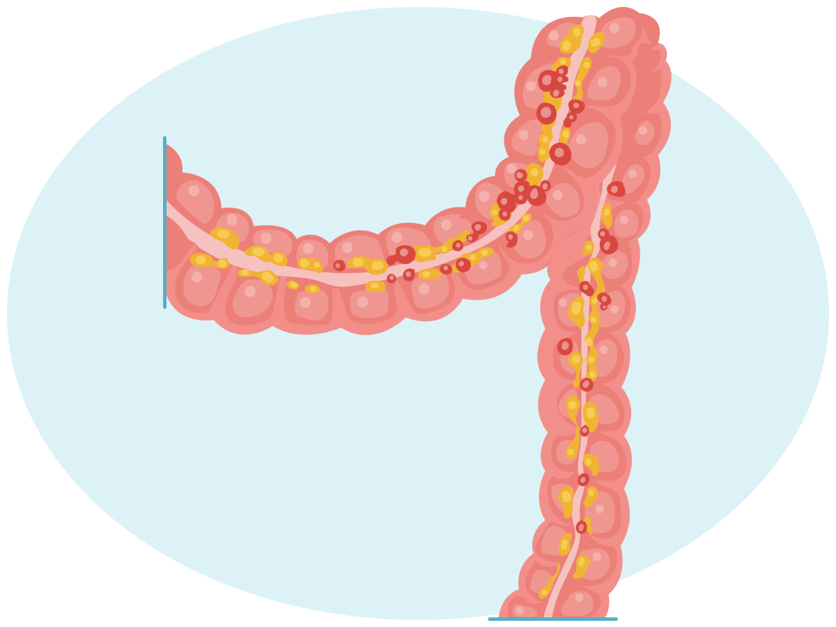
A diverticulum bulges outward through weak spots in the colon. Diverticula can become inflamed or infected, a condition called diverticulitis.
Diverticulitis- Diverticulitis develops when one or more of the small sacs or pouches in the colon, called diverticula, become inflamed.
Irritable Bowel Syndrome
Irritable bowel syndrome (IBS) is a term used to describe recurrent abdominal pain and changes to bowel elimination patterns that can include diarrhea, constipation, or a mixture of both. IBS with constipation (IBS-C) occurs when most of the daily bowel movements are hard or lumpy. IBS with diarrhea (IBS-D) occurs when most of the daily bowel movements are loose and watery. IBS with mixed bowel habits (IBS-M) is a combination of both diarrhea and constipation elimination throughout the day. IBS occurs when the gut and brain do not communicate as expected, which makes the intestines oversensitive (pain with normal stretching) and can cause irregular movement (too fast or too slow). Changes in gut bacteria, mild immune activation, and a leaky intestinal lining, along with stress and certain foods, can trigger or worsen manifestations. Thus, IBS is a gut–brain interaction disorder rather than damage to bowel tissue. Risk factors for IBS include a family history, female gender, stressful events, postinfectious onset after a GI infection, coexisting digestive disorders, and conditions such as anxiety, depression, and fibromyalgia.
irritable bowel syndrome (IBS)- A gastrointestinal condition characterized by abdominal pain and changes to bowel elimination patterns that can include diarrhea and/or constipation.
Findings depend on the kind of IBS, but may include abdominal pain, bloating, mucus in the stool, and a sense of incomplete bowel emptying. Treatment starts with diet and lifestyle. Clients should be encouraged to increase their intake of soluble fiber (such as psyllium) and, when manifestations persist, to make simple dietary changes to limit foods that trigger manifestations. Nurses should also promote healthy habits, such as reducing stress, getting enough sleep, and staying active. Some clients may benefit from therapies that help manage the connection between stress and the gut (relaxation or cognitive behavior strategies) and from medications chosen based on whether the main problem is constipation, diarrhea, or both. Routine probiotics or long-term gluten-free diets are not consistently helpful. A short, structured trial of reducing certain fermentable carbohydrates (lowFODMAP) and then adding foods back stepwise has stronger support when guided by manifestation response.
Bowel Obstruction
Bowel obstruction is a partial or complete blockage of the small or large bowel. Manifestations include nausea, vomiting, abdominal pain and distention, and severe constipation. Prompt diagnosis is a critical factor in reducing morbidity and mortality. Computed tomography (CT) is the preferred imaging to identify the site/cause and guide treatment decisions. Hospital admission and early surgical assessment are recommended to determine the obstruction’s type, severity, and cause. Initial supportive care includes IV fluids, electrolyte correction, and placing a nasogastric (NG) tube when the client has persistent vomiting, profound distention, or has a high risk of aspiration.
bowel obstruction- A partial or complete blockage of the small or large bowel.
A mechanical blockage—most commonly postsurgical adhesions, and less commonly hernias that trap a loop of bowel, tumors narrowing the lumen, inflammatory strictures (Crohn disease), twisting, other causes, such as volvulus or gallstone ileus—creates a transition point where bowel proximal (above) to the blockage distends with fluid and gas while the distal (below) bowel decompresses. The rising intraluminal pressure first impairs venous return (bowel wall edema). The swelling squeezes arterial blood vessels, preventing the bowel wall from getting enough blood or oxygen. This leads to mucosal ischemia and, if uncorrected, necrosis and perforation. Distention and mucosal injury also cause large third-space fluid/electrolyte losses (worsening dehydration) and allow bacteria from the bowel lumen to cross an injured mucosal barrier and enter the peritoneal cavity or bloodstream, which can progress to peritonitis and sepsis. Hence, the need for rapid imaging, resuscitation, and early surgical decision making.
Ileus
Ileus, also known as paralytic/functional ileus, is a condition where the nonmechanical flow of intestinal contents decreases or stops in the absence of a physical blockage. Ileus can be caused by several factors, including recent abdominal or retroperitoneal surgery, trauma, severe illness or infection (sepsis/peritonitis), medications (especially opioids), electrolyte disturbances (hypokalemia), and postoperative complications (pneumonia/abscess). Manifestations include an inability to tolerate food, nausea and vomiting, slow-onset abdominal distension and bloating, and inability to pass flatus. Bowel sounds are diminished or absent. Imaging helps distinguish ileus from a mechanical bowel obstruction. With an ileus, x-rays or CT scans usually show generalized intestinal swelling, with air seen throughout the bowel, even into the colon and rectum, without a clear area where the bowel is blocked. In a mechanical obstruction, imaging typically shows a more obvious blocked area where the bowel changes from swollen to collapsed, indicating a true blockage.
ileus- A condition where the flow of intestinal contents decreases or stops.
Treatment of ileus requires the active involvement of health care professionals, who ensure that the client receives nothing by mouth (NPO); decompress the stomach with an NG tube, when needed; provide IV fluid therapy; correct electrolytes; and address the underlying cause (reduce opioids, treat infection). However, if the client cannot tolerate an oral diet after 7 days, the provider will prescribe total parenteral nutrition (TPN).
Ulcerative Colitis
Ulcerative colitis (UC) is a chronic idiopathic inflammatory disease that causes continuous mucosal inflammation and ulcerations of the colon, typically beginning in the rectum and extending proximally. Onset is gradual, and the condition may worsen over time. However, clients also experience periods of remission that can last weeks or years. Risk factors include family history, Jewish ancestry, environmental influences, and an overactive intestinal immune system.
ulcerative colitis (UC)- A chronic disease that causes inflammation and ulcerations of the large intestine or colon.
Manifestations of UC include diarrhea with blood or pus, abdominal discomfort/pain, fatigue, nausea, fever, and iron-deficiency anemia. Interventions initially focus on medications to induce remission, followed by maintenance medications. If medications alone are unsuccessful at keeping a client in remission or cancerous lesions develop, surgery may be warranted. At times, surgery to remove the entire colon is required.
Crohn’s Disease
Crohn’s disease, like ulcerative colitis, is a chronic inflammatory bowel disease that causes full-thickness inflammation anywhere in the GI tract, but it most commonly affects the end of the small intestine and/or the colon, unlike ulcerative colitis, which is limited to the colon. Crohn’s disease begins gradually and can worsen over time. However, clients can experience periods of remission. Risk factors for developing Crohn’s disease include a family history of Crohn’s disease, autoimmune disorder, and tobacco use, although no definitive cause has been identified. Crohn’s disease can cause intestinal obstruction (from strictures), fistulas (tunnels that pass from the wall of the intestine to another organ), abscesses, ulcers, fissures or tears in the anus, malnutrition, and inflammatory processes elsewhere in the body.
Crohn’s disease- A chronic disease that causes inflammation in the GI tract but commonly affects the small intestine.
Manifestations of Crohn’s disease include diarrhea, abdominal pain or cramping (often right lower-quadrant when ileum is involved), weight loss, anemia (iron or B12 deficiency), fatigue, fever, joint pain (arthralgias), nausea/vomiting, and painful bumps under the skin. Care aims to stop a flare and then keep remission. Flares are treated with short courses of corticosteroids to quickly reduce inflammation and are not intended for long-term use. For maintenance, providers use steroid-sparing medicines, such as immunomodulators, and, when the disease is moderate–severe or refractory, biologic/targeted therapies. Surgery treats complications (strictures/obstruction, fistulas, abscess, perforation) or medication failure, often followed by a plan to reduce recurrence. Across all care, support includes nutrition (iron/B12 when the ileum is involved).
Fecal or Bowel Diversions
Like urinary diversions, fecal diversions, also known as bowel diversions, may be required when either part or all of the intestinal tract is not functioning properly. Surgery is performed to create a temporary or permanent diversion to redirect intestinal contents to exit through a stoma on the abdominal wall. Clients may require fecal diversion if they have colon or rectal cancer, traumatic injury to the intestine, inflammatory bowel disease such as Crohn’s disease or UC, an obstruction, or diverticular disease.
Incontinent diversions drain continuously into an external appliance/pouch (ileostomy, colostomy), whereas continent diversions store stool in an internal reservoir that the patient empties on demand, either from the anus after a J pouch or via intermittent catheterization of a Kock pouch (continent ileostomy). This section will cover the ileostomy, colostomy, J pouch, and Kock pouch.
Ileostomy
An ileostomy is a temporary or permanent fecal diversion that uses the terminal end of the small intestine, called the ileum, to form a stoma. Permanent ileostomies are created when the entire colon, including the rectum and anus, must be removed or bypassed. The surgeon redirects the ileum through a surgically created opening (stoma or ostomy) in the abdominal wall to allow for continuous drainage of stool (typically liquid). If the ileostomy is temporary, once the colon has healed, the stoma can be reversed by removing the ileum from the abdominal wall and reattaching it to the colon so bowel contents can continue to pass through the colon.
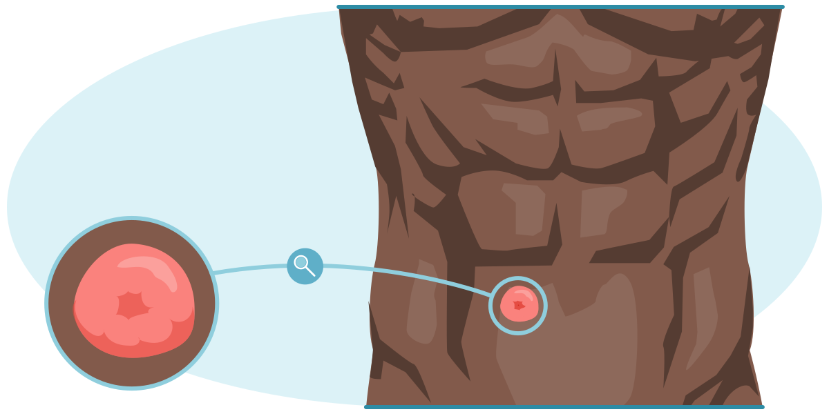
Ileostomy stoma- An ileostomy is a fecal diversion that uses the terminal end of the small intestine to form a stoma.
Colostomy
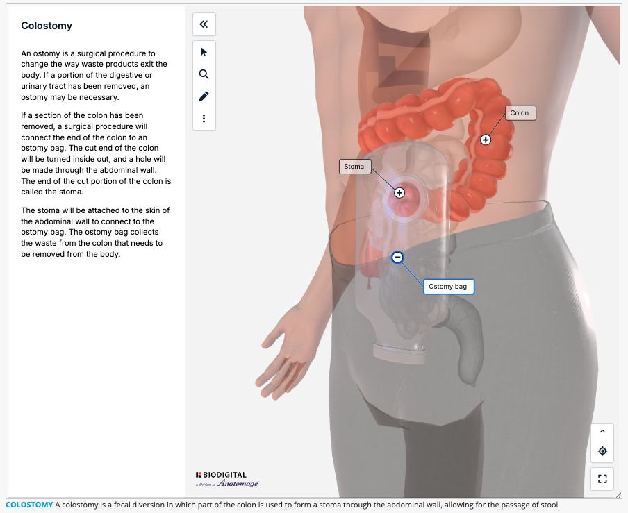
A colostomy is a fecal diversion in which part of the colon is used to form a stoma through the abdominal wall, allowing for the passage of stool. These diversions can be temporary, to allow the colon to heal and rest, or permanent. Once the colon is healed, the stoma may be reversed by removing the colon from the abdominal wall and reconnecting it to the rest of the colon, so that stool exits through the anus. Output consistency varies by location: ascending, liquid; transverse, loose/soft; descending/sigmoid, firmer.
Colostomy irrigation is another method to remove stool from the colon. This technique is especially beneficial for clients with permanent colostomies who had expected bowel function prior to the colostomy procedure. To irrigate the colostomy, warm water is inserted into the colon via the stoma site to evacuate feces/gas on a schedule. This acts as a type of bowel training, helping to prevent passage of stool at other times and reducing the client’s need to wear a colostomy pouch. Colostomy irrigation can also help prevent constipation (Cleveland Clinic, 2021; Longo, 2025; Maria & Lieske, 2023; Schub & Woten, 2024). Refer to Skill: Irrigating a Colostomy.
J-Pouch
A J-pouch is an internal ileal reservoir connected to the anus after removal of the rectum and colon. Body waste collects in the reservoir. Then, instead of passing through the colon and rectum, it directly passes through the anus during a bowel movement. Thus, it preserves sphincter function and continence without an external stoma. J-pouches are usually created with a staged approach that includes a temporary diverting ileostomy, so that the internal reservoir system can heal prior to its use. Once healed, the surgeon will reverse the ileostomy and connect the ileum to the J-pouch.
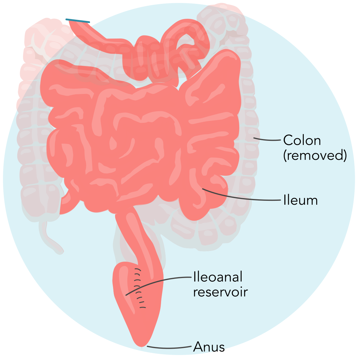
J-pouchA J-pouch is an internal ileal reservoir connected to the anus after removal of the rectum and colon.
Kock Pouch
A Kock pouch is a continent ileostomy system. Using the ileum, the surgeon forms an internal reservoir with a nipple-valve mechanism to maintain continence. Stool is emptied intermittently by inserting a catheter through the ileostomy stoma to empty the reservoir. The catheter is removed once emptying is complete. Thus, an external appliance need not be worn continuously.
Complications of Fecal Diversion
Depending on the kind of fecal diversion used, clients need to take appropriate measures to avoid complications. Complications may include peristomal skin irritation; stoma issues, such as hernias, prolapse, or narrowing (stenosis); blockages; diarrhea; bleeding; electrolyte imbalances; infections; and leakage. Vigilant assessment, correct pouch fit, and timely referral mitigate risks.
Ostomy Care for Urinary and Fecal Diversions
When surgical diversions are created, a wound, ostomy, and continence (WOC) nurse will often work with the clients to teach them how to care for their stomas or pouches. These specialized nurses assist clients in maintaining skin integrity around stoma sites, changing dressings, and troubleshooting common issues as they adjust to their new diversion. WOC nurses are also a vital resource in assisting clients in obtaining supplies for dressing changes and maintaining stoma health. Refer to Skill: Providing Ostomy Care and Skill: Draining an Ostomy Pouch.
Diagnostic Tests of the Gastrointestinal System
Clients may present with findings that indicate a problem with the GI tract. GI manifestations may include complaints of bloating, bleeding, diarrhea, constipation, nausea and vomiting, abdominal pain, heartburn, incontinence, weight gain or weight loss, or difficulty with swallowing. When this occurs, a provider may recommend diagnostic testing.
A variety of diagnostic tests can be performed to assess the GI system. Diagnostic testing of the GI tract depends on the findings the client reports and can include celiac disease testing, colonoscopy, endoscopic retrograde cholangiopancreatography (ERCP), flexible sigmoidoscopy, lower GI series, upper GI series, or upper GI endoscopy.
Types of Diagnostic Tests of the Gastrointestinal System
Type of Test | What the Test Evaluates |
Celiac disease testing | Diagnoses celiac disease by blood tests and small-bowel biopsy via upper endoscopy |
Colonoscopy | Screening procedure for colon or rectal cancer; can also be used to evaluate for causes of GI bleeding, changes in bowel habits, abdominal pain, and unexplained weight loss; allows biopsy and polyp removal during the same procedure |
Endoscopic retrograde cholangiopancreatography (ERCP) | Diagnoses problems associated with pancreatic and bile ducts such as gallstones, infections, pancreatitis, and pancreatic masses |
Flexible sigmoidoscopy | Evaluates the rectum and left colon for causes for rectal bleeding, bowel habit changes, pain, weight loss, and for cancer screening |
Lower GI series (barium enema) | X-ray/fluoroscopy of the large intestine using barium to evaluate for bleeding, changes in bowel habits, chronic diarrhea, unexplained weight loss, abdominal pain, cancer, diverticula, fistulas, polyps, or ulcers |
Upper GI endoscopy (esophagogastroduodenoscopy [EGD]) | Visualizes the esophagus, stomach, and upper intestine to evaluate persistent heartburn, bleeding, nausea and vomiting, pain, issues with swallowing (dysphagia), unexplained weight loss, ulcers, cancer, precancerous conditions, celiac disease, narrowing of the esophagus, or blockages |
Upper GI series | X-ray/fluoroscopy with barium to evaluate for nausea and vomiting, abdominal pain, difficulties with swallowing (dysphagia), unexplained weight loss, cancerous growths, and injuries to the esophagus, reflux, hernias, scarring, or ulcers |
Stool Specimen Collection
Stool specimen collection by the nurse is an important procedure for determining the presence of hidden (occult) blood or abnormal bacteria in the digestive tract. In conjunction with diagnostic testing of the GI tract, a provider may order the nurse to obtain a stool specimen. The most common stool tests are described in this section.
Fecal Occult Blood Test
A fecal occult blood test (FOBT) checks for the presence of small amounts of occult blood in stool that cannot be seen with the naked eye. It is commonly used as part of colorectal cancer screening for adults who do not have manifestations and may also be ordered when clients report changes in bowel habits or possible bleeding. The test simply determines whether blood is present. It does not identify where the bleeding originates.
Clients may report blood in their stool or changes in bowel habits. Because certain foods and medicines can temporarily change stool color and mimic bleeding (beets can cause red stool; iron or bismuth can cause very dark/black stool), the nurse should ask about recent diet and medications before testing and still treat persistent red/black stools or red/black stools with warning signs as possible GI bleeding. Utilizing an FOBT to determine whether blood is present is usually the first step in investigating potential causes of these findings. However, when manifestations of active bleeding are present, providers often proceed directly to diagnostic evaluation to identify the source.
Once an FOBT is ordered, the nurse will educate the client on how to prepare and obtain a specimen according to the specific kit instructions and provider orders. Preparation varies by test type. Some guaiac-based tests (gFOBT) require short-term diet/medication adjustments, whereas fecal immunochemical tests (FIT) generally do not require diet changes. The nurse should follow the kit’s instructions and the provider’s orders. The two main types of fecal occult blood tests differ in how they detect blood and therefore have different preparation requirements. The fecal immunochemical test (FIT) uses antibodies to identify human hemoglobin and is generally more sensitive and specific than older guaiac-based methods. Because it targets human blood directly, it typically does not require clients to follow dietary restrictions before testing. In contrast, the guaiac-based fecal occult blood test (gFOBT) depends on a chemical color reaction involving the peroxidase activity of heme.
First, the nurse reviews dietary and medication restrictions required for the type of FBOT to avoid any issues with testing results. Some kits may require clients to temporarily avoid certain foods or medications that can interfere with this reaction. For instance, if a client consumes beets within the window of time that the test is to be performed, the results may indicate that there is blood in the stool when, in fact, no blood is present. This is called a false-positive result. In this case, the result is due to the chemicals used to screen for blood in stool. They identify the beet dye, not actual blood, in the sample. Other foods, such as red meat, broccoli, and turnips, can also affect the results of an FOBT. The nurse should also instruct the client to avoid medications such as aspirin and ibuprofen, as well as vitamin C supplements, because they may also lead to false positives.
false positive- A false positive test result incorrectly indicates that a condition or attribute is present.
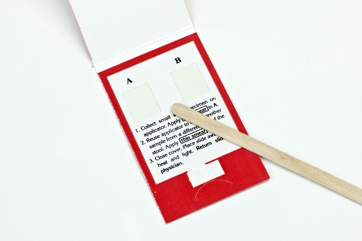
Depending on the type of FOBT used, the nurse instructs the client on how to obtain a specimen and provides the appropriate supplies so the collection can be done in the comfort of the client’s home. Collection supplies usually consist of a collection container that can be placed in the toilet; a collection spatula, such as a tongue depressor or collection spoon; the test container or test cards (also called hemoccult test slides); appropriate labels; and mailing containers.
Collecting a specimen is generally easy, given that it is done in the comfort of a client’s home. The client places the collection container in the toilet prior to a bowel movement, produces the bowel movement in the container, and then uses the collection spatula to place the specimen in the appropriate container as instructed for that kit. Next, the client affixes all the necessary labels to the specimen container and places it in the return or mailing container so it can be analyzed in a lab for the presence of blood. Depending on the results and the findings the client presents with, the provider may determine the need for further testing to rule out potentially serious conditions such as colon cancer. Refer to Skill: Obtaining Occult Blood Specimen.
© MedstockPhotos/Shutterstock
fecal occult blood testing- FOBT checks for the presence of small amounts of occult blood in stool that cannot be seen with the naked eye.
Stool Culture
A stool (fecal) culture is a laboratory test used to detect organisms in stool that may be the cause of severe or persistent diarrhea. A provider may order a stool culture if the client reports severe or persistent diarrhea and has recently traveled outside the United States to areas that may be prone to water contamination. A client may also become infected with bacteria that cause severe diarrhea by eating or drinking something that may have become contaminated; by consuming raw or undercooked eggs or meat, or unpasteurized milk; or by drinking untreated water from sources such as lakes or streams. At other times, a provider will investigate for the presence of fecal bacteria if the client reports recent or prolonged antibiotic use.
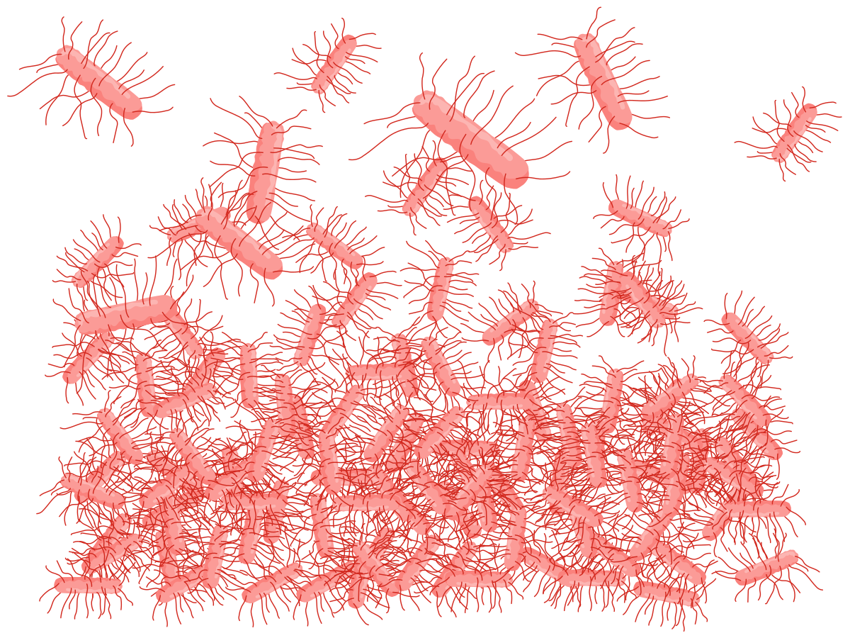
Stool cultureA stool culture is a laboratory test used to detect organisms, such as bacteria, in stool.
Once the provider orders a stool culture, the nurse will educate the client about how to prepare and obtain a specimen for analysis based on the specific kit and instructions provided. Unlike with the FOBT, there are no routine dietary or medication restrictions prior to obtaining a stool specimen. The nurse will provide the client with the appropriate collection supplies, which usually include a collection container that can be placed in the toilet, a collection spatula, such as a tongue depressor or collection spoon, the test container, appropriate labels, and mailing containers. Instruct the client to avoid contaminating the specimen with urine, toilet water, or tissue with the sample.
Collecting a specimen is generally easy, given that it is usually done in the comfort of the client’s home. The client places the collection container in the toilet prior to a bowel movement, produces the bowel movement in the container, and then uses the collection spatula to place the specimen in the appropriate container. The client then affixes all necessary labels to the specimen container, places it in the return or mailing container, and returns the specimen to the laboratory as soon as possible so it can be analyzed in a lab for the presence of infectious bacteria. Results usually take a few days. Depending on the results and the client’s presenting manifestations, the provider may determine the need for further testing, such as ova and parasite testing, gram stain, or fecal smear to rule out other nonbacterial pathogens.
Nursing Interventions to Facilitate Bowel Elimination Patterns
Alterations in bowel elimination are usually treatable or manageable. A few nursing interventions can be implemented to assist clients with a return to expected bowel elimination patterns. Nurses can assist clients with making lifestyle changes by providing bowel training and by providing them with agents to stimulate a bowel movement.
Lifestyle Changes
Nurses can review lifestyle changes that clients can make to help restore usual bowel elimination patterns, such as dietary modifications. Diets higher in fiber, such as those rich in fruits and vegetables, can help clients maintain expected bowel elimination patterns. Stress reduction techniques can be reviewed with clients, and clients can be instructed to listen to their bodies and use the bathroom when they feel the urge to have a bowel movement. Ensuring adequate hydration, staying active, and getting regular exercise are other lifestyle changes nurses can review with clients to promote usual bowel elimination.
Enema
An enema is the instillation of a liquid solution into the rectum and sigmoid colon through the anus to stimulate peristalsis and defecation or to empty the bowel in preparation for diagnostic testing, procedure, or surgery. Enema administration is not a sterile procedure. The nurse follows standard precautions and adheres to medication administration rights throughout the process. For safe and effective administration, the client is positioned in the left lateral (Sims’) position with the right knee flexed, which aligns the rectum and sigmoid colon to promote proper flow of the solution.
Several types of enemas are used clinically. The solution and volume selected are based on the purpose of the enema. A cleansing enema is instilled into the client, retained for a short time (commonly within about 15 min), and then expelled. In contrast, a retention enema requires the client to retain the fluid for 15 to 30 min, depending on the type of solution instilled, before expelling it. Enema solutions contain bowel irritants that stimulate the bowel. Common solutions used in enemas for medical purposes are tap water (hypotonic), mineral oil, 0.9% sodium chloride (isotonic), a sodium phosphate solution (hypertonic), and soap suds. Hypertonic preparations draw water into the lumen, thereby distending the colon. Large-volume hypotonic/isotonic solutions rely on volumetric stimulation. Because repeated hypotonic enemas (tap water) can shift fluid and electrolyte levels, solutions and volumes should be administered only as ordered, with monitoring for electrolyte imbalance and other adverse effects.
Medications
Other substances used to treat constipation prior to enema use are laxatives or stool softeners. Laxatives and stool softeners work more slowly than enemas and are intended for less severe forms of constipation. Clients taking medications that slow bowel transit may need to use laxatives or stool softeners to encourage bowel movements. Clients need to be instructed on proper laxative and stool softener use, as they can interact with certain medications. These agents may be harmful to clients who are pregnant or who are younger than 6 years of age. Use of these agents can complicate serious medical conditions such as appendicitis or bowel obstructions.
Laxatives – Classification, Mechanism of Action, and Therapeutic Response
Classification | Mechanism of Action | Therapeutic Response |
|---|---|---|
Bulk forming
| Softens stool by absorbing water/retaining water; increases stool bulk and promotes peristalsis. | Bowel movement in 12 to 72 hr |
Surfactant
| Softens stool by lowering stool surface tension to allow water and lipids to penetrate. | Bowel movement in 12 to 72 hr |
Stimulant
| Stimulates peristalsis and increases intestinal secretion; decreases water reabsorption. | Suppository: 15 to 60 min Oral: 6 to 12 hr |
Osmotic
| Retains water osmotically, softening the stool and promoting peristalsis. | Oral: Within 24 to 72 hr Rectal/enema: Acts rapidly, within minutes |
Rectal Tube
Fecal management systems and rectal tubes are devices placed into the rectum to prevent incontinent clients from soiling themselves. They are typically used in intensive care units to help protect the client’s skin from breakdown and infection when there is no bowel control. They are also used (albeit rarely) for clients with chronic fecal incontinence or diarrhea.
Bowel Training
Bowel training, like bladder training, assists clients with gaining control of their elimination schedule. Clients may be incontinent for a variety of reasons, including spinal cord injury, multiple sclerosis, surgery, laxative abuse, childbirth, or other emotional problems. Bowel training allows clients to gain control of their bowel movement schedule to avoid unexpected accidents and the embarrassment associated with such events. Bowel training may include the use of laxatives to assist with bowel stimulation. Clients must ensure they follow a high-fiber diet and stay adequately hydrated to promote proper bowel elimination.
Diversions and Ostomies
Diversions and ostomies are surgical interventions that are performed when nursing interventions are no longer sufficient to ensure safe elimination or protect renal function. Several types of diversions and ostomies are possible, and selection depends on the underlying pathology, client factors, and surgeon expertise.
When the usual flow of urine from the body is interrupted, surgery may be required to form a urinary diversion. Urinary diversions may be either temporary or permanent, depending on the causative factor. A diversion may be required if urine is blocked, if the urethra is damaged from either injury or a birth defect, because of tumors, or in case of bladder malfunction. Conditions such as bladder cancer, radiation damage, chronic infections, congenital disorders (spina bifida), neurological conditions, or severe incontinence often necessitate diversion when the bladder cannot function properly. Surgical interventions can create neobladders, ileal conduits, cutaneous reservoirs, or nephrostomies, depending on the client’s anatomy and health status and the surgeon’s experience.
Nursing interventions for clients with urinary diversions include teaching the client proper hand hygiene, stoma and peristomal care, and use of a collection pouch and barrier device, if applicable. Clients should be instructed to cleanse the peristomal skin with warm water. If soap is preferred, choose a mild, residue-free soap (no moisturizers, oils, or fragrance) to maintain skin barrier adhesion and reduce irritation. The opening of the wafer/skin barrier should be cut no more than about 3 mm (1/8 in) larger than the stoma to minimize leakage and protect skin.
If the client has a ureterostomy or ileal conduit, education is also needed regarding the collection pouch and barrier wafer, including how to empty the bag, how to measure the wafer to fit the stoma when it is replaced, and how to change out the system if it is a single-piece or two-piece device.
Other urinary diversions, such as continent cutaneous reservoirs, may require irrigation and clean intermittent catheterization, which may be scheduled (about every 4 hr) to maintain drainage and clear mucus, as instructed. Encourage adequate hydration (typically about 2 L/day unless contraindicated) to help flush mucus and reduce the risk of UTIs.
The nurse should also instruct the client to report findings such as pain with urination (for those voiding through a neobladder or performing clean intermittent catheterization) or changes in urine color or odor, as these may indicate infection; fever, chills, nausea/vomiting, back or abdominal pain, cloudy/foul urine, or increased confusion in older adults are concerning for UTI and warrant evaluation. Finally, changes in stoma color (dusky, cyanotic, or brown/black) or inability to pass a catheter or to drain a continent pouch require prompt provider notification due to the risk of ischemia/necrosis or reservoir overdistension.
Fecal or bowel diversions are surgical procedures that reroute intestinal contents to stomas formed on the abdominal wall. Such surgical interventions can be implemented to create ileostomies, J-pouches, Kock pouches, or colostomies.
Nursing interventions for clients with bowel diversions include many of the same tasks as are carried out when educating clients with urinary diversions. Clients should understand manifestations of possible problems, such as skin or pouch irritation, stoma issues (retraction/prolapse), blockages/obstructions, changes in stool output/consistency, and postoperative bleeding through the stoma or rectum.
Skin irritation from stool exposure is one of the most common findings reported by clients who have bowel diversions and is often due to a poorly fitted collection system (incorrectly sized barrier opening or inadequate seal). The nurse or wound, ostomy, and continence (WOC) nurse should teach peristomal skin cleansing (warm water; if soap is used, a mild, residue-free option) and accurate stoma measurement with cutting the barrier about 3 mm (1/8 in) larger than the stoma to reduce leakage and protect the skin. Clients should also be shown how and when to change/empty the pouch to prevent undermining. Clients should avoid perfumes, deodorants, and other scented products on or around the stoma to reduce irritation and preserve wafer adhesion; fragrance-free options are preferred.
In addition, the stoma itself can develop parastomal hernias, adhesions, or retraction, which may then cause obstruction (reduced or absent output, cramping pain, abdominal distention). These situations warrant prompt evaluation. Bowel obstruction and strangulation may rapidly progress to life-threatening complications and require urgent surgical assessment. Clients should be instructed to promptly report pain, bleeding, or any change in stoma color (dusky, cyanotic, brown/black), as these may indicate impaired perfusion, infection, or other complications requiring urgent evaluation.
Finally, teaching the client pelvic floor strengthening exercises, such as Kegel exercises, may be helpful with certain types of bowel diversions, such as an ileoanal reservoir, to help control incontinence and reduce seepage after surgery as bowel function adapts. Refer to Skill: Providing Ostomy Care and Skill: Draining an Ostomy Pouch.
Nasogastric Decompression
Nasogastric (NG) tubes are flexible tubes that are inserted into a nostril and advanced down to the stomach to remove gastric contents and decompress the upper GI area. NG tubes are most frequently placed for bowel obstructions to reduce abdominal distention and subsequent nausea and vomiting, thereby decreasing the risk of aspiration. NG tubes also may be used for enteral feedings and/or medication administration (called nasoenteric tubes), but these uses are distinct from decompression and are not performed at the same time.
When placing the tube, the nurse should encourage the client to sit upright, if possible, to reduce the chance of vomiting and aspiration during insertion. NG tube length should be measured from the tip of the client’s nose to the tragus of the ear, to the xiphoid process, which is a standard estimation method to approximate the required length. The tube should be advanced as the client swallows or sips water to facilitate passage through the oropharynx.
Do not use the NG tube until placement is verified. Current safety guidance supports a combination of evidence-based methods—typically abdominal/chest X-ray (the initial gold standard and required before using the tube for enteral medications or nutrition or any time placement is uncertain), pH testing of gastric aspirate (an acidic pH supports gastric placement, noting that acid-suppressing medications and continuous feeds can alter results), and capnography/capnometry (useful to detect airway placement by identifying CO₂, though absence of CO₂ does not confirm gastric location). These recommendations reflect ongoing patient-safety events when tubes are used before radiographic verification is obtained and read. Importantly, the traditional air-bolus “whoosh” test is unreliable and should not be used.
Once the tube's location is confirmed, it should be secured to the nose with tape or a commercial device, connected to suction as ordered, and monitored for migration by regularly checking the external tube length and tube markings, because movement can occur with coughing, vomiting, repositioning, or loosening of the securement device. Refer to Skill: Nasogastric Decompression.
Providing Skin Care to Incontinent Clients
Clients who are incontinent are at risk for impaired skin integrity as a result of increased skin moisture and prolonged contact with urine and stool. For this reason, clients’ skin should be kept clean and dry. Nurses play an important role in protecting clients’ skin from breakdown and infection. Clients who have impaired mobility are at increased risk of incontinence, and frequent skin assessments are important to maintain skin integrity. Nurses should observe for areas of redness that do not blanch or turn white when pressed, as well as peeling/irritation, blisters, open wounds, and areas of ulceration. These problems most often occur on the buttocks, hips, genitals, and perineum, where moisture and irritants are greatest.
To maintain skin integrity, it is important to cleanse the affected areas promptly after each episode of incontinence. The skin can be cleaned with mild, diluted soap and water, then rinsed thoroughly, or with a soap-free perineal cleanser designed to minimize dryness and irritation. Regardless of the cleansing method, the area should be gently patted dry rather than rubbed. Alcohol-containing products should be avoided because they can dry or damage the skin.
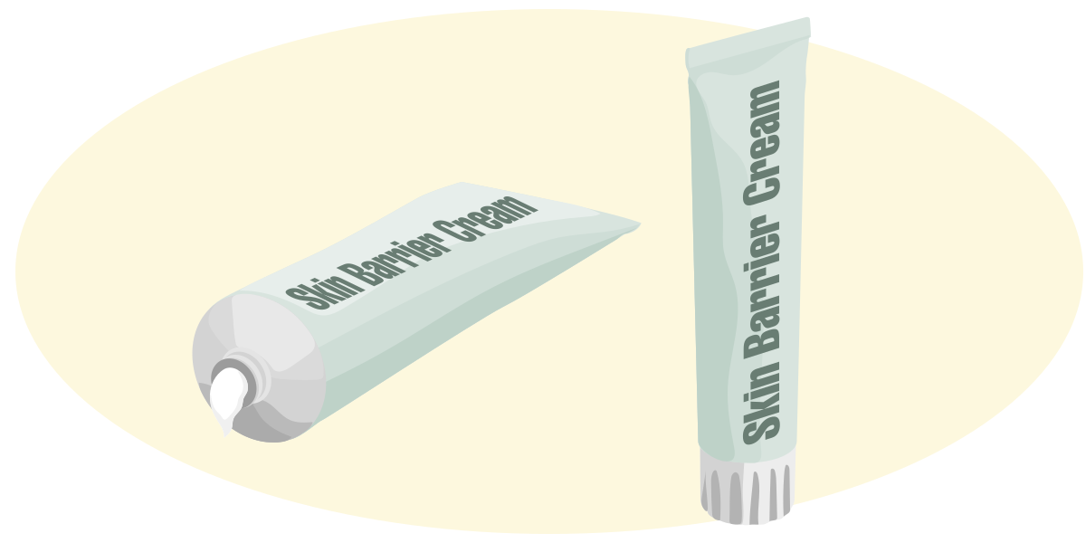
After cleansing, the skin should be moisturized using alcohol-free creams to support the natural skin barrier. Applying a moisture-barrier product, such as ointments or creams containing zinc oxide, lanolin, or petrolatum, or a clear, protective sealant helps shield the skin from further exposure to urine or stool. Even when barrier products or absorbent briefs are used, the barrier must be reapplied after every episode of incontinence to maintain effectiveness.
For clients who spend long periods in bed or in a wheelchair, nurses should also reinforce regular repositioning, timely changes of soiled linens or clothing, and adequate nutrition to support healthy skin. These strategies, combined with consistent cleansing and protection, help reduce the risk of skin breakdown and maintain comfort and dignity for clients living who have incontinence.
Getty Images/Stefan_Alfonso
Skin Barrier Cream- After cleansing skin, moisturize it using an alcohol-free cream to support the natural skin barrier.