Ch. 4: Cells, Membrane & Membrane Proteins, Protein Production
Things to Know
You should have an understanding of the following:
1. Review cell structures and their basic functions.
2. Nucleus:
A) DNA, nucleolus, nuclear pores
3. Mitochondria:
A) Internal structures
Contains own DNA → self-replication and protein synthesis
B) Make the connection to what you learned about cellular respiration from Ch. 3 and this organelle.
Mitochondria is the powerhouse of the cell; site of Intermediate Step, Krebs/Citric Acid Cycle, and ETC
4. Membranes:
A) Be able to describe the structure of a membrane (fluid mosaic model). How do phospholipids contribute to the characteristics of the membrane?
Phospholipids are the ‘sea’ and the proteins are the ‘icebergs’
B) What are the functions of the various membrane proteins?
Transport protein
Channel (leak)
Generally stay open, but can close
Carrier
Requires ATP to open
ID markers/Recognition
Cell Receptor Surfaces/ligands
Anchoring Sites
Cell Adhesion
Enzymes
5. Transport:
A) Differentiate between active and passive transport through a cell membrane.
Active
Req’s ATP to move against concen gradient (low → high)
Passive
No ATP req’d
B) What is the difference between simple diffusion, osmosis and facilitated diffusion?
Simple diffusion
Substances have their own concen gradient
Ions run along electrical gradients
Molecules must be small and have no electrical charge
Osmosis
Diffusion of water through a selectively permeable membrane
Facilitated Diffusion
Diffusion of a molecule with the help of a
Channel protein: ions
Carrier protein: small polar molecules
Polar = hydrophilic, meaning they can’t easily pass through phospholipid bilayer
C) What is meant by concentration gradient?
How the concentration of substances change over time (high → low = passive; low → high = active)
D) Tonicity: isotonic, hypertonic and hypotonic; crenation, hemolysis
Isotonic = same tonicity; no net movement
Hypertonic = one solution is more concentrated than the other; water follows solution with more solute; if ECF is more hypertonic than ICF, cell may crenate
Hypotonic = one solution is less concentrated than the other; water follows solution with more solute; if ECF is more hypotonic than ICF, cell may hemolyze
E) Know what exocytosis and endocytosis are. Know terms phagocytosis and pinocytosis.
Exocytosis — vesicle released out of cell
Fuses w/ membrane
Endocytosis — vesicle formation
Moves into cell
Phagocytosis = ingestion of substances
Pinocytosis = ingestion of liquid
Cells

The Cell Theory
Cells are the basic unit of life
Building blocks of the human body
All organisms are composed of cells
New cells are derived from existing cells
Common Characteristics of Cells
All cells perform the general functions necessary to sustain life
Divided by functions (skin cells will do one thing, heart cells will do other things, etc.)
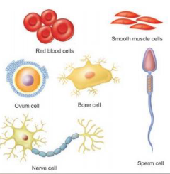
Cells vary in shape
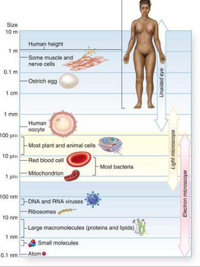
Cells vary in size
Leleimited due to transport
Choice is to die or divide
Some cells are amitotic
Plasma (Cell) Membrane
Forms the outer barrier
Phospholipids and proteins
Semi-permeable

Cytoplasm
Cellular contents located between the plasma membrane and the nucleus
Organelles
Cytosol
Jelly-like fluid that fills inside of a cell
Cytoskeleton
Nucleus
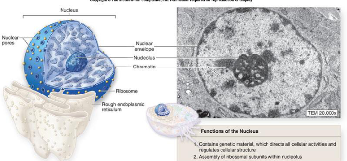
Cell’s control center
Controls protein synthesis
Nuclear envelope w/ pores, nucleolus (ribosomes), chromatin, and chromosomes
Nuclear envelope is around the nucleus
Ribosomes are responsible for protein production (transcription & translation); associated w/ RER
Each ribosome has a small and a large subunit
They exit from the nucleus via the nuclear pores
Organelles
Each type of organelle performs a different function for the cell
Collectively, the specialized functions of all organelles are essential for normal cellular structure and activities
Mitochondria
~powerhouse of the cell~
Produce large amounts of ATP (cellular respiration)
Double-membrane
Contains own DNA: self-replication & protein synthesis
Endoplasmic Reticulum
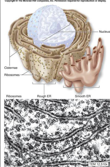
Synthesis of lipids, proteins, storage, and transport
Rough ER — ribosomes make proteins; granular appearance
Closest to nucleus
Smooth ER — synthesis of lipids; smooth, tubular appearance
Further from nucleus
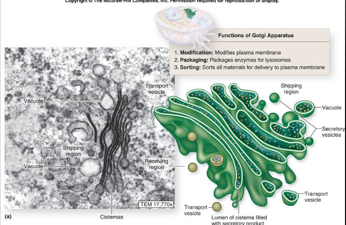
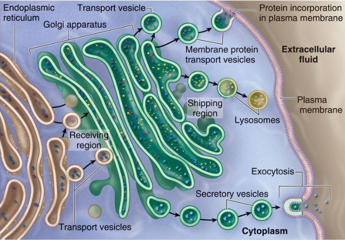
Golgi Apparatus
“FedEx of the cell”
Modifies & repackages proteins and lipids as vesicles
Can make glycoproteins, lipoproteins
Flattened stack of pancakes
Vesicles
Membrane-bound sacs produced by many organelles to transport products throughout the cell
Lysosomes, peroxisomes
Helps clean out waste
Cytoskeleton
Filamentous proteins which helps give the cell its shape and coordinates cellular movements
Microfilaments
Intermediate filaments
Microtubules
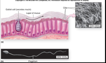
Cilia & Flagella
Appendages extending from the surface of some cells
Cilia work together to move materials or fluids along the surface of a cell (ex. respiratory cells)
Flagella act like a whip-like tail (ex. sperm)
Cell-to-Cell Junctions
Tight junctions
So tightly held together, nothing can get in between the membrane of one cell and another
Very common
Adherens junctions
Keeps cells close together, but allows for some stretch
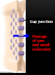
Gap junctions
Little channels between two membranes
Allows stuff to pass through from one cell to another
Desmosomes
Don’t allow for any stretch
Will allow some passage in between

Brief Overview of Cell Cycle
New cells must be made continuously in order for an organism to grow and replace its damaged cells
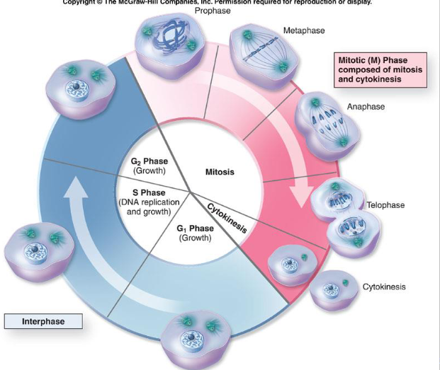
Mitosis (somatic cells) & cytokinesis
Phases: Interphase, prophase, metaphase, anaphase, telophase
Interphase — steady state of cell
G1 Phase: making enzymes for next step
S Phase: DNA replication and growth; will be in chromatin state
G2 Phase: Makes enzymes to be used in mitosis (PMAT)
No need to memorize G/S phases
Prophase: chromatin → chromosomes
Metaphase: lines up chromosomes at equatorial plate
Centrioles are poles
Anaphase: pulls chromosomes apart via centrioles
Telophase: Pinching of cytoplasmic membrane (cytokinesis); chromosomes decondense back into chromatin
Review the basics of what occurs in each phase
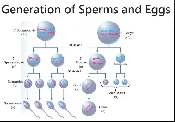
Meiosis (sex cells) & cytokinesis
Diploid → haploid
2 parent cell → 4 daughter cells
2 sets of phases: Meiosis I and Meiosis II
Review the basics of what occurs in each phase



Membrane & Membrane Proteins

The lipid bilayer gives the membrane it flexible and fluid characteristics
The membrane is self-healing when punctured
When vesicles fuse w/ membrane (exocytosis)
When bits of membrane pinch off and become vesicles (endocytosis)
Fluid Mosaic Model (def gonna be on test in some fashion)
“Protein icebergs floating in a sea of lipids'“
Fluid: lipids (phospholipids and cholesterol)
Mosaic: many different proteins
Peripheral and integral proteins
Membrane Proteins
Transport proteins
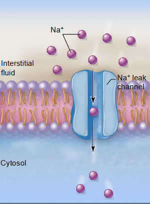
Channel (leak)
Generally stay open, but can close
Carrier
Typically needs ATP to open up
Identity markers/Recognition
Cell surface receptors/Ligands
Anchoring sites
Cell adhesion
Enzymes
Channel-mediated diffusion
Certain small water-soluble molecules can pass through
Non-water soluble (lipids) don’t need a protein; can just pass on through phospholipid bilayer
Specific to what it allows to pass through
Selective due to charge of molecule and channel protein
High → low concen
Carrier proteins
Can move larger molecules across the membrane by changing shape
Carrier proteins change shape to transport molecules across the plasma membrane
Specific shape for certain molecules or ions

Transport Through the Membrane

Lipid: can pass right thru phospholipid
Water: needs a carrier protein (can’t pass through phospholipid)
Active: requires ATP to move against concen gradient (low → high)
Passive Transport - no ATP
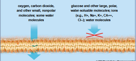
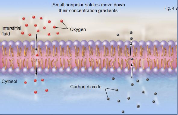
Simple diffusion
Each substance has its own concentration gradient
Ions run along electrical gradients
Through the lipid bilayer: molecules must be small w/ no electrical charge
Osmosis
Diffusion of water through a selectively permeable membrane
Most water molecules pass through special channels called aquaporins
A few water molecules can slip through the lipid bilayer
Osmotic pressure — pressure exerted by the mvmt of water across a semipermeable membrane
Caused by different in water concentration
The steeper the gradient, the greater the amount of water moved and the higher the osmotic pressure
0.9% cell, 10% Na solution (extremely hypertonic, water moves out readily)
0.9% cell, 1% Na solution (barely hypertonic; some water movement to outside, but not alot)
Facilitated diffusion (channel and carrier mediated)
Diffusion of a molecule with the help of a
Channel protein: used by ions
Ions are smaller; can kinda meander their way through
Carrier Protein: used by small polar molecules
Typically larger; need carriers
Passive, no energy required
Active Transport
Moves particles from low to high concen, against concen gradient
Requires ATP
Uses carrier proteins
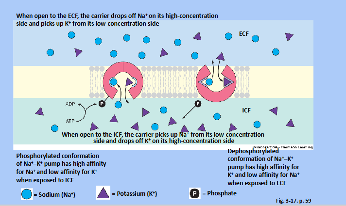
Ex. Sodium-Potassium pump (3 Na go out : 2 K go in)
Primary v. Secondary
Primary Active Transport
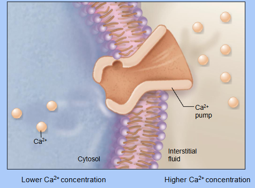
A carrier protein uses energy from ATP to pump molecules across a membrane AGAINST (up) their concen gradient
Ex. Calcium pump (an ion pump)


Secondary Active Transport
Involved 2 different substances
Ones moves w/ (down) its concen gradient through a channel protein (passive transport)
The flow of the 1st substance powers the pumping of a 2nd substance through the same channel protein (active transport)
The 2nd substance is moving against (up) its concen gradient
The energy for active transport here is not from ATP; it’s the kinetic energy from the flow of the 1st substance that powers the pump
Types
Symporter: substances move in the same direction
Even though they’re moving in the same direction, one is still powering the other
Antiporter: substances move in opposite directions
as
Vesicular Transport (a form of active transport)
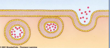
Endocytosis — vesicle formation
Phagocytosis, pinocytosis

Exocytosis — vesicle release out of cell
Can inc cell membrane


Protein Production
RNA & Protein Synthesis
DNA contains all the info needed to make proteins
RNA acts as intermediary and can be used over and over to make the same type of protein
Ribonucleic Acid (RNA)
3 parts
Sugar = ribose
Phosphate group
Nitrogenous base (4)
Adenine (A)
Guanine (G)
Cytosine (C)
Uracil (U)
Complimentary strands follow same base pair rule, except U replaces T (A-U)
Purines & pyrimidines
3 types of RNA
mRNA (messenger)
rRNA (ribosomal)
tRNA (transfer)
Stages of Protein Synthesis
2 stages
Transcription — assemble RNA from DNA template
Translation — RNA and ribosomes assemble amino acids into a long chain protein
Transcription
Takes place in the nucleus
DNA unzips and one strand acts as template for making RNA
RNA polymerase assembles nucleotide bases to region of DNA template
Starts at a promoter site that signals RNA growth, DNA sections are called triplets
RNA grows, then detaches from DNA and DNA re-zips
RNA is modified by snipping out unnecessary sections & RNA leaves the nucleus
Translation
Takes place in the cytoplasm in 3 parts
Nuclear pores allow ribosomes to come out
Initiation — strand of mRNA attaches to two ribosomal units to make complex
rRNA will be helping by ‘taping’ ribosome complex together
Start codon (AUG) is the start point
P and A sites, tRNA (anticodon)
Brings complimentary base
Elongation — complex slides over mRNA while tRNA brings in amino acids, making a long chain polypeptide
Termination — stop codon reached and protein synthesis is finished; complex fall apart
mRNA & tRNA can be used repeatedly, protein goes off

Mutations
Can occur in either DNA or RNA
Can be caused by mutagens, accidental exposures, or spontaneously
Can cause no effect, be harmful, or cause the protein to not be made
Types
Base pair substitution (point mutation)
Frame shift mutations
