Lecture_SecretionSystems_PvU_2025
Protein Machines in the Gram-negative Cell Envelope
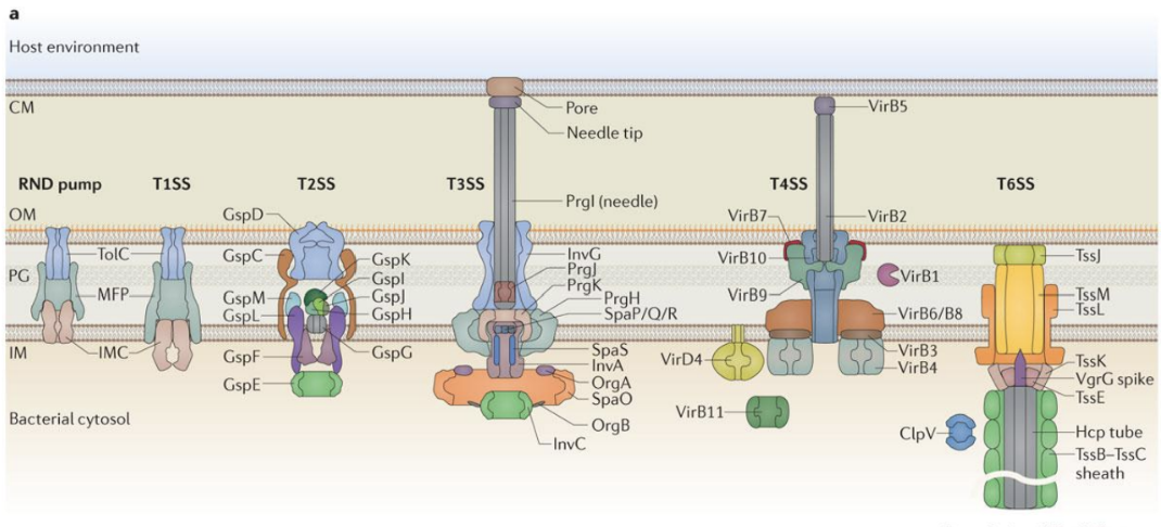
Classification: Secretion systems classified I-VI (currently at XI) based on their order of discovery.
Adapted Structures: Evolved from macromolecular structures on bacterial surfaces:
Pili (II)
Flagella (III)
Conjugation systems (IV)
Secretion Mechanisms
Two-step secretion (T2SS and autotransporters - T5SS):
Involves:
Cytoplasmic membrane: Sec/Tat pathway utilizing a signal peptide
Periplasmic chaperones
T5SS transports directly through autotransporter, T2SS through chaperone
Energy Requirement: No energy used at the OM; energy derived from protein folding.
One-step Secretion:
Involves: T1SS, T3SS, T4SS
Cytoplasmic chaperones for targeting to secretion machinery
Motifs that recognize machinery/chaperones in cytosol that guide substrate
ATP hydrolysis drives the process.
Structural Biology Techniques for Investigating Secretion Mechanisms
Techniques Discussed:
X-ray crystallography
Electron microscopy (EM)
Examination of different secretion systems:
Type I Secretion System
Type III Secretion System (injection needle)
Type VI Secretion Systems (T6SS) - identified through bioinformatics and structural biology tools.
X-ray Crystallography
Classical technique for determining protein structure:
Requires purification of proteins in large quantities.
Crystallization through trial and error methods.
Obtaining diffraction patterns to determine structures.
Limitations of Crystallography
Challenges with membrane proteins:
Hydrophobic surfaces,
Low solubility in crystallization solutions.
Issues with purification and high aggregation tendency.
Electron Microscopy
Overview: Detects electron-dense materials;
Needs heavy metal staining for biological samples.
Introduction of cryo-EM to avoid radiation damage and reduce staining needs, suitable for various samples.
3D Tomography: Allows for detailed visualization of structures.
Cryo-EM and Single Particle Analysis
Preparation of purified samples on grids, followed by imaging.
Grouping structures and fitting protein structures effectively.
Advances in Cryo-EM Techniques
Enhanced electron detectors allow for better resolutions and techniques like plunging samples in liquid ethane for preservation.
Tomography: Achieves high-resolution structures in native conditions,
Used for Cryo-EM imaging of Type III complexes.
Type I Secretion System (T1SS)
Key Components:
Transporters: ABC transporter facilitates translocation via trimeric b-barrels in OM.
Adaptor Protein: Provides a link between the inner membrane complex and the OM channel.
Functionality: Secretes various proteins including toxins and enzymes.
Multidrug Efflux Pump in E.coli
Role: TolC used as the OMP channel, specific transporters based on systems.
Substances: Pumps out antibiotics and harmful chemicals, effectively countering threats.
Type III Secretion in Yersinia Species
Linked to various diseases:
Y. pestis: Bubonic plague
Y. enterocolitica: Enteric diseases, diarrhea
Y. pseudotuberculosis: Enteric diseases
Mechanism: Type III secretion system facilitates interaction with host cells, promoting pathogen survival.
Type III and Type VI Secretion Systems
Type III: Yersinia outer proteins modulate host processes, secreted via T3SS, inducing apoptosis and anti-inflammatory responses.
Type VI Secretion: Identified in V. cholerae, essential for inter-species competition. Type VI is an inverted bacteriophage injection machine
Molecular Ruler for Needle Length
YscP Function: Regulates length based on protein repeat changes; correlated with virulence.
Importance: Correct needle length required for optimal interaction with host cells and evasion of immune responses.
Summary of Secretion Systems
Type I: Solved structures of trimeric OMPs and mechanisms unveiled through crystallography and cryo-EM.
Type III: Needle length precision ensures effective interaction mechanisms.
Type VI: Derived from bacteriophage systems, reflecting complexity in bacterium-host interactions and competition for environmental niches.