5. Secondary Glaucoma
Pseudoexfoliation Syndrome (PXF, PEX)
Pseudoexfoliative material (PXM) from abnormal basement membrane of aging cells in the anterior segment
Affected structures: lens, zonules, iris, ciliary body (CB), trabecular meshwork (TM)
PXM deposits in the TM block aqueous outflow → pseudoexfoliative glaucoma
More common in females : male ratio (2:1)
Classified as a secondary glaucoma when outflow is impeded by PXM
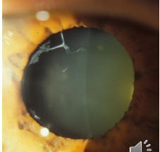
Pseudoexfoliation (clinical signs on anterior segment)
PXM on the anterior lens surface with a clear ring at the pupil- lens contact zone; rubbing off PXM
PXM and pigment may be visible on the corneal endothelium → can mimic keratic precipitates (KP)
PXM and clumps of pigment seen on the posterior TM with gonioscopy and at Schwalbe's line (Sampaolesi's line)
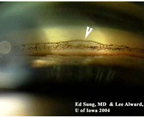
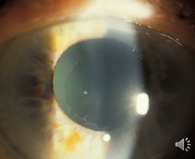
Pigment Dispersion Syndrome (PDS)
Pigment from posterior iris is lost due to mechanical rubbing of the iris on the zonules caused by the iris curvature
Pigment deposits in the TM, blocking aqueous outflow → pigmentary glaucoma
About of patients with PDS develop glaucoma
More common in males (ratio ≈ )
Associated with myopia: deep anterior chamber (AC) and convex curvature of the iris
Typically presents in individuals in the mid-30s or older
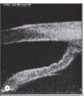
Pigment dispersion signs (PDS)
Pigment deposition on the corneal endothelium, usually in a vertical band centrally (Krukenberg spindle)
Peripheral iris transillumination with slit-like defects
Pigment dusting on the peripheral iris
Heavy pigment seen on the posterior TM on gonioscopy
Figure reference (Pigment Dispersion)
(a) Krukenberg spindle; (b) pigment granules on iris surface and partial loss of the pupillary ruff; (c) radial slit-like transillumination defects; (d) homogeneous trabecular hyperpigmentation
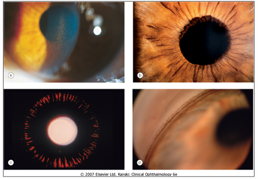
Rubeosis Iridis
Neovascularization of the iris and angle → neovascular glaucoma (secondary open-angle glaucoma; can be closed-angle if fibrovascular tissue contracts, causing anterior synechiae)
Common causes:
diabetes mellitus (proliferative diabetic retinopathy)
central retinal vein occlusion (100-day glaucoma)
any retinal ischemia leading to neovascular response
Management: refer promptly; treat the underlying cause (e.g., panretinal photocoagulation, PRP) and manage the glaucoma
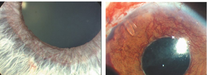
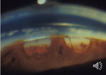
Vessels proliferate in the angle to form a fibrovascular membrane → trabecular blockage
Fibrovascular membrane may contract and pull the peripheral iris over the TM → progressive angle closure
Inflammation of the anterior segment (uveitis) and glaucoma
Cells and flare in uveitis may lead to inflammatory/uveitic glaucoma
Two mechanisms: with pupil block and without pupil block
With pupil block
Angle-closure glaucoma with pupil block: inflammatory adhesion (posterior synechiae) at the pupil margin causes pupil block
Iris bombe can occur, leading to angle closure
Without pupil block
Closed-angle glaucoma: inflammatory cells/flare cause peripheral iris sticking to TM and cornea (anterior synechiae) closing the angle
Open-angle glaucoma: inflammatory material in the anterior chamber may block TM, causing rise in IOP
IOP often falls as inflammation subsides
Peripheral anterior synechiae (PAS)
Abnormal adhesions between the iris and cornea or TM contributing to angle closure risk
Fuchs’ heterochromic iridocyclitis (Fuchs’ uveitis)
Idiopathic, unilateral (≈90%), chronic uveitis
Often resistant to steroids
Common complication: cataract; glaucoma in about of patients
Rare overall: accounts for about of all uveitis cases
Typically diagnosed in the 30s–40s
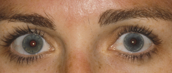
Additional signs
Affected eye shows lighter, paler iris and iris atrophy
Cataract formation
HOODD (presentation on slides; not a diagnostic criterion here)
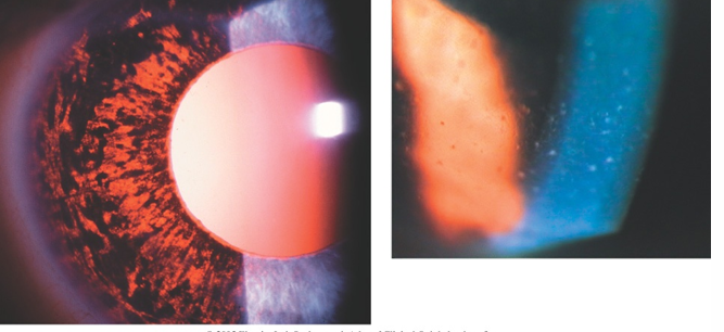
Posner-Schlossman syndrome ( glaucomatocyclitis)
Recurrent attacks of unilateral acute iritis with secondary open-angle glaucoma
The eye often appears relatively white; open angle on gonioscopy
Aqueous shows few cells with fine central keratic precipitates (KP)
Elevated IOP thought to be from trabeculitis
Typical age range:
IOP elevation lasts for hours to days
Symptoms: mild discomfort, halos, slight blur; corneal edema
IOP can reach during attacks
Phacolytic glaucoma (lysis/burst of lens capsule)
Occurs with mature or hypermature cataract
Soluble lens proteins leak through the capsule and obstruct TM
Acute IOP elevation
Redness, pain, intense aqueous flare
White particles in aqueous humor; macrophages loaded with lens protein
Classic example of lens-induced glaucoma
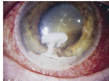
Phacomorphic glaucoma (morphology/shape)
Acute secondary angle closure due to lens enlargement (intumescent lens)
Mechanisms:
Pupillary block
Lens pushing the iris forward
Both cause irido-corneal contact and angle closure
Acute secondary angle closure • Caused by intumescent (expanding) lens
Traumatic glaucoma
Hyphema: blockage of TM by red blood cells
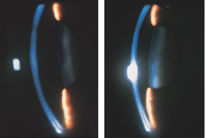
Angle recession: rupture of the inner circular/facial aspect of ciliary body between iris root and scleral spur due to blunt trauma
Rise in IOP secondary to trabecular damage Glaucoma may develop months or years later Gonioscopy
Iridodialysis: iris separation from ciliary body
Rise in IOP secondary to trabecular damage Glaucoma may develop months or years later Gonioscop
Management and treatment notes (post-traumatic and steroid-related)
Steroid-related pathways require monitoring of intraocular pressure (IOP) for hyphaema
IOP-lowering medications may be used for non-inflammatory glaucoma management while addressing the inflammation
Gonioscopy should be performed after blood clears
Iridocorneal Endothelial (ICE) syndrome
ICE is a group of three overlapping disorders:
Progressive iris atrophy
Iris nevus (Cogan-Reese) syndrome
Chandler syndrome
Characterized by abnormal corneal endothelium that proliferates and migrates across the angle to the iris
Can cause corneal decompensation and glaucoma (angle-closure due to synechiae) or open-angle glaucoma
Believed to have a viral association
Differential diagnosis includes Axenfeld anomaly (autosomal dominant, bilateral)
ICE signs
Typically unilateral in middle-aged women
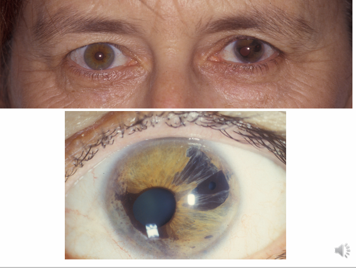
Corectopia (displaced pupil)
Pseudopolycoria (false extra pupils)
Iris atrophy
Corneal endothelial abnormalities with a hammered-silver appearance
Broad peripheral anterior synechiae extending to Schwalte’s line
Glaucoma develops in about of cases
Sturge-Weber Syndrome
Port-wine stain (naevus flammeus) of the face, following the distribution of the trigeminal nerve
Ocular involvement: conjunctival, episcleral, and choroidal haemangiomas
Glaucoma more common when lids and conjunctiva are involved (≈ )
Mechanism unknown:
in children, raised IOP may be due to malformation of the trabecular meshwork
in adults, raised episcleral venous pressure contributes
Glaucoma in Sturge-Weber: reduced aqueous outflow through episcleral veins due to the haemangioma

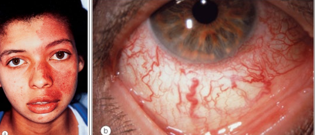
Carotid-Cavernous Fistula
Fistula = abnormal communication between an artery and a vein
Blood within the venous system becomes arterialised; venous pressure rises, drainage altered
Communication between the carotid artery and the cavernous sinus
Arterial blood enters the sinus; venous blood cannot drain effectively
Glaucoma from raised episcleral venous pressure (EVP)
Types
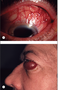
Direct: carotid artery flows directly to cavernous sinus through a defect in the wall of the intracavernous section
often high-flow
commonly trauma-related or spontaneous rupture of a intracavernous aneurysm or atherosclerotic artery
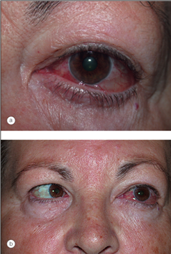
Indirect: intracavernous carotid is intact
arterial blood reaches cavernous sinus via meningeal branches of external or internal carotids
slower flow
congenital malformations or spontaneous rupture
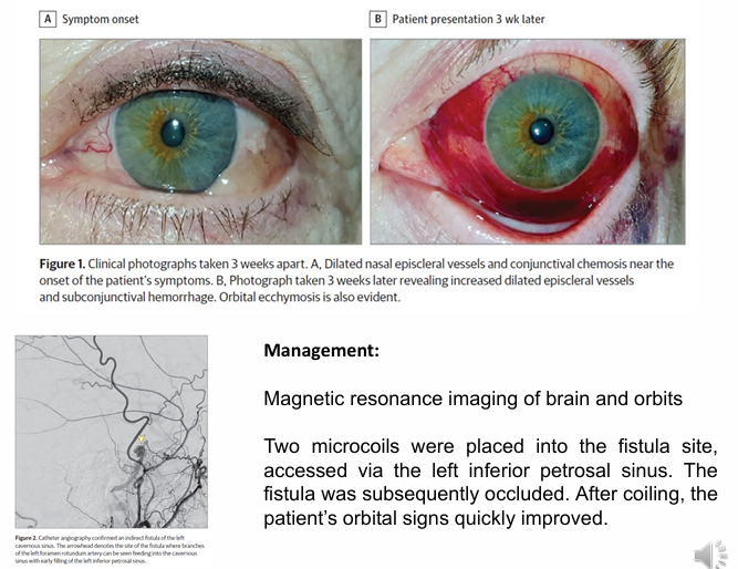
Fistula - Imaging and management notes
Management can include imaging (e.g., MRI) and endovascular intervention (e.g., coil embolization) for fistulas
Example: MRI of brain/orbits with endovascular coiling to occlude the fistula and rapid clinical improvement
Intraocular tumours and angle-related pathologies
Intraocular tumours can involve the iris or ciliary body or anything affecting the anterior chamber angle
Case example (scan data)
70-year-old female with sore eye for 10 weeks; VA R = 6/7.5, L = 6/6; IOP: R = 39 mmHg, L = 12 mmHg
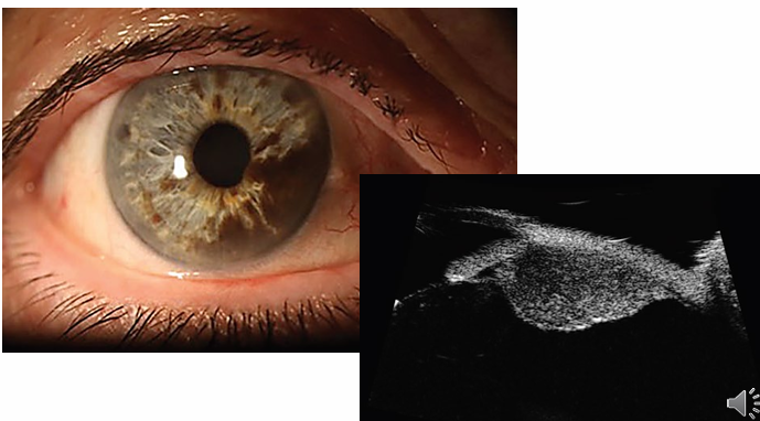
Steroid-induced glaucoma and routes of administration
Steroid-induced glaucoma: elevated IOP due to increased outflow resistance from steroids
Upregulation of glucocorticoid receptors in trabecular meshwork
suppression of phagocytic activity → deposition of material in the angle
Risk increases with duration and dose (penetration of tissue)
IOP rise typically occurs 2–4 weeks after starting steroids
Management: cease steroid if possible; consider non-steroidal anti-inflammatory alternatives; initiate glaucoma therapy when needed
Steroid routes and their implications
Topical ocular preparations: drops or ointment applied to the eye/eyelids
Periocular: subconjunctival, sub-Tenon’s, or retrobulbar injections
Intravitreal: about of patients develop an IOP spike within 2–4 weeks
Dermatologic: steroids applied to the skin around the eyelids (long-term use)
Systemic: oral steroids; rare, may include inhaled or nasal steroids