Lecture 8: Size Exclusion, Ion Exchange, and Affinity Chromatography
Chromatography Overview
Historical Origins:
1900: Russian-Italian botanist Mikhail Tsvet developed a liquid-adsorption column containing calcium carbonate () while studying chlorophyll to separate plant pigments. He coined the term “chromatography” from the Greek for “color writing.”
1952: The Nobel Prize was awarded for the development of Partition Chromatography.
Core Terminology:
Analyte: The specific molecule being separated.
Immobilized Phase (Stationary Phase): The solid matrix or beads inside the column.
Eluent (Mobile Phase): The buffer or solvent that flows through the column.
Basic Process: Sample analyte is loaded onto the column (), and then eluted with solvent sequentially to separate components based on physical or chemical properties.
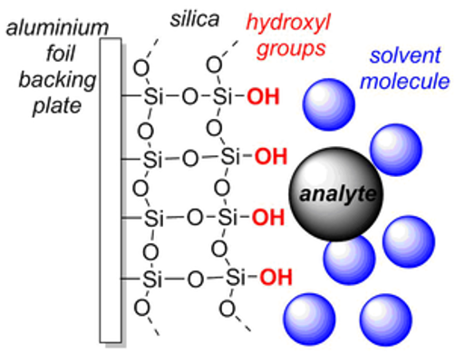
The Stationary Phase (The Mud): This is the solid material packed inside the column that stays completely still.
The Analyte (The Runners): This is your mixture of different chemicals. They start together at the starting line (the top of the column).
The Eluent/Mobile Phase (The Wind): This is the liquid solvent you pour in. It acts like a strong wind blowing the runners down the track.
Liquid Chromatography (LC) for Separation
Mobile phase = solvent, stationary phase = solid, gel, or resin
Analyte sample is loaded on top and separates in column based on affinity/interaction with column

General Purpose: Separates metal ions and organic compounds in solution.
Operating Pressure: Operates at low pressure, typically .
High-Performance Liquid Chromatography (HPLC):
Development: Created in the 1970s using high-pressure pumps (up to ) to increase efficiency.
Mechanism: High pressure allows for smaller beads, which provides increased surface area for separation; tighter packing and smaller particles create narrower, more restricted pathways for the solvent (mobile phase) to flow through.
Sensitivity: Can identify and quantify compounds present in parts per trillion.
Components: Columns, beads, and samples must withstand extreme pressures.
Applications: Pharmaceuticals, environmental samples, forensics, industrial chemicals, nucleotides, peptides, and clinical analysis (e.g., drugs in urine, vitamin deficiency in blood serum).
Protein Limitation: HPLC is rarely used for protein purification because high pressure often causes denaturation, though some low-pressure variants exist.
Other Chromatography Types:
Gas Chromatography (GC): Used for volatile compounds. Mobile phase = gas; stationary phase = solid adsorbent.

Thin Layer Chromatography (TLC): A rapid method to monitor reactions or check purity of organic compounds. Mobile phase = solvent such as acetone or alcohol; Stationary phase = paper, cellulose, or silica.

Protein Purification Components and Troubleshooting
Chromatography for protein purification;
FPLC = Fast Protein Liquid Chromatography
LPLC = Low Pressure Liquid Chromatography
System Components:
Column and beads/gel matrix.
Buffers and gradient mixers.
Peristaltic or piston pumps.
Conductivity meter, UV detector (measuring ), and fraction collectors (automated spiral or serpentine types).
Column Maintenance and Troubleshooting:
Many different types: size exclusion, ion exchange, affinity, hydrophobic
Loading: Columns have a maximum load; larger/longer columns offer higher resolution.
Air Bubbles: Buffers must be degassed and kept at to prevent bubbles.
Cracks: Occur if the column runs dry.
Clogs: Caused by insoluble particles or microbial growth. Clean with . Store columns in absolute ethanol () or sodium azide () to prevent growth.
Proteins may not elute, charge/hydrophobic interactions with beads
Gradient mixer for the eluting agent/eluent (NaCl or imidazole): built into the chromatogram, increases concentration of eluent in column
Fraction Collector:
Measuring absorbance at ().
Collect the fractions from the column by hand or using an automated fraction collector (spiral or serpentine)
Fractions can be collected by time or by drop number (to prevent variations)
Some can collect 384 fractions plus flow-through and washes
Detecting the proteins
A of the individual fractions (chromatogram)
Bradford or Lowry protein assay
The biological activity of aliquots from the individual fractions can be assayed (CDNB assay, GFP fluorescence)
SDS-PAGE (size)
Western blot (antibodies)
For enzymes: specific activity (units/mg of protein) increases as purification techniques are used
Size Exclusion Chromatography (SEC)
History:
1940s: Starch columns used for amino acid/protein analysis.
1956: Grant Lathe and Colin Ruthven demonstrated size separation on starch columns in London.
Arne Tiselius: Discovered dextran production by bacteria on beets. His student, Stellan Hjert, developed acrylamide.
1959: Jerker Porath and Per Flodin cross-linked dextran to show protein separation by size.
Mechanism: Also called gel filtration or gel permeation chromatography.
Separates by size and shape.
Mobile phase = solvent; Stationary phase = a packing of porous particles

Separating molecules based on size (MW); Best for molecules with at least a -fold difference in molecular weight ().
Desalting (buffer exchange)
Removing ammonium sulfate after a precipitation

The sample is applied in a narrow band at the top of the column and then washed through the column across the mobile phase
Band broadening as goes down the tube (injecting <10 ml helps reduce this), therefore recovery may be <80%
The Gel Matrix:
Polymers: Dextran, Acrylamide, Agarose.
Exclusion Limits: G-50 beads excludes proteins above roughly ; G-100 excludes above
Process:
Very large molecules are completely excluded, elute in the void volume (first fractions)
Small proteins usually enter the beads, elute later
Buffer and salts enter the gel/beads, elute last in the column volume


SEC vs. SDS-PAGE:
SEC is preparative ( (cold temperature), native protein), whereas SDS-PAGE is analytical (denatured, boiled).
Large proteins move fastest in SEC but slowest in SDS-PAGE.
Peak Shape/Resolution
Larger/longer columns will provide better resolution, as seen by individual peaks
Peaks should follow normal distribution
Bead Shape/Resolution
Sephadex (separation Pharmacia dextran) beads
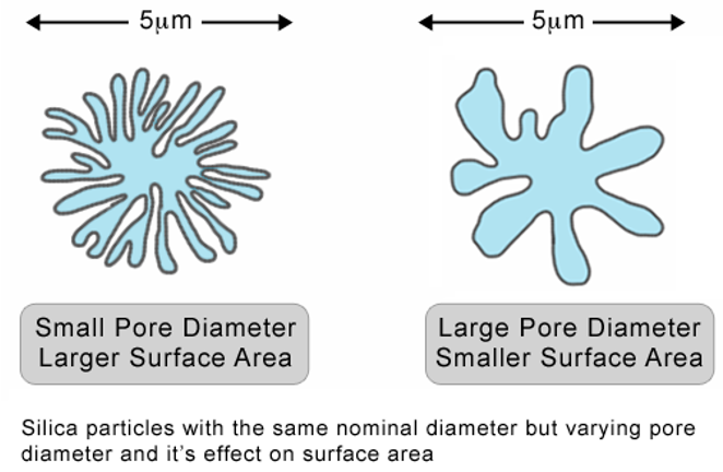
Beads have exclusion limits; G-50 should exclude proteins above 50 kDa, but this rating is based on dextran isolation (not globular proteins) so may not be perfect,
G-50 beads work best from 1.5-30 kDa; G-100 from 4-150 kDa
Flow rate and viscosity affect resolution due to bead interactions
Beads typically 20-300 mm, smaller beads increase surface area but also pressure
smaller beads mean more total surface area (SA) because they pack more tightly together and inherently have a higher surface-to-volume ratio.
reducing the "interstitial volume" (the empty space between the beads). This forces the fluid mobile phase into narrower
As a sphere gets smaller, its volume decreases cubically (r³) while its surface area decreases only quadratically (r²). This means that smaller beads yield a significantly higher ratio of surface area per unit of volume.
Stokes Radius
the effective radius a molecule has as it tumbles rapidly in solution (i.e. the radius of a hard sphere that diffuses at the same rate as the molecule)
A long extended molecule has a larger Stokes radius than a compact molecule of the same molecular mass, and interacts differently with the beads

Applications
SEC is compatible with physiological conditions (pH, salts) and can be used with most soluble proteins
Separates proteins of different molecular weights
Separates monomers from dimers, trimers, tetramers, etc.
Separates proteins from aggregates or degraded products, which often form during protein purification
Removes salts via a buffer exchange: since the salts/buffer from the sample take the longest path, the proteins transfer into the buffer that the column is equilibrated in.
SEC is often a final polishing step to remove aggregates and act as buffer exchange into the final solution

SEC columns are used analytically for studying:
Protein purity
Protein folding
Protein interactions, mutation analysis
Quaternary structure (oligomeric state)
Mw with a calibration (standard) curve
Ion Exchange Chromatography (IEC)
Fundamentals: Separates proteins based on molecular net charge.
Proteins have positive and negative side chains that vary with pH.
Ions immobilized on a matrix bind charged protein groups; bonds are broken by changing ionic strength () or .

Types of Exchangers:
Anion Exchangers: Positive beads bind negative proteins. Counterion is Chloride ().
Cation Exchangers: Negative beads bind positive proteins. Counterion is Sodium ().
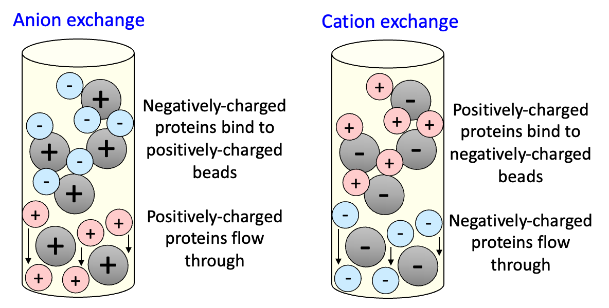
Anion Exchange
Positive and neutral flow through
In order to get the anion out of column, add Cl- (NaCl) to compete with (+) charged column
Cation Exchange
Negative and neutral flow through
In order to get the cation out of column, add Na+ (NaCl) to compete with (-) charged column

In an anion exchange, as NaCl is added, proteins desorb from the column and into the buffer
Proteins elute based on net charge; weakly-binding proteins elute first (in this case positive and neutral charged)
At a certain level of ionic strength, all the sample components are fully desorbed and move down the column with the same speed as the mobile phase
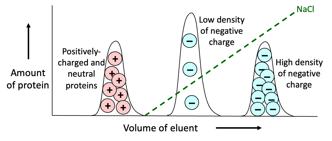
Absorbed proteins are eluted in order of least to most strongly bound molecules by increasing strength or varying pH
PH and pI Elution:
Isoelectric Point (pI): The where net charge is zero.
Further apart pI of protein is from pH of environment/buffer = greater charge difference = stronger binding
Elution Mechanism:
Proteins with lower charge difference (weaker binding) elute first
Typically uses a gradient from low salt to high salt elution buffer (e.g., ).
Fractions collected are based on salt gradient
Protein Charge
Proteins carry both (+) and (–) charged groups
Protein charge is highly pH dependent, affected by the pKa of termini, side chains
pH > pI: protein will have net negative (-) charge, use anion exchange
pH < pI: protein will have net positive (+) charge, use cation exchange
At low pH, there is a lot of H+ in solution and less on the protein H+ diffuses on to amino acid, creating a (+) charge.
As pH increases there is less H+ protons in solution H+ diffuses off the protein, creating a (-) charge
N-terminus;
C-terminus;
Choice of an IEX column depends on the following:
pH range where the protein of interest is stable
Solubility test: aliquot or dialyze sample of protein into various pH and salt concentrations, look for stability or precipitation
If the protein is most stable at a pH above its pI, the protein will be negatively-charged (–), and an anion exchange column should be used
If it is most stable at a pH below its pI, the protein will be positively-charged (+), and a cation exchange column should be used
pH must be at least one unit away from pI; further away from pI means stronger binding
Both anion and cation exchange can be used, but you must change the pH and remove the salt from the sample (via SEC or dialysis) after the first column before the second IEX can be used
IEX Material
Cellulose and cross-linked dextran (Sephadex, separation Pharmacia dextran) or agarose (Sepharose, separation Pharmacia agarose) are used for the IEX beads
Anion Exchange uses (+) charged material; Strong Quaternary ammonium (Q) antimicrobial found in fabric softener and hair conditioner OR weak Diethylaminoethyl (DEAE)
Cation exchange uses (-) charged material: Strong Sulfonic acid (S) OR weak carboxymethyl (CM)
Hydrophobic and Affinity Chromatography
Hydrophobic Interaction Chromatography (HIC):
Stationary phase: Inert hydrophobic groups (ethyl, propyl).
Binding: High salt (e.g., ammonium sulfate) enhances hydrophobic interactions.
Elution: Reducing salt concentration or using detergents. Least hydrophobic proteins elute first, more hydrophobic proteins elute later
Reverse-Phase Chromatography:
Type of HIC for small molecules/peptides on HPLC.
Uses long-chain hydrocarbons (C8-C18) on silica inside of column
Performed under high pressure (HPLC); eluted with acetonitrile or methanol

Affinity Chromatography:
Highest selectivity. Exploits specific biological interactions.
Affinity chromatography involves purifying proteins through complementary interactions
Stationary phase = an immobilized ligand chemically coupled to a dextran, acrylamide, or agarose bead/matrix that binds with high specificity to a single protein
The target protein binds to the ligand, and impurities wash away
Types: Enzyme/substrate, hormone/receptor, antibody/antigen, avidin/biotin, metal/histidine.

Elution: Usually involves a gradient of free ligand solution to compete with the column-bound ligand for the target protein
Can be a mobilized substrate, mobilized antigen, mobilized receptor, histidine ring (for metals)

Gene Fusion and Tagging Systems
Gene Fusion Technique: Fusing the gene of interest to a “tag” for purification, which is encoded in the vector used for cloning

GST (Glutathione-S-transferase): Binds to glutathione beads; eluted with reduced glutathione ().
MBP (Maltose-binding protein): For solubility; binds to amylose; eluted with maltose.
GFP (Green Fluorescent Protein): For tracking;
CBP (Calmodulin-binding), Thioredoxin, NusA, GST, MBP, and Chitin-binding all increase solubility of protein
Vector Design: Contains a promoter, the tag, a linker, and the multiple cloning site (MCS).
Linker;
allow for flexibility and folding
contains restriction enzyme and protease cut sites

Construct a fusion gene using restriction enzymes and transform it into bacteria; example = P53 gene put in right after MBP tag in the multiple cloning site (MCS)
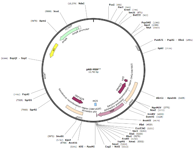
Grow bacteria, induce expression of the fusion gene, centrifuge and lyse cells
Run lysate on amylose affinity column, where the MBP-target protein will be bound (elute with maltose)
Cleave the fusion protein with a protease at the linker, separating MBP from the target protein
Rerun sample on the amylose column to separate the target protein (P53) from MBP (tag) as MBP binds to amylose column.
The tag may need to be cleaved off and removed, which can take time and degrade target protein
Immobilized metal affinity chromatography (IMAC): a type of affinity chromatography based on the chelation of metals such as nickel or cobalt by histidine (sample cannot have any EDTA)
The gene of interest is modified to encode a polyhistidine tag, usually six histidines in length, at either the N- or C-terminus of the protein. The protein with histidine will bind to nickel which are bound to beads on column.
Columns are eluted with imidazole, which competes with histidine for the immobilized nickel
His-tags are Encoded in Vectors or Primers
Comes in the vector

or in the primer

Magnetic His Beads
Magnetic beads can be used as an alternative to chromatography
Fast, inexpensive, but no gradient

Intein-Mediated Purification:
Chitin is sugar found on insects exoskeleton
Allows for inducible on-column cleavage using DTT, separating the target protein from the chitin-binding domain (CBD) on the column without having to elute with chitin
HaloTag: Covalent linkage to a ligand on the column allows for strong binding; target is released via protease cleavage.

HaloTag is tagged with Histag, so once tag is cleaved, the target protein and HaloTag can be run through a His affinity column and flow through should have target protein.