Structure and Function
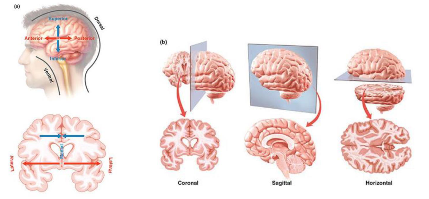
Referencing Location Within The Brain
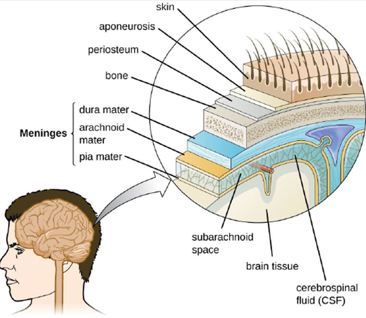
Layers of Brain Tissue
Cerebrospinal Fluid (CSF) and Ventricular System
CSF:
Ultra-filtrate of blood, primarily produced by the choroid plexus within the ventricles.
Supports the weight of the brain, providing buoyancy (approx 150gapprox150g effective weight from 1400g1400g actual weight) and protecting it from mechanical shock.
Homeostatic regulation of the Central Nervous System (CNS) by maintaining a stable chemical environment.
Clearance of metabolic waste products, transporting them away from brain tissue.
Nutrient circulation, delivering essential substances to the brain.
Age-related size increase, where the volume of CSF and ventricles tends to expand with age.
Ventricular system:
A network of four fluid-filled cavities (two lateral ventricles, third ventricle, and fourth ventricle) deep within the brain.
Produces, contains, and circulates CSF throughout the brain and spinal cord, ensuring continuous CNS protection and metabolic support.
Nervous System
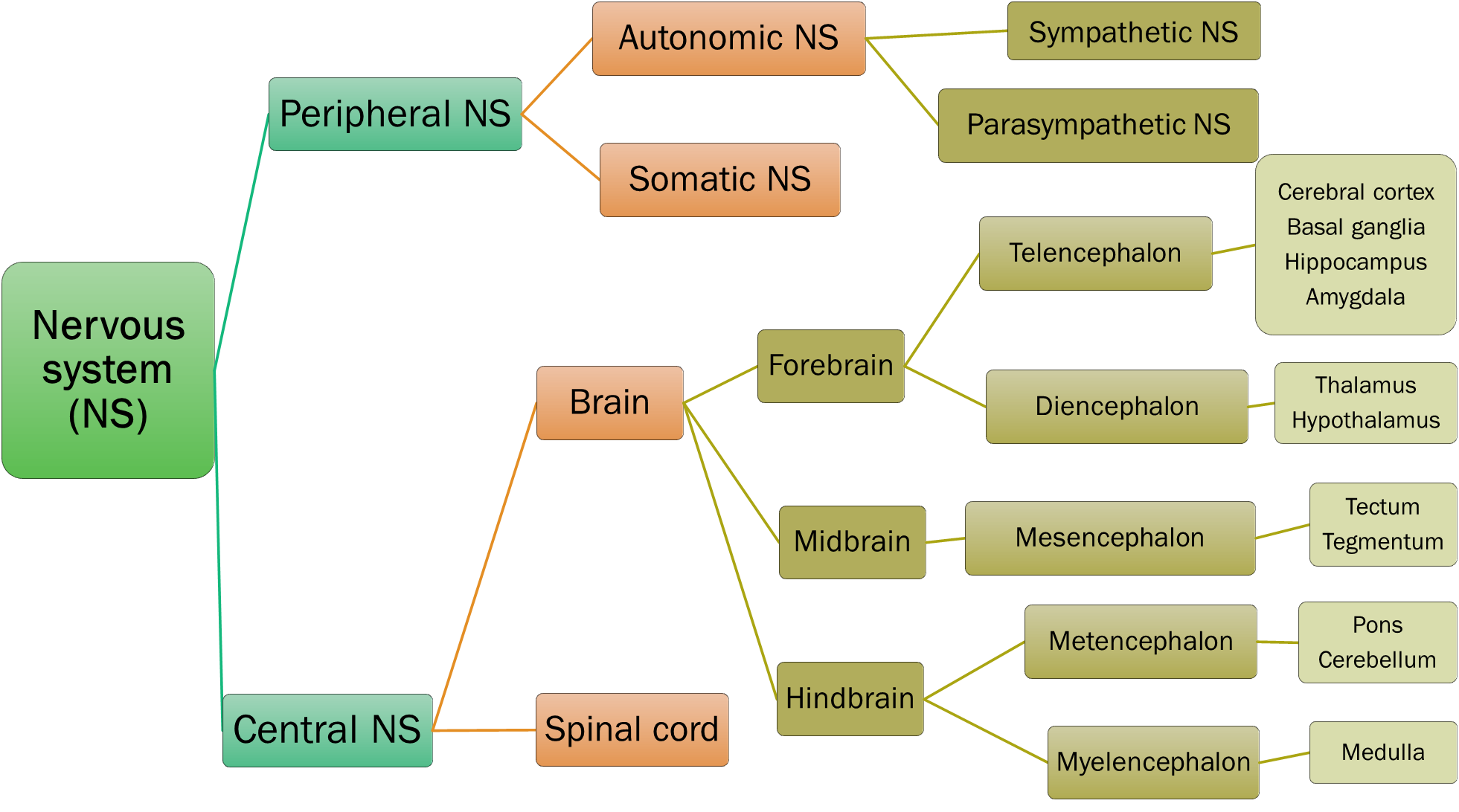
CNS
Brain → 6 subdivisions (major components of the encephalon):
Cerebrum: The largest and most superior part, responsible for higher-level functions.
Diencephalon: Located between the cerebrum and brainstem, essential for sensory relay and autonomic control.
Midbrain: Superior portion of the brainstem, involved in motor control, sensory processing, and arousal.
Pons: Middle portion of the brainstem, connecting the cerebrum and cerebellum, involved in sleep, respiration, and sensory information from the face.
Medulla Oblongata: Inferior portion of the brainstem, regulating vital autonomic functions like heart rate and breathing.
Cerebellum: Posterior to the brainstem, crucial for motor coordination, balance, and fine-tuning movements.
(Last three in the brainstem: Midbrain, Pons, and Medulla Oblongata, which together form a critical conduit for nerve signals and house many cranial nerve nuclei.)
The Cerebrum
Largest component of the brain, constituting about 85%85% of its total weight.
Composed of two cerebral hemispheres (left and right), separated by the medial longitudinal fissure.
The outermost part is the cerebral cortex, a layer of grey matter responsible for conscious thought, memory, language, and perception.
Most evolutionarily new part of the brain (neocortex), highly developed in humans.
Contains approximately 94% of all neural tissue in the brain.
Only 2% of body weight but receives 16% of blood supply, reflecting its high metabolic demand (20% of the body's oxygen and glucose consumption).
Divided along the midline by the medial longitudinal fissure.
Consists of 4 primary cerebral lobes, each associated with distinct functions:
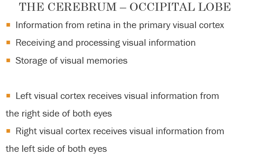
Occipital lobe:
Primarily houses the visual cortex, responsible for processing visual information such as interpreting colour, form, and motion.
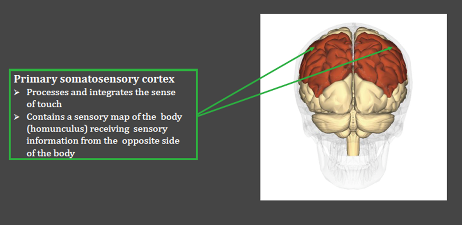
Parietal lobe:
Involved in attending to stimuli (internal or external), spatial awareness, processing somatosensory information (touch, temperature, pain), and integrating sensory input.
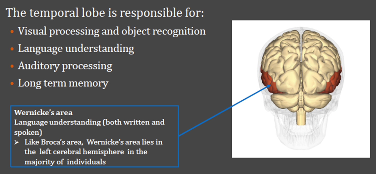
Temporal lobe:
Crucial for identifying the nature of auditory stimuli (hearing), processing memory, language comprehension (Wernicke's area), and emotional responses.
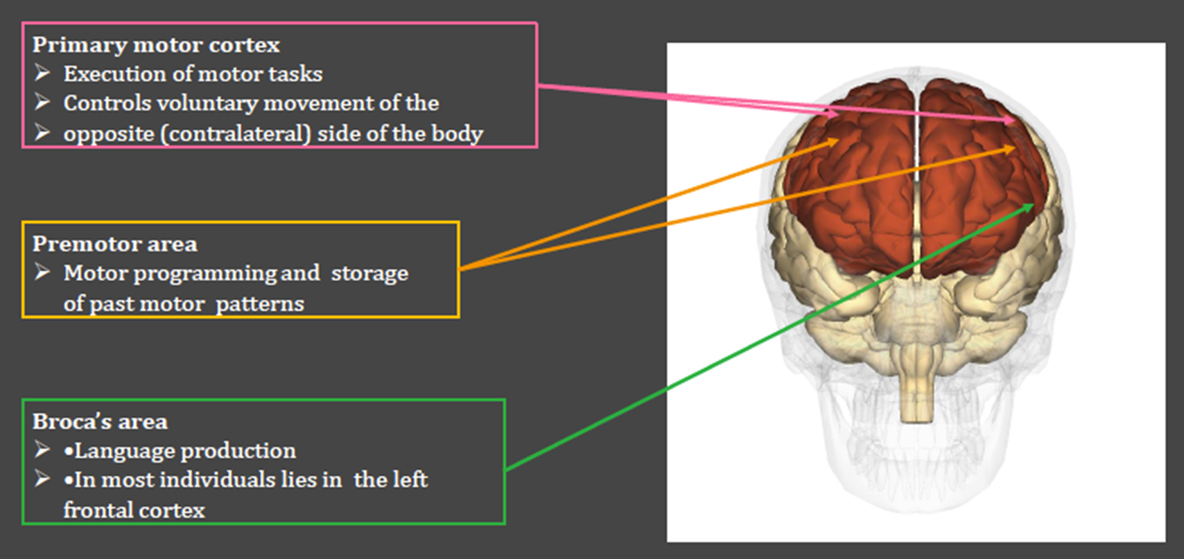
Frontal lobe:
Responsible for selecting and planning appropriate behavioural responses, executive functions, decision-making, problem-solving, voluntary movement (primary motor cortex), and speech production (Broca's area).
Association Cortex:
Regions of the cerebral cortex that are not directly involved with primary sensory experiences or motor responses.
Responsible for linking up information from various sensory modalities, higher-order cognition, language processing, abstract thought, and constructing a coherent/unitary understanding of the world.
Includes prefrontal cortex, temporal-parietal junction, and other areas.
The Cerebral Cortex
A thin sheet of grey matter tissue, typically 2−4mm2−4mm thick, forming the outer layer of the cerebrum.
Primarily composed of grey matter (neuron cell bodies, dendrites, unmyelinated axons, glial cells).
Extensively convoluted (folded) into ridges and grooves, which significantly increases its surface area (up to 2500 cm22500cm2) to fit within the restricted cranial volume. This convolution allows for a much larger number of neurons.
Outward bulge: gyrus (plural gyri) - a ridge on the cerebral cortex.
Inward fold: sulcus (plural sulci) - a groove or furrow on the cerebral cortex.
Larger or deeper sulci are often called fissures (e.g., longitudinal fissure, lateral fissure).
Irregular in shape and vary in configuration and location between individuals, though major gyri and sulci are consistent across most brains.
Diencephalon
Located superior to the brainstem, forming the central core of the forebrain.
Key components include:
Thalamus: Major communication and relay centre for sensory information.
Hypothalamus: Regulator of internal body states and basic drives.
Pituitary gland: Master endocrine gland, controlled by the hypothalamus.
Pineal gland: Important for circadian rhythms.
Thalamus:
Often referred to as the "gateway to the cortex" due to its role as the major communication centre for almost all sensory information (except olfaction) ascending to the cerebral cortex.
Plays a vital role in relaying information from the cerebellum and basal ganglia to the primary motor cortex, influencing motor control and coordination.
Involved in pre-processing of initial auditory and visual sensory information before it reaches the respective cortical areas.
Has a significant role in arousal, awareness, consciousness, and cognition by regulating information flow to the cortex.
Hypothalamus:
Contains many small nuclei, regulating basic survival instincts, such as feeding, fighting, fleeing, and sexual reproduction.
Central to maintaining homeostasis, controlling body temperature, fluid balance, blood pressure, and sleep-wake cycles.
Produces hormones that control the pituitary gland.
Pituitary gland:
An endocrine gland composed of anterior and posterior lobes.
Crucial for endocrine regulation and metabolic functions, secreting hormones that regulate growth, metabolism, reproduction, and stress response.
Pineal gland:
A small endocrine gland located deep in the brain.
Critical for regulating circadian rhythms (sleep-wake cycles) by secreting the hormone melatonin.
Brainstem
Contains Midbrain, Pons, and Medulla Oblongata.
Serves as a crucial bridge between the spinal cord and the cerebral hemispheres, relaying both motor and sensory pathways.
Site of cranial nerves (nuclei for CN III through CN XII originate here), controlling critical functions like eye movement, facial sensation, taste, hearing, balance, and swallowing.
Regulates essential involuntary functions necessary for survival, such as breathing, heart rate, blood pressure, and consciousness.
Basal Ganglia
A group of subcortical nuclei (not a single structure) deeply situated within the cerebral hemispheres.
Key components include:
Caudate nucleus: Involved in goal-directed behaviour and learning.
Putamen: Plays a role in motor control and learning.
Globus Pallidus (not explicitly listed but a key component):
Nucleus accumbens: Part of the reward system, involved in motivation and pleasure.
Substantia nigra: Located in the midbrain, critical for dopamine production, which is essential for smooth movement (degeneration leads to Parkinson's disease).
Subthalamic nuclei: Involved in modulating motor pathways.
Primarily involved in the control of posture, voluntary movement, motor learning, and executive functions.
Limbic System
Historically defined by James Papez (1937) as a circuit of interconnected brain structures involved in emotion and motivation: the “Papez” circuit.
However, modern understanding (e.g., LeDoux, 2003) recognizes that the concept of a single "Limbic System" is somewhat redundant and oversimplified, with various structures contributing to emotion and memory in complex ways.
Involved in a wide range of emotional responses: anger, pleasure, affection, fear, and also plays a role in memory formation (especially emotionally charged memories) and olfaction.
Amygdala
An almond-shaped structure located within the temporal lobe, part of the limbic system.
Plays a central role in processing and remembering emotional experiences, particularly fear.
Crucial for memory consolidation, decision-making based on emotional significance, and generating appropriate emotional responses.
Hippocampus
A seahorse-shaped structure located in the medial temporal lobe, also part of the limbic system.
Crucial in memory processes, especially the formation of new declarative memories (facts and events) and spatial memory.
Essential for learning and transferring information from short-term to long-term memory.
The Cerebellum
Located at the back of the brain, under the occipital and temporal lobes; means "little brain" in Latin.
Contains more neurons than every other region of the brain combined, packed into a relatively small volume.
Characterized by small neurons firing weak action potentials, allowing for precise fine-tuning of motor commands.
Primary role is in smooth coordinated body movement, balance, posture maintenance, and motor learning (e.g., learning to ride a bike).
Increasingly recognized for its role in language processing, attention, and cognitive functions.
Corpus Callosum
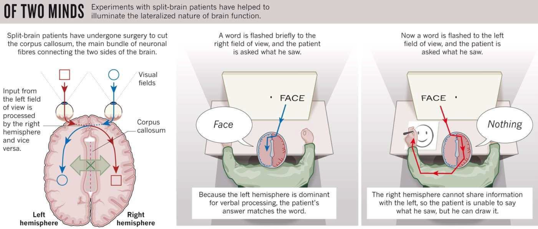
A large, C-shaped nerve fibre bundle connecting the two cerebral hemispheres, located beneath the cerebral cortex.
Research by Sperry & Gazzaniga significantly contributed to understanding brain lateralization (specialization of functions in each hemisphere) by studying "split-brain" patients where the corpus callosum was severed. This provided evidence for hemispheric specialization in functions like language and spatial processing.
Imaging Methods
Structural imaging methods: techniques used to visualize brain anatomy and detect structural abnormalities.
CT (Computed Tomography): Fast structural scans using X-ray technology. Provides cross-sectional images of the brain, useful for detecting large-scale structural changes like haemorrhages, tumours, or fractures quickly.
MRI (Magnetic Resonance Imaging): Provides high-resolution anatomical images based on a strong magnetic field and radio waves. Different tissues in the human brain (e.g., grey matter, white matter, CSF) have different water densities and molecular properties, resulting in distinct appearances on MRI scans, allowing for detailed visualization of soft tissues.
DTI (Diffusion Tensor Imaging): An MRI-based technique that quantifies the relative diffusivity of water molecules in bundles of axons (white matter tracts) to determine their location, orientation, and integrity. Used to map the brain's white matter connections and study neurological disorders affecting these tracts.
Functional imaging methods: techniques used to measure or infer neural activity in the brain.
PET (Positron Emission Tomography): One of the first functional imaging methods. It involves injecting a radioactive tracer (e.g., 2-deoxy-D-glucose or 2-DG, a glucose analogue) into the bloodstream. Active brain regions consume more glucose, accumulating the tracer, which then emits positrons. The PET scanner detects these emissions, revealing the location of neural activity in a living brain. Used to study metabolic activity, blood flow, and receptor binding.
fMRI (Functional Magnetic Resonance Imaging): Measures changes in blood flow (hemodynamic response) and oxygenation (BOLD signal - Blood-Oxygen-Level Dependent) to detect neural activity during specific tasks. It provides a non-invasive, dynamic, 3D map of brain regions involved in functions like memory, language, and movement with good spatial resolution.
EEG (Electroencephalography): Records the brain's electrical activity using electrodes placed on the scalp. It measures synchronized synaptic activity of millions of neurons, detecting changes in brain waves. EEG helps diagnose epilepsy, sleep disorders, and other brain issues by showing abnormal brain wave patterns, and has excellent temporal resolution.
TMS (Transcranial Magnetic Stimulation): A non-invasive brain stimulation technique that uses magnetic fields to generate electric currents in specific nerve cells (neurons) in the brain. This can transiently excite or inhibit brain activity. It is used both for research (to study causality in brain-behaviour relationships) and for therapeutic purposes, such as improving symptoms of major depression and other neurological conditions.
tDCS (Transcranial Direct Current Stimulation): A non-invasive brain stimulation technique that uses a weak, constant direct current delivered via electrodes placed on the scalp to modulate brain activity. It can increase or decrease neuronal excitability, making it a tool for studying and potentially enhancing cognitive functions and for therapeutic applications.
Neuroplasticity
The intrinsic process of structural and/or functional adaptive reorganization of the nervous system, occurring throughout the lifespan, in response to intrinsic (e.g., genetic factors, disease) or extrinsic (e.g., environment, learning, injury) stimulation.
Synaptic Plasticity: Changes in the strength and number of synaptic connections between neurons, underlying learning and memory (e.g., Long-Term Potentiation (LTP) and Long-Term Depression (LTD)).
Structural Plasticity: Changes in the physical structure of neurons and glia, including neurogenesis (birth of new neurons), changes in dendritic arborization, and axonal sprouting.
Functional Plasticity: Changes in the way brain regions are engaged or reorganized to perform specific functions, often seen after injury or intensive training.
Examples: recovery after brain injury (e.g., stroke), learning new skills (repeated stimulation leads to strengthening of neural pathways), degeneration (loss of plasticity in diseases like Alzheimer's), and re-organisation after sensory deprivation or motor skill acquisition.