3.8 Gel Electrophoresis
Gel Electrophoresis - technique used to separate DNA fragments based on their size
Sample of DNA extracted from tissue and exposed to one or more restriction enzymes - cut DNA at specific recognition sites
Sample then placed into small plastic tube
DNA in sample will contain many different sized fragments
If we could see them, they would be easily sorted
Too small to be seen and must be detected in another way
Agarose gel used as “filter” to separate fragments based on size
Technique can also be used to separate other organic molecules such as proteins
DNA fragments placed in “wells” at one end of agarose gel
Electric current placed across gel, causing fragments to move away from well
Occurs because phosphate groups in DNA carry a negative charge, which is attracted to positive electrode
Smaller fragments move through gel more easily than larger fragments - travel farther along gel
Fragments of same size move at same speed and group together
Groups of fragments are still not visible
Stain used to make DNA fragments visible
Commonly used stain - ethidium bromide - inserts itself among complimentary base pairs of DNA and fluoresces under U.V. light
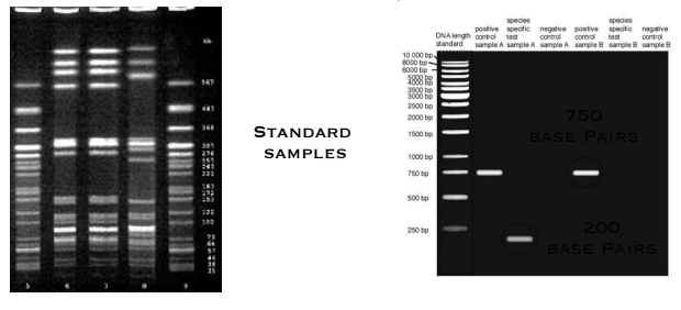
Result is pattern of bands indicating groups of same sized fragments
Length of these fragments can be determined by comparing position of each band to a standard sample of DNA fragments