Lecture 5: Jaw, Occiput, Neck, Clavicle, Scapula, Brachial Plexus
Osteology: Bones of the Head and Neck
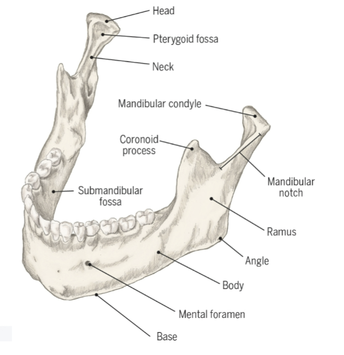
Mandible: In the vernacular, “Jawbone”
Mandibular condyle forms part of the TMJ (temporomandibular joint)
Floor of the mouth~ several tongue and other skeletal muscles form the floor of the mouth
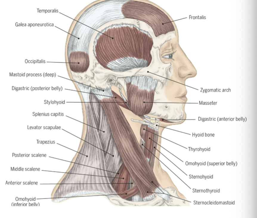
Muscles of the head and neck
The external or superficial muscles of mastication are the Masseter and Temporalis. Deep to those muscles are the Lateral Ptyergoid muscles which come off of the wings of the sphenoid bone.
The strap muscle and digastric muscle raise and lower the hyoid bone and larynx.
The hyoid bone and larynx are connected through membrane, so when one moves the other moves as well.
Cervical Rotation
Rotation of the neck that expands the visual field and helps with occupational performance. Ex: turning your head to the side when going to change lanes while driving.
Plane of Cervical Rotation: Transverse plane
Axis of Cervical Rotation: Vertical axis
Prime movers of Cervical Rotation:
Sternocleidomastoid
Trapezius (upper)
Levator scapulae
Splenius capitis
Splenius cervicis
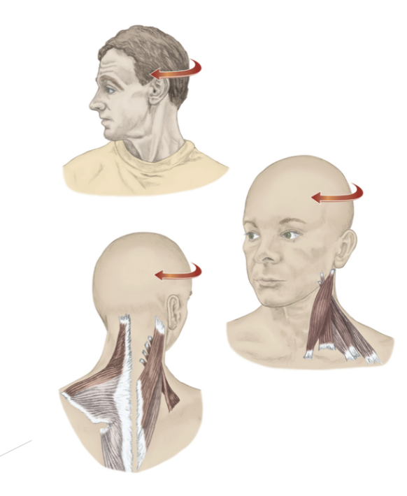
Cervical rotation can happen due to C1 moving around the the Dens of C2. the dens of C2 is actually the body of C1 that did not form properly
Triangles of the Neck
The neck is often categorized into zones in the shape of triangles
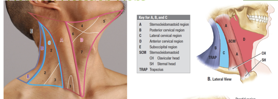
The triangle we will focus on in this course is the lateral cervical triangle and knowing the importance of the Sternocleidomastoid.
The Sternocleidomastoid protects the Carotid artery, Internal jugular vein, and the Vagus nerve.
The lateral cervical triangle (c in the picture above) holds the spinal accessory nerve. Thus radical neck surgeries within the lateral cervical triangle run the risk of damaging the spinal accessory nerve.
*Call Back* If the Spinal Accessory nerve- CN XI (or any nerve) is cut, then all the muscles it innervates will become flaccid. The Spinal Accessory nerve innervates the Trapezius so if its cut then the affect side will have a drooping shoulder and muscle atrophy will occur.
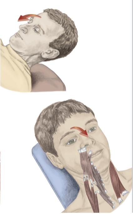
Cervical Flexion
Neck flexion occurs with the assistance of gravity when standing and against gravity when lying supine. Ex: Lying in bed and look at someone who has entered your room. (lifting head up and forward, bring chin towards the chest)
Plane of Movement of Cervical Flexion: Sagittal plane
Axis of Cervical Flexion: Frontal axis
Prime movers of Cervical Flexion~
Sternocleidomastoid
Anterior scalene
Longus capitis
Longus colli
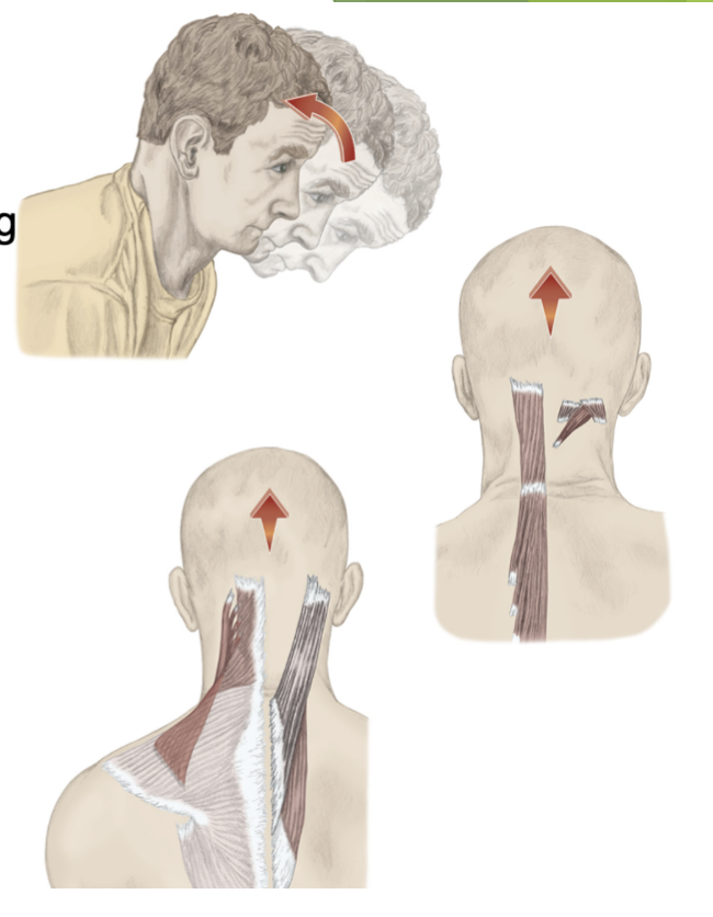
Cervical Extension
Neck extension occurs with the assistance of gravity when standing and against gravity when lying prone. Ex: lying on your stomach watching TV.
Plane of Movement of Cervical Extension: Sagittal plane
Axis of Cervical Extension: Frontal axis
Prime movers of Cervical Extension~
Trapezius (upper fibers)
Levator scapulae (upper fibers)
Splenius capitis
Splenius cervicis
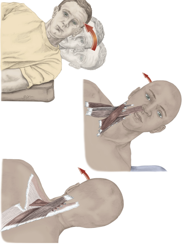
Cervical Lateral Flexion
Lateral flexion of the neck occurs with the assistance of gravity when standing and against gravity when the head is positioned horizontally. Ex: lying on your side and lifting your head up and towards your shoulder
Plane of Movement of Cervical Lateral Flexion: Frontal plane
Axis of Cervical Lateral Flexion: Sagittal axis
Prime movers of Cervical Lateral Flexion~
Trapezius (upper fibers)
Levator scapulae
Sternocleidomastoid
Scalenes (with ribs fixed)
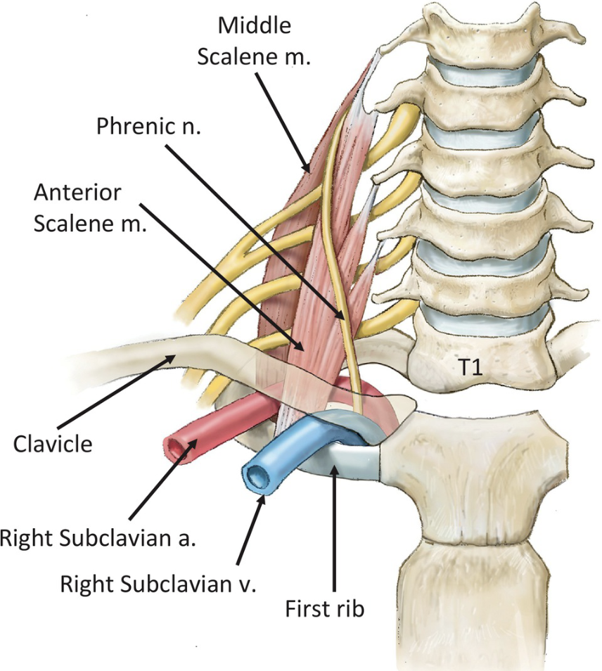
Scalene Muscles
There are 3 Scalenes~ Anterior, Middle, and Posterior
The anterior and middle Scalene muscles insert onto the first rib, and the posterior Scalene inserts on the second rib
*The Brachial Plexus and Subclavian artery pass through the anterior and middle Scalene through the Scalene Interval*
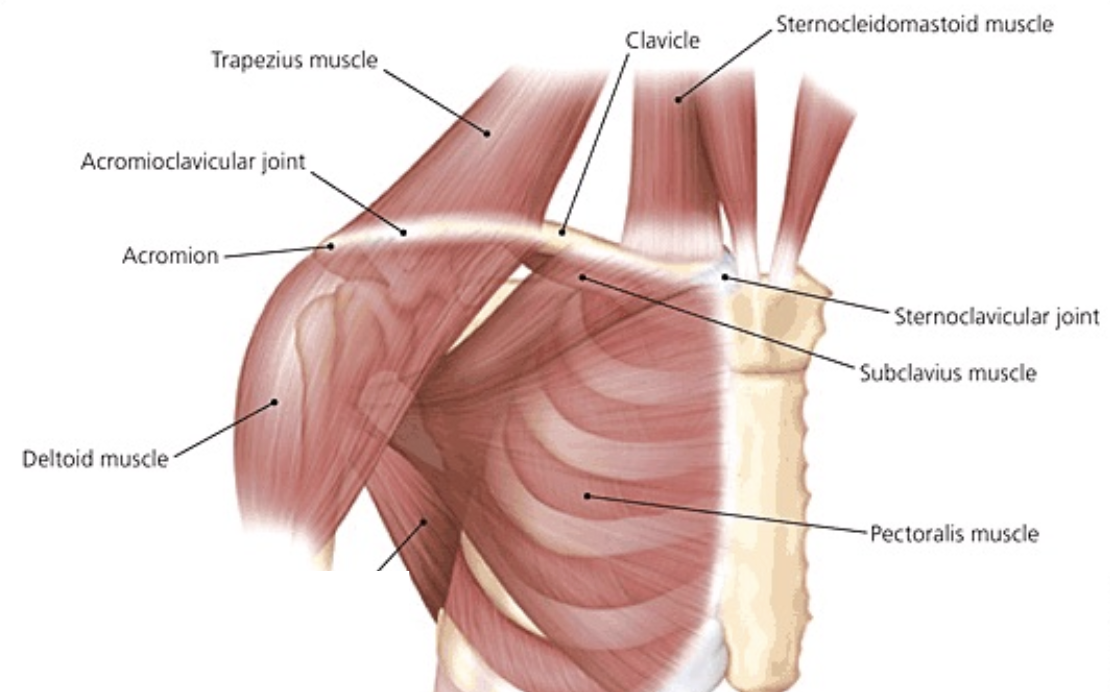
Clavicle
The clavicle bone attaches at the sternum and acromion.
The clavicle and acromion do not meet perfectly, the clavicle is slight higher than the acromion.
The clavicle pushes the upper extremities (UEs) away from the trunk, which allows more ROM.
The clavicle is held in place with 4 strong clavicular ligaments and is an attachment site of 5 muscles.
Muscles Attaching to the Clavicle
Trapezius
Deltoid
Sternocleidomastoid
Subclavius- this muscle is small and helps anchor and depress the clavicle
Pectoralis major
Clavicular Ligaments
There are 4 clavicular ligaments are~ Acromioclavicular, Coracoclavicular, Costoclavicular, and Sternoclavicular ligaments
The clavicular ligaments aim to support and stabilize the clavicle
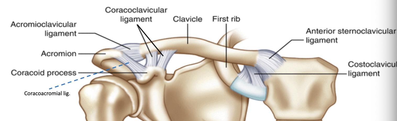
Acromioclavicular Joint (AC)~ the synovial joint where the clavicle and acromion connect.
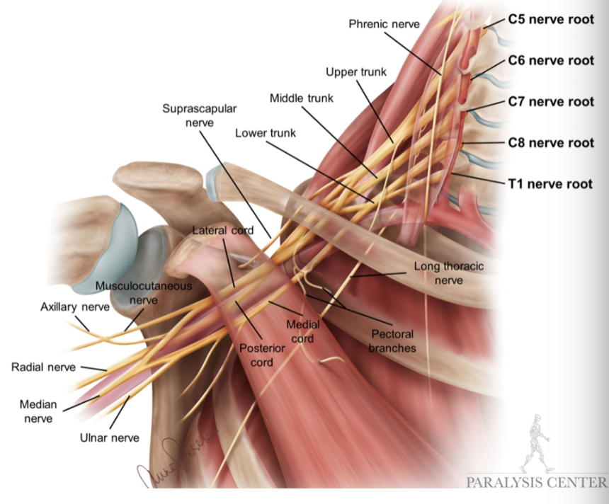
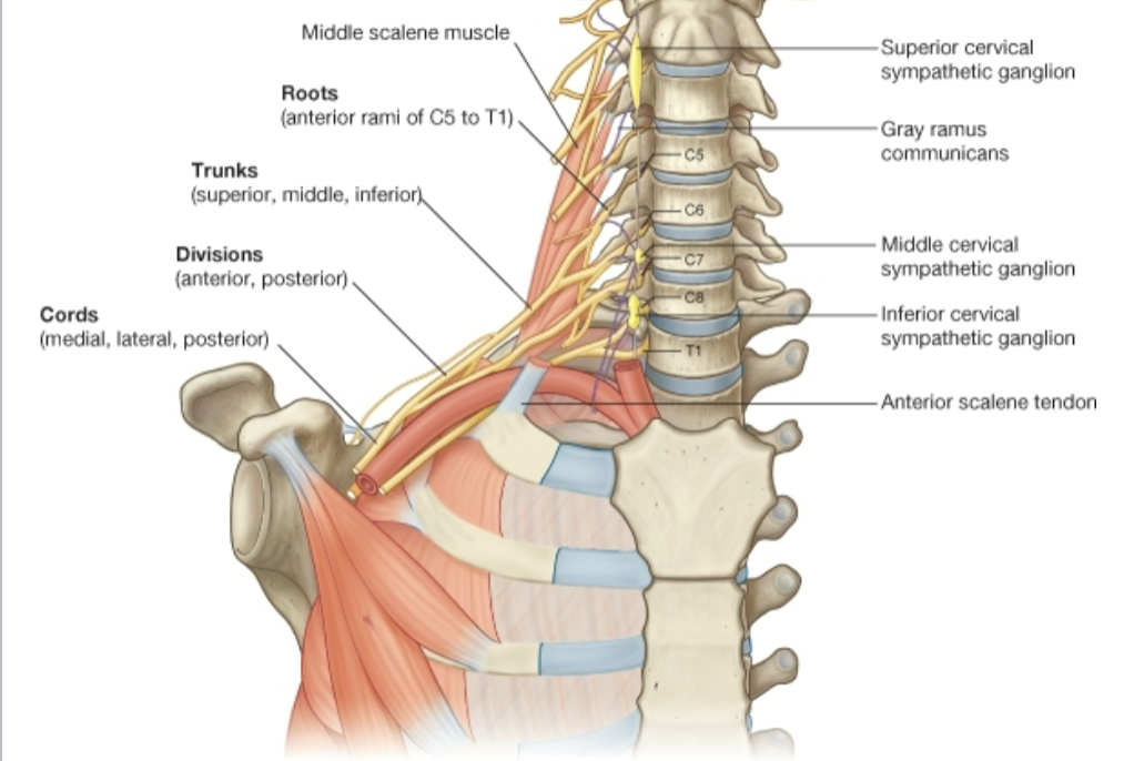
Brachial Plexus
The roots of the brachial plexus emerge between the anterior and middle scalene muscles.
It innervates at least 32 muscles in the shoulder and upper extremity
It bundles some of the axons together for structural strength and bundles some axons together for functional purposes.
Roots~ Ventral Rami of Spinal Nerve (Recall: Ventral rami is the front of the body and Dorsal rami is the back of the body)
The brachial plexus is a network of ventral primary rami that combine to form peripheral nerves of the upper extremity.
The brachial plexus is formed by the union of the ventral rami of C5 through C8 and the greater part of the ventral rami of T1.
The brachial plexus 5 main branches:
Axillary
Musculocutaneous
Radial
Median
Ulnar
Brachial Plexus Sections/Categories
The brachial plexus is sectioned into 5 portions: Root, Trunk, Division, Cord, Branches
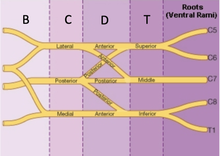
Roots- formed by the ventral rami of C8-T1, the roots usually pass through the scalene interval.
Divisions & Cords- accompanies by the axillary artery
The axillary artery is a continuation of the subclavian artery.
Muscles Innervated by the Brachial Plexus Nerves
Dorsal scapular nerve- Levator scapulae, Rhomboid major, and Rhomboid minor
Suprscapular nerve- Supraspinatus and Infraspinatus
Long thoracic nerve- Serratus anterior
Lateral pectoral- Pectoralis major
Medial pectoral- Pectoralis major and Pectoralis minor
Upper subscapular nerve- Subscapularis
Lower subscapular nerve- Subscapularis and Teres major
Thoracodorsal nerve- Latissimus