knowt logo

Foot Anatomy

Sole/plantar aspect: the bottom of the foot

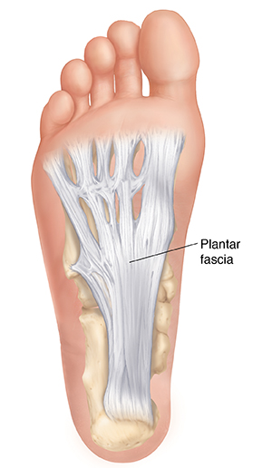
Plantar aponeurosis

a thick band of fibrous tissue which extends from heel to toes

Posteriorly: attached to medial tubercle of calcaneus

Anteriorly: divides into 5 slips that pass to all toes

Laterally: on each side, attached to metatarsal bones by medial and lateral plantar intermuscular septa

plantar aponeurosis

Sole muscles

2 groups of muscles:

  • extrinsic muscles: found in the lower leg, act to dorsiflex, plantarflex, invert and evert the foot

  • intrinsic muscles: found within the foot and primarily act to move the toes and support foot arches to maintain foot structure

Intrinsic muscles

Divided into 4 layers:

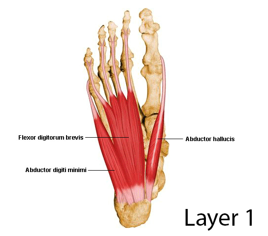
First layer

  • most superficial

  • immediately underneath plantar fascia

Contains 3 muscles:

  1. Abductor hallucis

  2. Flexor digitorum brevis

  3. Abductor digiti mini

All the muscles are supplied by medial plantar nerve, except abductor digiti minimi (supplied by lateral plantar nerve)

First layer of sole

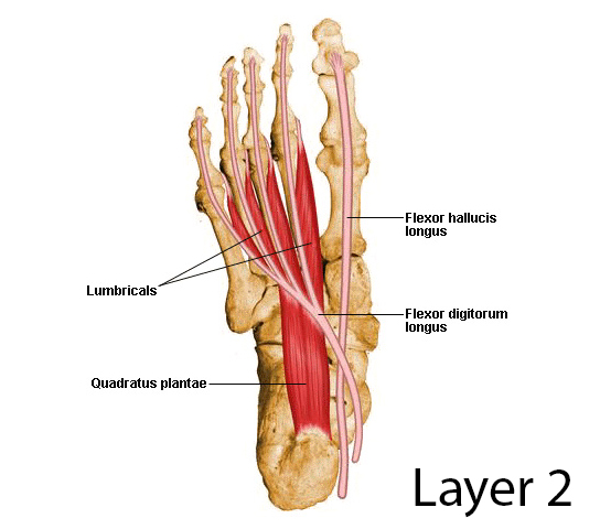
Second layer

  • deep to 1st layer

Contains 2 muscles and 2 tendons:

  1. Quadratus plantae (flexor digitorum accessories)

  2. 4 Lumbrical muscles (1st lumbrical muscle supplied by medial plantar nerve)

  3. Tendon of flexor digitorum longus (crosses superficial to FHL to reach insertion)

  4. Tendon of flexor hallucis longus

All the muscles are supplied by lateral plantar nerve, except 1st lumbrical muscle (supplied by medial plantar nerve )

second layer of sole

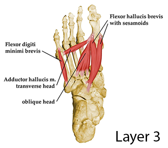
Third layer

  • deep to 2nd layer

  • 2 sesamoid bones develop in tendon of adductor hallucis

Contains 3 muscles:

  1. Flexor hallucis brevis

  2. Adductor hallucis (originates by 2 heads: oblique and transverse)

  3. Flexor digiti minimi brevis

All the muscles are supplied by lateral plantar nerve, except flexor hallucis brevis (supplied by medial plantar nerve )

third layer of sole

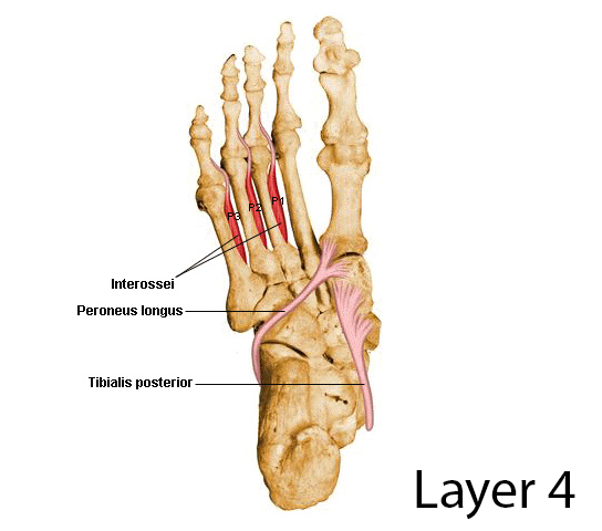
Fourth layer

  • deepest layer

Contains 2 muscles and 2 tendons:

  1. Plantar interossei

  2. Dorsal interossei

  3. Tendon of fibularis longus

  4. Tendon of tibialis posterior

All muscles are supplied by lateral plantar nerve

fourth layer of sole

Sole Nerves and Vessels

Plantar nerves

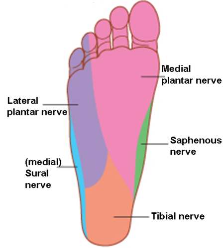
The sole is innervated by 2 terminal branches of tibial nerve:

  • medial plantar nerve: largest and accompanies medial plantar artery

  • lateral plantar nerve: smallest and accompanies lateral plantar artery

Medial plantar nerve

  • arises beneath the flexor retinaculum

  • passes forward deep to the abductor hallucis muscle

Branches:

  1. Muscular:

    • abductor hallucis

    • flexor digitorum brevis

    • 1st lumbrical muscle

    • flexor hallucis brevis

  2. Cutaneous

    • medial 2/3 of the sole

cutaneous innervation of sole

Lateral plantar nerve

  • arises beneath the flexor retinaculum

  • passes forward deep to the abductor hallucis muscle

  • reachs base of 5th metatarsal bone, and divides into 2 branches:

    • superficial

    • deep

Branches:

  1. Muscular:

    • abductor digiti minimi

    • 2nd, 3rd, 4th lumbrical muscle

    • adductor hallucis

    • flexor digiti minimi brevis

    • all interossei (4th layer)

  2. Cutaneous

    • lateral 1/3 of the sole

Plantar vessels

Medial plantar artery

  • smallest

  • terminal branch of posterior tibial artery

  • arises beneath flexor retinaculum

  • passes forward deep to abductor hallucis muscle

Lateral plantar artery

  • largest

  • terminal branch of posterior tibial artery

  • arises beneath flexor retinaculum

  • passes forward deep to abductor hallucis and flexor digitorum brevis muscles

  • on reaching base of 5th metatarsal bone, it curves medially to form deep plantar arch

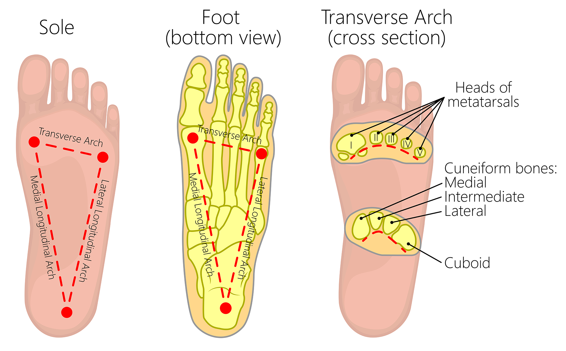
Arches of Foot

  • 3 arches:

    • 2 longitudinal (medial + lateral)

    • 1 transverse

  • main function: to support body weight and act as a lever to propel the body

Medial longitudinal arch

Formed by:

  • calcaneus

  • talus

  • navicular

  • 3 cuneiform bones

  • medial 3 metatarsal bones

Keystone bone: talus

Supporting structures:

  • plantar aponeurosis

  • muscles:

    • tendons of tibialis anterior

    • tibialis posterior

    • flexor hallucis longus

    • fibularis longus

    • intrinsic muscles

  • ligaments:

    • spring

    • deltoid

    • long and short

    • plantar and interosseous

Lateral longitudinal arch

  • flatter than medial arch

  • lies on the ground during standing position

Formed by:

  • calcaneus

  • cuboid

  • lateral 2 metatarsal bones

Keystone bone: cuboid

Supporting structures:

  • muscles:

    • tendons of fibularis longus, brevis, and tertius

    • intrinsic muscles

  • ligaments:

    • long and short

    • plantar and interosseous

Transverse arch

Formed by:

  • cuboid

  • 3 cuneiforms

  • bases of metatarsal bones

Keystone bone: intermediate cuneiform

Supporting structures:

  • muscles:

    • tendons fibularis longus (most important)

    • slips of tibialis posterior tendon (pulling tarsal bone together)

    • transverse head of adductor hallucis (drawing metatarsal bones)

  • ligaments:

    • long and short

    • plantar and interosseous

LA

Foot Anatomy

Sole/plantar aspect: the bottom of the foot

Plantar aponeurosis

a thick band of fibrous tissue which extends from heel to toes

Posteriorly: attached to medial tubercle of calcaneus

Anteriorly: divides into 5 slips that pass to all toes

Laterally: on each side, attached to metatarsal bones by medial and lateral plantar intermuscular septa

plantar aponeurosis

Sole muscles

2 groups of muscles:

  • extrinsic muscles: found in the lower leg, act to dorsiflex, plantarflex, invert and evert the foot

  • intrinsic muscles: found within the foot and primarily act to move the toes and support foot arches to maintain foot structure

Intrinsic muscles

Divided into 4 layers:

First layer

  • most superficial

  • immediately underneath plantar fascia

Contains 3 muscles:

  1. Abductor hallucis

  2. Flexor digitorum brevis

  3. Abductor digiti mini

All the muscles are supplied by medial plantar nerve, except abductor digiti minimi (supplied by lateral plantar nerve)

First layer of sole

Second layer

  • deep to 1st layer

Contains 2 muscles and 2 tendons:

  1. Quadratus plantae (flexor digitorum accessories)

  2. 4 Lumbrical muscles (1st lumbrical muscle supplied by medial plantar nerve)

  3. Tendon of flexor digitorum longus (crosses superficial to FHL to reach insertion)

  4. Tendon of flexor hallucis longus

All the muscles are supplied by lateral plantar nerve, except 1st lumbrical muscle (supplied by medial plantar nerve )

second layer of sole

Third layer

  • deep to 2nd layer

  • 2 sesamoid bones develop in tendon of adductor hallucis

Contains 3 muscles:

  1. Flexor hallucis brevis

  2. Adductor hallucis (originates by 2 heads: oblique and transverse)

  3. Flexor digiti minimi brevis

All the muscles are supplied by lateral plantar nerve, except flexor hallucis brevis (supplied by medial plantar nerve )

third layer of sole

Fourth layer

  • deepest layer

Contains 2 muscles and 2 tendons:

  1. Plantar interossei

  2. Dorsal interossei

  3. Tendon of fibularis longus

  4. Tendon of tibialis posterior

All muscles are supplied by lateral plantar nerve

fourth layer of sole

Sole Nerves and Vessels

Plantar nerves

The sole is innervated by 2 terminal branches of tibial nerve:

  • medial plantar nerve: largest and accompanies medial plantar artery

  • lateral plantar nerve: smallest and accompanies lateral plantar artery

Medial plantar nerve

  • arises beneath the flexor retinaculum

  • passes forward deep to the abductor hallucis muscle

Branches:

  1. Muscular:

    • abductor hallucis

    • flexor digitorum brevis

    • 1st lumbrical muscle

    • flexor hallucis brevis

  2. Cutaneous

    • medial 2/3 of the sole

cutaneous innervation of sole

Lateral plantar nerve

  • arises beneath the flexor retinaculum

  • passes forward deep to the abductor hallucis muscle

  • reachs base of 5th metatarsal bone, and divides into 2 branches:

    • superficial

    • deep

Branches:

  1. Muscular:

    • abductor digiti minimi

    • 2nd, 3rd, 4th lumbrical muscle

    • adductor hallucis

    • flexor digiti minimi brevis

    • all interossei (4th layer)

  2. Cutaneous

    • lateral 1/3 of the sole

Plantar vessels

Medial plantar artery

  • smallest

  • terminal branch of posterior tibial artery

  • arises beneath flexor retinaculum

  • passes forward deep to abductor hallucis muscle

Lateral plantar artery

  • largest

  • terminal branch of posterior tibial artery

  • arises beneath flexor retinaculum

  • passes forward deep to abductor hallucis and flexor digitorum brevis muscles

  • on reaching base of 5th metatarsal bone, it curves medially to form deep plantar arch

Arches of Foot

  • 3 arches:

    • 2 longitudinal (medial + lateral)

    • 1 transverse

  • main function: to support body weight and act as a lever to propel the body

Medial longitudinal arch

Formed by:

  • calcaneus

  • talus

  • navicular

  • 3 cuneiform bones

  • medial 3 metatarsal bones

Keystone bone: talus

Supporting structures:

  • plantar aponeurosis

  • muscles:

    • tendons of tibialis anterior

    • tibialis posterior

    • flexor hallucis longus

    • fibularis longus

    • intrinsic muscles

  • ligaments:

    • spring

    • deltoid

    • long and short

    • plantar and interosseous

Lateral longitudinal arch

  • flatter than medial arch

  • lies on the ground during standing position

Formed by:

  • calcaneus

  • cuboid

  • lateral 2 metatarsal bones

Keystone bone: cuboid

Supporting structures:

  • muscles:

    • tendons of fibularis longus, brevis, and tertius

    • intrinsic muscles

  • ligaments:

    • long and short

    • plantar and interosseous

Transverse arch

Formed by:

  • cuboid

  • 3 cuneiforms

  • bases of metatarsal bones

Keystone bone: intermediate cuneiform

Supporting structures:

  • muscles:

    • tendons fibularis longus (most important)

    • slips of tibialis posterior tendon (pulling tarsal bone together)

    • transverse head of adductor hallucis (drawing metatarsal bones)

  • ligaments:

    • long and short

    • plantar and interosseous