Anatomy of the Pelvic Region
Anatomy of the Pelvic Region
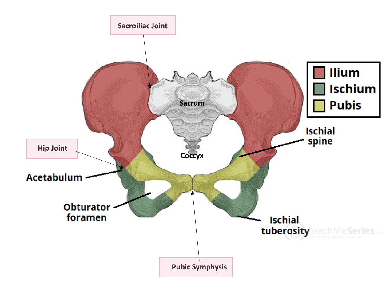
Bones of the Hip:
Ilium (red), Ischium (green), Pubis (yellow)
Key articulations:
Sacroiliac joint (sacrum to ilium)
Pubic symphysis (joining pubis)
Hip joint (acetabulum for femu)
Ilium Features:
Lateral view: acetabulum indicates lateral side
Medial view: no acetabulum, identify posterior (greater sciatic notch) vs anterior (iliac crest, iliac spines)
Pubis Features:
Superior ramus, inferior ramus, body
Obturator foramen visible
Ischium Features:
Superior ischial ramus, body of ischium, inferior ischial ramus
Ischial spine and ischial tuberosity noted
Sacrum Structures:
Anterior surface: sacral promontory, anterior sacral foramina, transverse ridges
Dorsal surface: median sacral crest, lateral sacral crest, intermediate sacral crest, posterior sacral foramina
Coccyx:
Remnants of tail bone (caudal eminence), varying number of fused bones
Features: coccygeal conure, transverse processes, articular facet
Sexual Dimorphism of Pelvis:
Female: wider pubic angle (>100°), broader/flatter ilium, wider pelvic inlet/outlet
Male: narrower pelvic angle, less concave sacrum and coccyx
Musculature of Pelvic Region:
Gluteal Group:
Gluteus maximus, medius, minimus
Actions: hip extension and lateral rotation (maximus); abduction/medial rotation (medius & minimus)
Lateral Rotator Group:
Muscles: Piriformis, Gemellus superior, Obturator internus, Gemellus inferior, Quadratus femoris
Action: lateral rotation, some abduction/adduction (Mnemonic: p-go-g-q)
Iliopsoas Group:
Muscles: Iliacus, Psoas major
Action: hip flexion
Conclusion: Overview of the anatomy, significant structures, and muscle groups in the pelvic region.