membranes - lecture 2
Barrier function of membrane:
1) To prevent/restrict spontaneous diffusion of different molecules in and out of cells or between different cellular compartments.
2) To control the amounts of different metabolites and ions inside and outside of cells (or within cellular compartments) for maintaining cellular homeostasis.
3) To regulate cellular signaling
transport in the cell Passive Transport Across the Membrane:
Movement without energy input.
Simple diffusion of molecules along a concentration gradient.
No involvement of cellular energy (ATP).
Passive Transport Through Channels and Transporters:
Utilizes specific channels or transporters.
Facilitated diffusion of substances across the membrane.
Direction follows concentration gradient.
Active Vesicular Transport (Exocytosis, Endocytosis, Pinocytosis):
Energy-dependent process.
Involves vesicles budding off or merging with the membrane.
Exocytosis: Release of substances outside the cell; endocytosis: uptake of substances into the cell; pinocytosis: nonspecific uptake of fluids and solutes.
Active Transporter-Mediated Transport:
Requires energy (ATP).
Relies on specific membrane proteins (transporters or pumps).
Moves substances against the concentration gradient.
The kinetics of diffusion and transporter-mediated transport differ in several key aspects:
vmax (Maximum Transport Rate):
Diffusion: There's no defined vmax. The rate of diffusion depends on the concentration gradient.
Transporter-Mediated: It has a vmax as transporters can only work at a certain maximum rate, saturating when all transporters are in use.
KM (Michaelis Constant):
Diffusion: No KM as it's not enzyme-mediated. It's solely dependent on the concentration gradient.
Transporter-Mediated: KM exists, indicating the substrate concentration at which the transporter works at half its maximum velocity.
Concentration of Transported Molecules:
Diffusion: Moves molecules from high to low concentration until equilibrium is reached.
Transporter-Mediated: Can move molecules against the concentration gradient using energy, maintaining a concentration gradient.
Type of Transport:
Diffusion: Can be simple (non-facilitated) or channel-mediated (facilitated) depending on the presence of channels aiding movement.
Transporter-Mediated: Involves specific transport proteins that facilitate the movement of molecules.
Energy Dependency:
Diffusion: No energy input required, relies on kinetic energy.
Transporter-Mediated: Requires energy (ATP in the case of active transporters) to actively move substances across the membrane.
Regulation and Specificity:
Diffusion: Generally nonspecific and regulated by the concentration gradient.
Transporter-Mediated: Highly specific, regulated, and selective for certain molecules or ions.
Membrane is permeable for small hydrophobic molecules, but impermeable for ions
hydrophobic molecules - 02 co2 steroids hormones - high permability
small uncharged poalr moelcuels - water urea glycerol - selectively so
large uncharged polar molecules - glucose sucros - very little
ions - all not permeable
channels vs transporters
Mechanism:
Channels: Form open pathways across the membrane, allowing passive movement of specific ions or molecules based on size and charge.
Transporters: Actively bind to specific molecules, undergo conformational changes, and transport these substances across the membrane, often against their concentration gradient.
Selectivity:
Channels: Are often selective based on size and charge, allowing specific ions or molecules to pass through. Some are highly specific (e.g., potassium channels), while others are less selective (e.g., ion channels for multiple ions).
Transporters: Exhibit high specificity for the molecules they transport. They often bind to specific substrates and undergo structural changes to facilitate their transport.
Mode of Operation:
Channels: Tend to remain open or have regulated states (open/close) controlled by various stimuli such as voltage, ligands, or mechanical forces.
Transporters: Undergo conformational changes to transport molecules. They can exhibit different states: open to the outside, open to the inside, or undergoing a cycle of conformational changes to transport substances.
Energy Utilization:
Channels: Generally facilitate passive movement down the electrochemical gradient and do not consume cellular energy (except for some gated channels that might require energy to change states).
Transporters: Often require energy, particularly in active transport, to move molecules against their concentration gradient. This energy is usually derived from ATP hydrolysis or from the energy stored in ion gradients.
Regulation:
Channels: Regulation is primarily through opening or closing states, controlled by factors like voltage, ligands, or mechanical forces.
Transporters: Regulation involves substrate binding, conformational changes, and often, post-translational modifications or interactions with other cellular components.
Speed of Transport:
Channels: Tend to allow rapid movement of ions or molecules through the open pathway.
Transporters: Generally have a lower transport rate due to the binding, conformational changes, and transportation steps involved.
passive transport
channels
transporters
diffusion
active
transporters
steps of transporters- outwards open -occuluded - inward open
Energy mediated transport:
1) Coupled transporters - energy is stored in concentration gradient to
couple uphill transport of one solute with downhill transport of another.
2) ATP-driven pumps - couple uphill transport to the hydrolysis of ATP.
3) Light- or redox-driven pumps – couple uphill transport to an input of
energy from light (eg bacterhorodopsin) or from redox reactions
(cytochrome C oxidase).
there are transporters that used secondary moelcules to bring molecules into the cell - there are two types
ymporters and antiporters
- symporters bring both molecules from the outside while anitport bring the desired in by the release of the secondary moelcule
an example of a symporter is na + symporter used to bring in glucose from the extrcellular space
structure of transporters
Transporter Structure: Inverted Repeats
LeuT: Bacterial Leu/Na+ symporter
Resembles mammalian Serotonin transporters structurally
Features repeated amino acid sequences arranged in opposite directions
Forms transmembrane structure
Facilitates substrate binding and transport (e.g., leucine and sodium ions)
Enables substrate recognition and conformational changes for transport
Reflects a similar mechanism as mammalian Serotonin transporters in neurotransmitter reuptake.
Transcellular Transport Overview
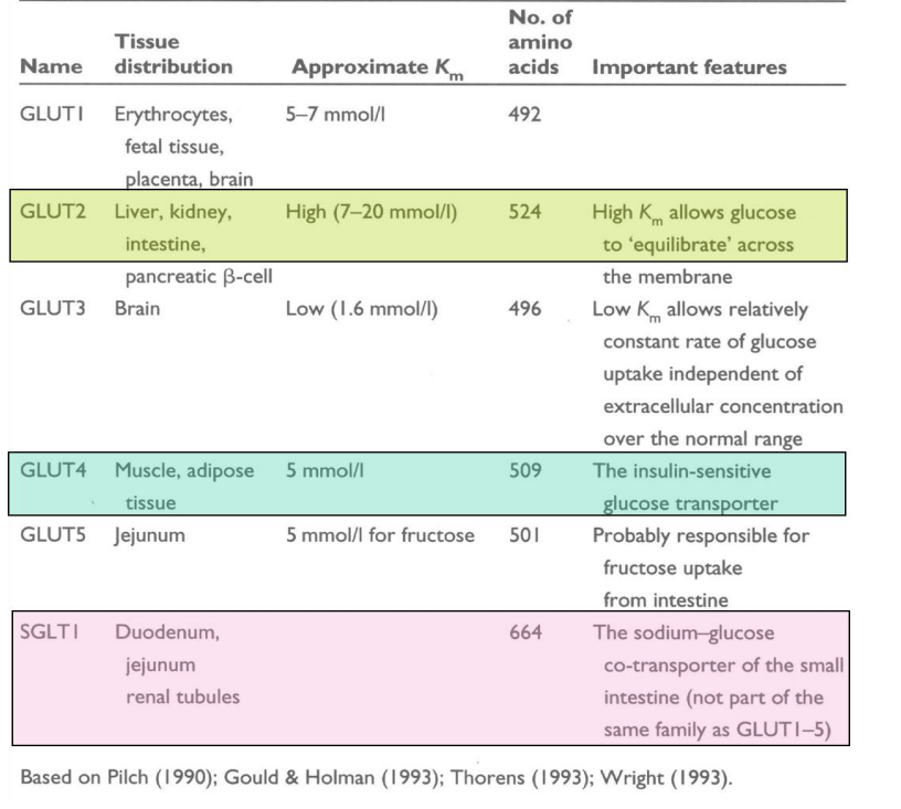
SGLT1 (Sodium-Glucose Linked Transporter 1)
Facilitates active transport of glucose and sodium ions across the membrane.
Found in the small intestine and kidney tubules.
Couples glucose transport to sodium ion movement, utilizing energy from the sodium gradient.
Crucial for glucose absorption in the gut and glucose reabsorption in the kidneys.
GLUT2 (Glucose Transporter 2)
Acts as a facilitative glucose transporter.
Found in the liver, pancreas, and intestines.
Facilitates bidirectional transport of glucose based on concentration gradients.
Plays a role in glucose sensing and release in pancreatic cells and glucose uptake in the liver.
Glucose Transporter – SGLT2
• SGLT2 is a low-affinity, high capacity sodium-dependent glucose
transporter located in the proximal tubule in the kidneys.
• It is responsible for 90% of glucose reabsorption.
• Inhibition of SGLT2 leads to the decrease in blood glucose due to the
increase in renal glucose excretion.
• Drugs in the SGLT2 inhibitors class include empagliflozin, canagliflozin,
dapagliflozin, ipragliflozin
ATP-driven pumps
1) P-type pumps (P-type ATPases) – phosphorylate themselves during
pumping cycle. Maintain Na, K and Ca gradient
2) ABC transporters (ATP-binding cassette transporters)- pump small
molecules (ions, amino acids, sugar, lipids)
3) V-type pump (V-type ATPase)– turbine like machine composed of
multiple subunits used to transfer H+ into different vesicles

Passive Transport Across the Membrane:
Movement without energy input.
Simple diffusion of molecules along a concentration gradient.
No involvement of cellular energy (ATP).
Passive Transport Through Channels and Transporters:
Utilizes specific channels or transporters.
Facilitated diffusion of substances across the membrane.
Direction follows concentration gradient.
Active Vesicular Transport (Exocytosis, Endocytosis, Pinocytosis):
Energy-dependent process.
Involves vesicles budding off or merging with the membrane.
Exocytosis: Release of substances outside the cell; endocytosis: uptake of substances into the cell; pinocytosis: nonspecific uptake of fluids and solutes.
Active Transporter-Mediated Transport:
Requires energy (ATP).
Relies on specific membrane proteins (transporters or pumps).
Moves substances against the concentration gradient.
The kinetics of diffusion and transporter-mediated transport differ in several key aspects:
vmax (Maximum Transport Rate):
Diffusion: There's no defined vmax. The rate of diffusion depends on the concentration gradient.
Transporter-Mediated: It has a vmax as transporters can only work at a certain maximum rate, saturating when all transporters are in use.
KM (Michaelis Constant):
Diffusion: No KM as it's not enzyme-mediated. It's solely dependent on the concentration gradient.
Transporter-Mediated: KM exists, indicating the substrate concentration at which the transporter works at half its maximum velocity.
Concentration of Transported Molecules:
Diffusion: Moves molecules from high to low concentration until equilibrium is reached.
Transporter-Mediated: Can move molecules against the concentration gradient using energy, maintaining a concentration gradient.
Type of Transport:
Diffusion: Can be simple (non-facilitated) or channel-mediated (facilitated) depending on the presence of channels aiding movement.
Transporter-Mediated: Involves specific transport proteins that facilitate the movement of molecules.
Energy Dependency:
Diffusion: No energy input required, relies on kinetic energy.
Transporter-Mediated: Requires energy (ATP in the case of active transporters) to actively move substances across the membrane.
Regulation and Specificity:
Diffusion: Generally nonspecific and regulated by the concentration gradient.
Transporter-Mediated: Highly specific, regulated, and selective for certain molecules or ions.
In summary, while diffusion occurs passively driven by concentration gradients and doesn't involve specific proteins, transporter-mediated transport is more selective, regulated, and can move molecules against their concentration gradient using energy. Transporter-mediated transport follows kinetics similar to enzyme kinetics with vmax and KM, while diffusion does not follow such defined parameters.
Cellular transporters and channels are both integral to moving substances across cell membranes, yet they differ in their mechanisms, selectivity, and modes of operation:
Mechanism:
Channels: Form open pathways across the membrane, allowing passive movement of specific ions or molecules based on size and charge.
Transporters: Actively bind to specific molecules, undergo conformational changes, and transport these substances across the membrane, often against their concentration gradient.
Selectivity:
Channels: Are often selective based on size and charge, allowing specific ions or molecules to pass through. Some are highly specific (e.g., potassium channels), while others are less selective (e.g., ion channels for multiple ions).
Transporters: Exhibit high specificity for the molecules they transport. They often bind to specific substrates and undergo structural changes to facilitate their transport.
Mode of Operation:
Channels: Tend to remain open or have regulated states (open/close) controlled by various stimuli such as voltage, ligands, or mechanical forces.
Transporters: Undergo conformational changes to transport molecules. They can exhibit different states: open to the outside, open to the inside, or undergoing a cycle of conformational changes to transport substances.
Energy Utilization:
Channels: Generally facilitate passive movement down the electrochemical gradient and do not consume cellular energy (except for some gated channels that might require energy to change states).
Transporters: Often require energy, particularly in active transport, to move molecules against their concentration gradient. This energy is usually derived from ATP hydrolysis or from the energy stored in ion gradients.
Regulation:
Channels: Regulation is primarily through opening or closing states, controlled by factors like voltage, ligands, or mechanical forces.
Transporters: Regulation involves substrate binding, conformational changes, and often, post-translational modifications or interactions with other cellular components.
Speed of Transport:
Channels: Tend to allow rapid movement of ions or molecules through the open pathway.
Transporters: Generally have a lower transport rate due to the binding, conformational changes, and transportation steps involved.
In essence, while both channels and transporters facilitate the movement of substances across cell membranes, they differ in their selectivity, mechanism of action, energy usage, and regulation. Channels provide a rapid and passive route for ions or molecules, while transporters are more selective, specific, and often require energy to transport substances across the membrane.
Transporters like LeuT, a bacterial Leu/Na+ symporter akin to mammalian Serotonin transporters, exhibit a structural pattern called "inverted repeats." This configuration involves repeated sequences of amino acids that fold into a transmembrane structure. These repeats, arranged in opposite directions, form the framework for transporter proteins. In LeuT, this arrangement facilitates substrate binding and transport, notably for leucine and sodium ions. This structural motif aids in substrate recognition, guiding the conformational changes necessary for substrate transport across the membrane, a process similar to how mammalian Serotonin transporters operate in neurotransmitter reuptake.
Transporter Structure: Inverted Repeats
LeuT: Bacterial Leu/Na+ symporter
Resembles mammalian Serotonin transporters structurally
Features repeated amino acid sequences arranged in opposite directions
Forms transmembrane structure
Facilitates substrate binding and transport (e.g., leucine and sodium ions)
Enables substrate recognition and conformational changes for transport
Reflects a similar mechanism as mammalian Serotonin transporters in neurotransmitter reuptake.
Transcellular Transport Overview
SGLT1 (Sodium-Glucose Linked Transporter 1)
Facilitates active transport of glucose and sodium ions across the membrane.
Found in the small intestine and kidney tubules.
Couples glucose transport to sodium ion movement, utilizing energy from the sodium gradient.
Crucial for glucose absorption in the gut and glucose reabsorption in the kidneys.
GLUT2 (Glucose Transporter 2)
Acts as a facilitative glucose transporter.
Found in the liver, pancreas, and intestines.
Facilitates bidirectional transport of glucose based on concentration gradients.
Plays a role in glucose sensing and release in pancreatic cells and glucose uptake in the liver.
P-Type ATPase: Ca2+-ATPase
Function: Moves calcium ions across membranes against their concentration gradient.
Structure:
Consists of a single polypeptide chain with multiple domains.
Contains transmembrane segments that anchor it in the membrane.
Has cytoplasmic domains responsible for ATP binding and hydrolysis.
Contains phosphorylation sites crucial for its activity.
Exhibits a Ca2+-binding domain that regulates ion transport.
Mechanism:
Undergoes conformational changes driven by ATP hydrolysis.
Cycles between phosphorylated and dephosphorylated states to transport calcium ions.
ATP binding and hydrolysis power the conformational changes necessary for ion transport.
Cellular Role: Critical in maintaining low cytosolic calcium levels, crucial for various cellular processes like muscle contraction, signaling, and enzyme regulation.
P-Type ATPase: Na+/K+ Pump
Function: Actively transports sodium ions out of cells and potassium ions into cells against their respective concentration gradients.
Structure:
Single polypeptide chain forming an α-subunit.
Contains transmembrane domains anchoring it within the membrane.
Comprises cytoplasmic domains for ATP binding, hydrolysis, and phosphorylation.
Has specific binding sites for sodium and potassium ions.
Mechanism:
Alternates between phosphorylated and dephosphorylated states through ATP hydrolysis.
Binding of ATP phosphorylates the pump, inducing conformational changes that allow ion binding and transport.
Transitions through multiple conformations (E1 and E2 states) during the transport cycle.
Cellular Significance:
Vital for maintaining the electrochemical gradients necessary for cell function.
Essential in nerve impulse transmission, muscle contraction, and regulating cell volume.
Accounts for a significant portion of the cell's energy expenditure.
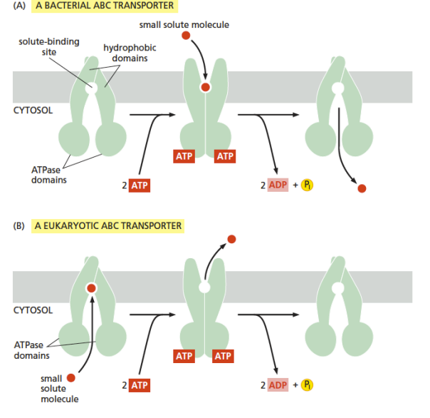
Importers:
Uptake Systems: Facilitate the uptake of nutrients, ions, and other essential molecules from the environment into the cell.
Examples:
Bacterial Periplasmic Binding Protein-Dependent Systems: These systems involve high-affinity binding proteins that capture substrates outside the cell. Upon binding, they interact with integral membrane transporters and the ATPase component to facilitate substrate transport into the cell.
Prokaryotic Importers: Involved in the uptake of essential nutrients like sugars, amino acids, and ions. For instance, the maltose/maltodextrin transporter in bacteria.
Exporters:
Efflux Systems: Expel toxins, waste products, or substances not required by the cell to the external environment.
Examples:
Multidrug Efflux Pumps: Help bacteria resist antibiotics and other harmful compounds by pumping them out of the cell. For instance, the E. coli AcrAB-TolC system.
Heavy Metal Efflux Systems: Aid in removing toxic metals from the cell to maintain homeostasis.
Substrate Specificity:
Broad-Specificity Transporters: Can transport a wide range of substrates, contributing to multidrug resistance or versatility in nutrient acquisition.
Narrow-Specificity Transporters: Specialized in transporting specific substrates, ensuring precise uptake or efflux of particular molecules required for cellular processes.
Each type of microbial ABC transporter plays a crucial role in microbial survival, adaptation to environmental challenges, and maintaining intracellular homeostasis by regulating the passage of substances across the cell membrane.
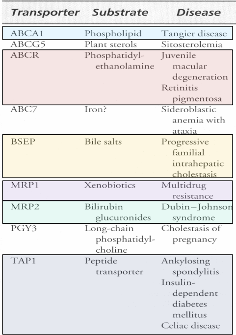
P-glycoproteins (MDR1) [ABCB1]:
Function: Multidrug Resistance Transporter.
Role: Efflux pump expelling various drugs and toxins out of cells, contributing to drug resistance in cancer cells.
Structure: ABC transporter with broad substrate specificity.
Clinical Significance: Impacts the effectiveness of chemotherapy in cancer treatment.
Cystic Fibrosis Transmembrane Regulator (CFTR) [ABCC7]:
Function: Chloride Transporter.
Role: Facilitates chloride ion transport across cell membranes.
DF508 Mutation: Most common mutation in cystic fibrosis (70% of patients).
Impact: Results in defective CFTR protein folding, affecting chloride transport, leading to thick, sticky mucus production and respiratory issues in cystic fibrosis patients.
These ABC transporters, P-glycoproteins (MDR1) and Cystic Fibrosis Transmembrane Regulator (CFTR), are crucial in cellular functions and have significant clinical implications. While P-glycoproteins contribute to drug resistance, CFTR dysfunction due to the DF508 mutation is central to the pathogenesis of cystic fibrosis, affecting chloride ion transport and leading to severe health complications.
 Vesicular Type ATPase (V-Type ATPases)
Subunit Composition:
A3B3C(stalk)DEab2(c)8-12
Structure:
V0V1 and stalk; V1 ~12nm diameter on cytoplasmic side.
Conformational Changes:
A&B subunits undergo conformational changes during function.
Functional Subunits:
Asubunit (70kDa) is the ATP-binding subunit responsible for ATP hydrolysis.
C subunits facilitate H+ transport.
Catalytic Process:
No covalent modification during catalysis.
ATP-binding motif: GXXGKS/T.
Location:
Found in intracellular bacterial membranes and in endosomal/lysosomal membranes of eukaryotes.
Aquaporins (AQP1) in Oocyte Swelling Assay
Experimental Setup:
Oocyte swelling assay comparing cells expressing AQP1 cDNA versus control cells.
AQP1 Function:
A transmembrane protein facilitating water transport across cell membranes.
Assay Objective:
Measures changes in oocyte volume when expressing AQP1 versus control cells.
Observations:
AQP1-expressing cells likely show increased water permeability, leading to faster oocyte swelling compared to control cells.
Significance:
Validates AQP1's role in facilitating water movement across membranes, evidenced by altered oocyte swelling in the presence of AQP1 cDNA.
Aquaporins operate via a proton exclusion mechanism
ion channels
voltage gated
ligand gated - extracellular ligand
ligand gated intracellular ligand
mechianically gated
five slides left
Vesicular Type ATPase (V-Type ATPases)
Subunit Composition:
A3B3C(stalk)DEab2(c)8-12
Structure:
V0V1 and stalk; V1 ~12nm diameter on cytoplasmic side.
Conformational Changes:
A&B subunits undergo conformational changes during function.
Functional Subunits:
Asubunit (70kDa) is the ATP-binding subunit responsible for ATP hydrolysis.
C subunits facilitate H+ transport.
Catalytic Process:
No covalent modification during catalysis.
ATP-binding motif: GXXGKS/T.
Location:
Found in intracellular bacterial membranes and in endosomal/lysosomal membranes of eukaryotes.
You
Channels: Aquaporins-Oocyte Swelling Assay: +AQP1 cDNA versus Control- brief iverview in notes
ChatGPT
ChatGPT
Aquaporins (AQP1) in Oocyte Swelling Assay
Experimental Setup:
Oocyte swelling assay comparing cells expressing AQP1 cDNA versus control cells.
AQP1 Function:
A transmembrane protein facilitating water transport across cell membranes.
Assay Objective:
Measures changes in oocyte volume when expressing AQP1 versus control cells.
Observations:
AQP1-expressing cells likely show increased water permeability, leading to faster oocyte swelling compared to control cells.
Significance:
Validates AQP1's role in facilitating water movement across membranes, evidenced by altered oocyte swelling in the presence of AQP1 cDNA.
You
make twenty mcqs on the infomation from this chat with five answer options
ChatGPT
ChatGPT
Sure, here are twenty multiple-choice questions based on the information discussed:
Which transporter type moves substances against their concentration gradient?
a) Passive transport
b) Diffusion
c) Active transport
d) Facilitated diffusion
e) Osmosis
What type of structural motif characterizes transporters like LeuT and mammalian Serotonin transporters?
a) Beta-sheets
b) Inverted repeats
c) Alpha-helices
d) Glycoproteins
e) Coiled-coils
Which transporter actively couples glucose transport to sodium ion movement?
a) GLUT2
b) SGLT1
c) P-glycoprotein
d) ABC transporter
e) CFTR
Which type of ATPase is crucial for maintaining low cytosolic calcium levels?
a) P-Type ATPase: Na+-K+ pump
b) P-Type ATPase: Ca2+-ATPase
c) V-Type ATPase
d) ABC Transporter
e) Aquaporin
What is the most common mutation in cystic fibrosis, affecting the CFTR protein?
a) DF508
b) ABCB1
c) V0V1
d) A3B3C
e) GXXGKS/T
Which transporter contributes to multidrug resistance in cancer cells?
a) CFTR
b) GLUT2
c) P-glycoproteins (MDR1)
d) SGLT1
e) Aquaporins
What structural motif characterizes the ATP-binding subunit in vesicular type ATPases?
a) Beta-sheets
b) Alpha-helices
c) ATP-binding motif (GXXGKS/T)
d) Coiled-coils
e) Inverted repeats
What technique compares oocyte swelling in cells expressing AQP1 cDNA versus control cells?
a) PCR analysis
b) Western blotting
c) Immunofluorescence
d) Oocyte swelling assay
e) Microarray analysis
Which ATPase type transports sodium ions out of cells and potassium ions into cells?
a) V-Type ATPase
b) P-Type ATPase: Ca2+-ATPase
c) P-Type ATPase: Na+-K+ pump
d) ABC Transporter
e) Aquaporin
What is the primary role of P-glycoproteins (MDR1)?
a) Chloride transport
b) Multidrug resistance
c) Glucose uptake
d) Sodium transport
e) Water permeability
Which transporter facilitates chloride ion transport and is impacted by the DF508 mutation in cystic fibrosis?
a) SGLT1
b) ABCB1
c) CFTR
d) GLUT2
e) V-Type ATPase
What structure in vesicular type ATPases anchors them within the membrane?
a) Alpha-helices
b) ATP-binding motif (GXXGKS/T)
c) Transmembrane domains
d) Coiled-coils
e) Inverted repeats
Which transporter type allows bidirectional transport of glucose based on concentration gradients?
a) SGLT1
b) ABC Transporter
c) GLUT2
d) P-glycoproteins (MDR1)
e) V-Type ATPase
What is the functional subunit responsible for ATP hydrolysis in vesicular type ATPases?
a) A subunit
b) C subunit
c) B subunit
d) E subunit
e) D subunit
Which assay measures changes in oocyte volume when comparing AQP1-expressing cells versus control cells?
a) PCR analysis
b) Western blotting
c) Immunofluorescence
d) Oocyte swelling assay
e) Microarray analysis
What is the primary function of aquaporins?
a) Sodium transport
b) ATP hydrolysis
c) Water transport
d) Glucose uptake
e) Multidrug resistance
Which transporter actively moves substances using energy from ATP hydrolysis?
a) GLUT2
b) Aquaporins
c) Passive transport
d) ABC Transporter
e) Diffusion
What structural motif characterizes the ATP-binding subunit in vesicular type ATPases?
a) Beta-sheets
b) Alpha-helices
c) ATP-binding motif (GXXGKS/T)
d) Coiled-coils
e) Inverted repeats
What technique compares oocyte swelling in cells expressing AQP1 cDNA versus control cells?
a) PCR analysis
b) Western blotting
c) Immunofluorescence
d) Oocyte swelling assay
e) Microarray analysis
What is the primary role of P-glycoproteins (MDR1)?
a) Chloride transport
b) Multidrug resistance
c) Glucose uptake
d) Sodium transport
e) Water permeability
Sure, here's the answer key for the questions:c
b
b
b
a
c
c
d
c
b
c
c
c
a
d
c
d
c
d
b