Vision
Sensation & Perception
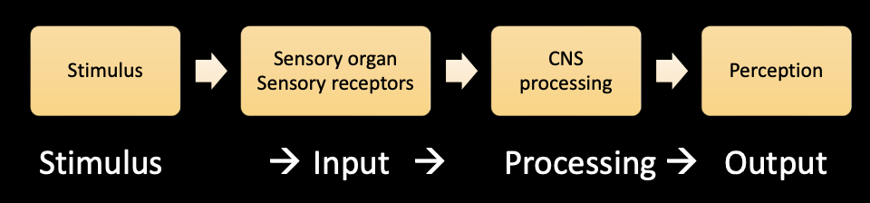
- sensation: specialized bervous system cells detect stimuli from the environment
- sensory receptors: specialized neurons that detect a variety of physical events (typically located outside of the PNS)
- sensory receptors for light are in the retina (CNS)
- sensory transduction: converts environmental stimuli into membrane potentials (receptor potentials)
- Perception: conscious experience and interpretation of information from the senses
- involves neurons in the CNS
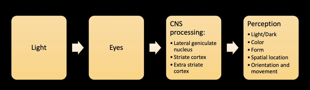
Stimulus: Light
- light: travels as a wave, type of light is determined by wavelength
- wavelength: inversely related to frequency of wave oscillations
- photoreceptors: specialized cells in the eye that detect light
- electromagnetic radiation: wavelength between 380-760nm (visible to humans)
- 3 dimensions of light: hue, brightness, saturation
- hue: determined by wavelength (roygbiv)
- brightness: intensity of light
- saturation: relative purity of light (purest. radiation is one wavelength)
- white: radiation contains all visible wavelengths
- colors with intermediate amounts of saturation consist of diffeent pixtures of wavelength
Sensory Organ: Eyes
- orbits: bony pockets in the front of the skull
- sclera: white outer layer of most of the eye (opaque)
- opaque: prevents light from entering
- cornea: outer layer at the front of the eye (transparent)
- pupil: opening in the iris, the size depends on how much light can enter the eye
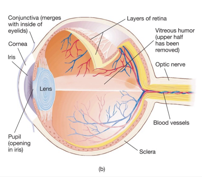
- lens: located just behind the eye, consists of a series of transparent onion like layers
- accommodation: shape of the lens changes permitting the eye to focus images on near or distant objects on the retina
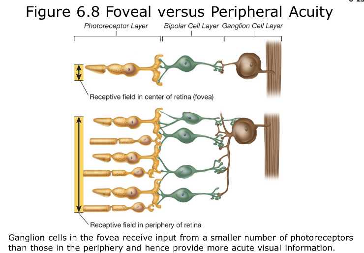
- retina: inner lining of the posterior surface of the eye, consists of the fovea, and 3 cellualr levels (photoreceptors {rods/cones}, bipolar cell layer, ganglion cell layer)
- fovea: centeral region of the retina, mediates the most acute vision of birds and higer animals, populated by colot sensitive cones
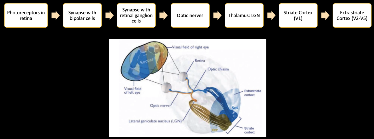
- optic disk: at the back of the eye, where axons conveying visual information gather anf leave the eye through the optic. nerve (theres a blind spot at the back of the disk because there are no receptors there)
- retina and optic nerve are parts of the CNS
- optic nerve: axons of retinal ganglion cells bundle together to convey information to the dorsal lateral geniculate nucleus
- optic chaism: optic nerves join at the base of the beain to form an x shaped chiasm
- each hemisphere recievces information from the contralateral visual field
Retinal Cells
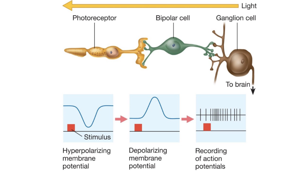
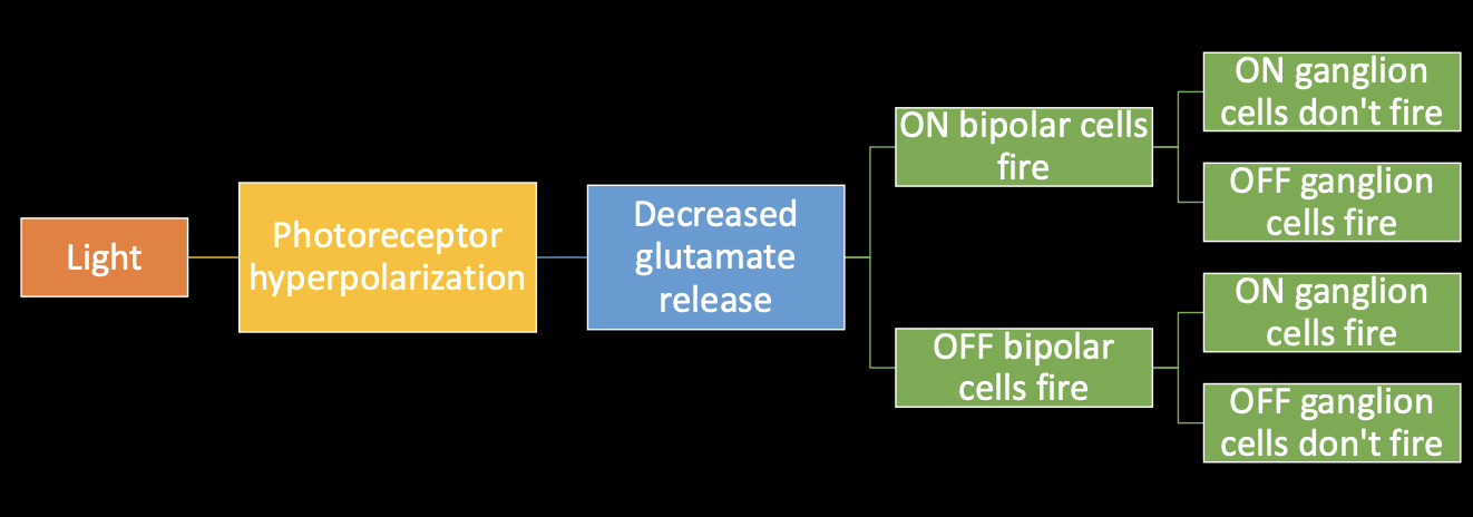
- Photoreceptors: sensory receptors that transduce photic energy into electrical potentials, located behind 2 translucent layers of cepps (bipolar and ganglion cell layers), darkness detectors (depolarize to darkness, repolarize to light, release glutamate without APs)
- rods: 120 million, not gound in the fovea, dimly lit environments because they are nore sensitive to light, provide poor acuity, do not detect different colors
- cones: 6 million, populate the fovea, daytime vision (produce mist of the info about our eenvironment), produce highest acuity
- photopigments: found outside of the photo receptors
- 2 parts of the pigment molecule: an opsin and a retinal
- rhodopsin: 10 million photopigment molecules in human rods
- transduction: light strikes photoreceptors, photopigments in the photoreceptors split, cascade of intracellular events that hyperpolarize the receptor membrane
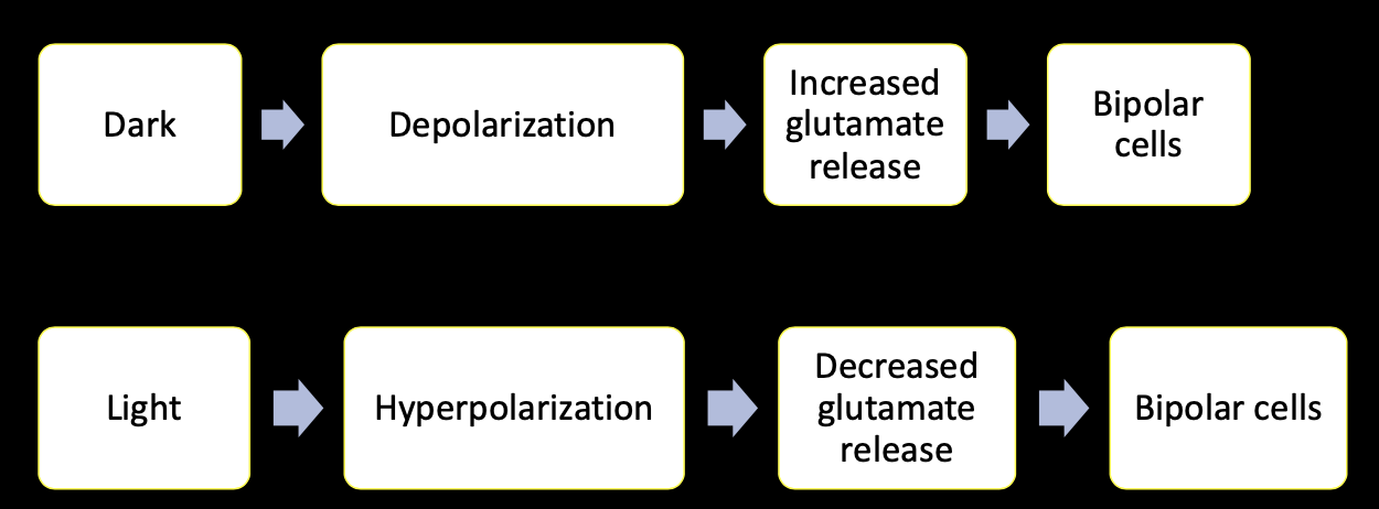
- bipolar cells: populate the middle later of the retina, recieve glutamate from photoreceptors and send information to ganglion cells
- 2 types of bipolar cells: On center (hyperpolarized by glutamate) and off center Idepolarized by glutamate)
- ganglion cell: neuron in the retina that recieves visual information from bipolar cells
- on/off information from bipolar cells determine the rate of ganglion cell firing
- axons give rise to the optic nerve
- Receptive field: place a visual stimulus must be located to produce a respinse in that neuron (what an individual neuron in the visual system sees)
- central vision: receptive fiels is the fixation point, the point where the eye is looking (photoreceptor in the fovea, acute {1 photoreceptor per ganglion cell})
- peripheral vision: receptive field is on one side (photoreceptor in the periphery of the retina {less percise many receptors converge on 1 ganglion cell})
Brain Regions Involved in Visual Processing
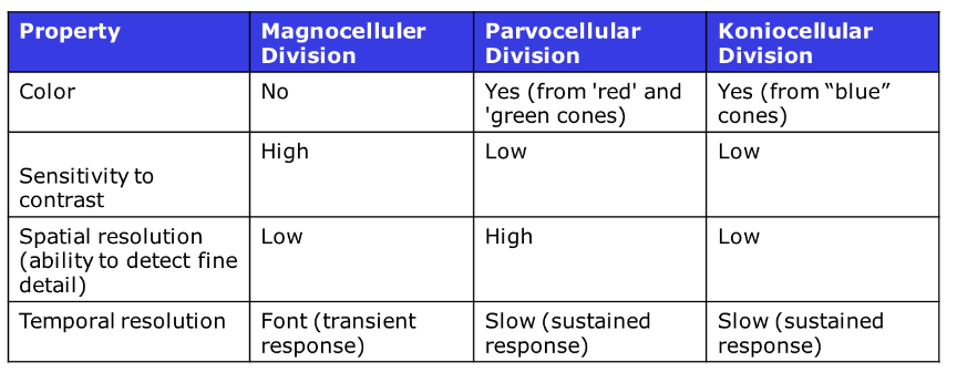
- lateral geniculate nucleus: region of the thalamus that recieves and sends visual information
- 6 layers of neurons: magnocellular (2 innermost layers of cell bodies), parvocellular (outer 4 layers of smaller cell bodies), koniocellular (beneath magnocellular and parvocellular layers)
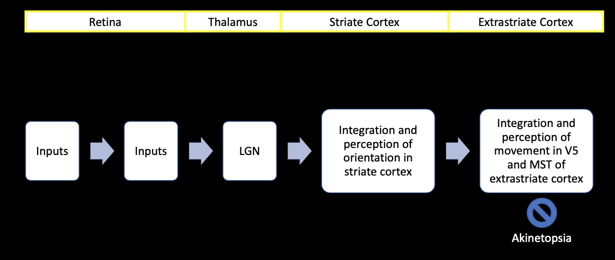
- striate cortex: primary visual cortex or V1, located in the occipital lobe, named for striated or stripped appearance from fark staning layers of cells
- function of striate cortex: visual perception, neurons respond to specific features/controls, combines information from several sources to detect features larger than those detected by a single ganglion cell (allows for detection of features larger than the receptive field)
- striate cortex anatomy: contains a map of the contralateral half of the visual field (map disproportionately favors 25% of the analysis of information from the fovea), composed of layers of neurons, cytochrome oxidate blobs, modules
- layers composed of nuclei of cell bodies and dendritic trees
- results in light and dark laters of tissue when died with a cell body stain
- cytochrome oxidase blob: process info from color sensitive ganglion cells (in layer 2, 3 and faintly in 5,6), most neurons respond to orientation, spatial frequency, movement, retinal disparity
- modules: striate cortex is divided into 2500 modules containing 150,000 neurons, each module is responsible for analyzinf a small portion of the visual field, consist of 2 segments of neurons (inputs from 1. left eye and 2. right eye, cytochrome blob → color, interblob regions→ orientation, movement, retinal disparity
- extrastriate cortex: visual association cortex or V2-V5, located in the occipital lobe surrounding the striate cortex, visual perception of objectsm visual scenes, combines the information from modules
- extrastriate cortex anatomy: V2-V5 contain maps of the visual fields, regions are specialized to respond to different orientations, movements, spacial frequencies, retinal disparitiesm colors.
- aranged higherarchally meaning that most information traces outward from the striate cortes
- hierarchical information passage: info reaches striate cortex, information is analyzed and passes onto the next region nad so on and so forth
- extrastriate cortex pathways: afte V2 pathways diverge into the dorsal strea, and ventral stream
- dorsal stream: where and how, projects to the posterior parietal cortex, guides navigation and movement towards objects, vision for action
- ventral stem:what, projects inferior to the temporal cortex, recognizes objects and people by color, size, texture
Visual Perception
- aspects of visual perception: color vision, form, spatial location, orientation, movement
- blindsight: in cortically blind patients, they can’t “see” due to a lesion in the visual cortex, but they can respind to stimuli, lots of variability
Color Perception
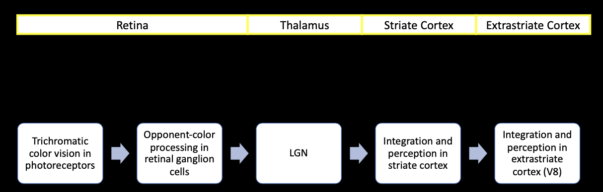
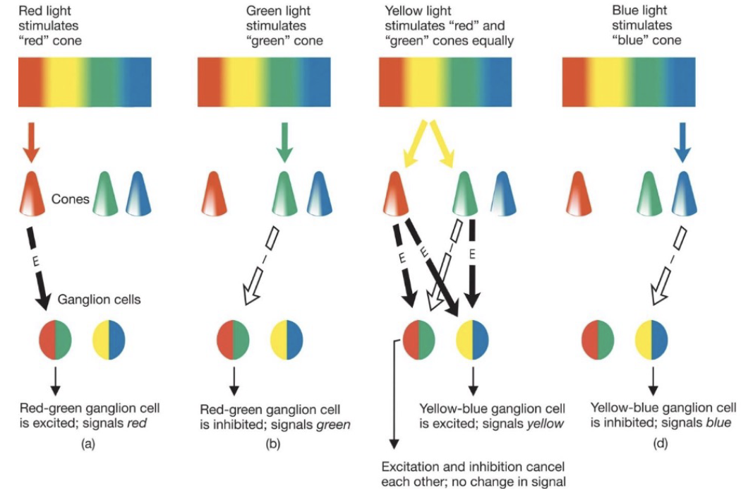
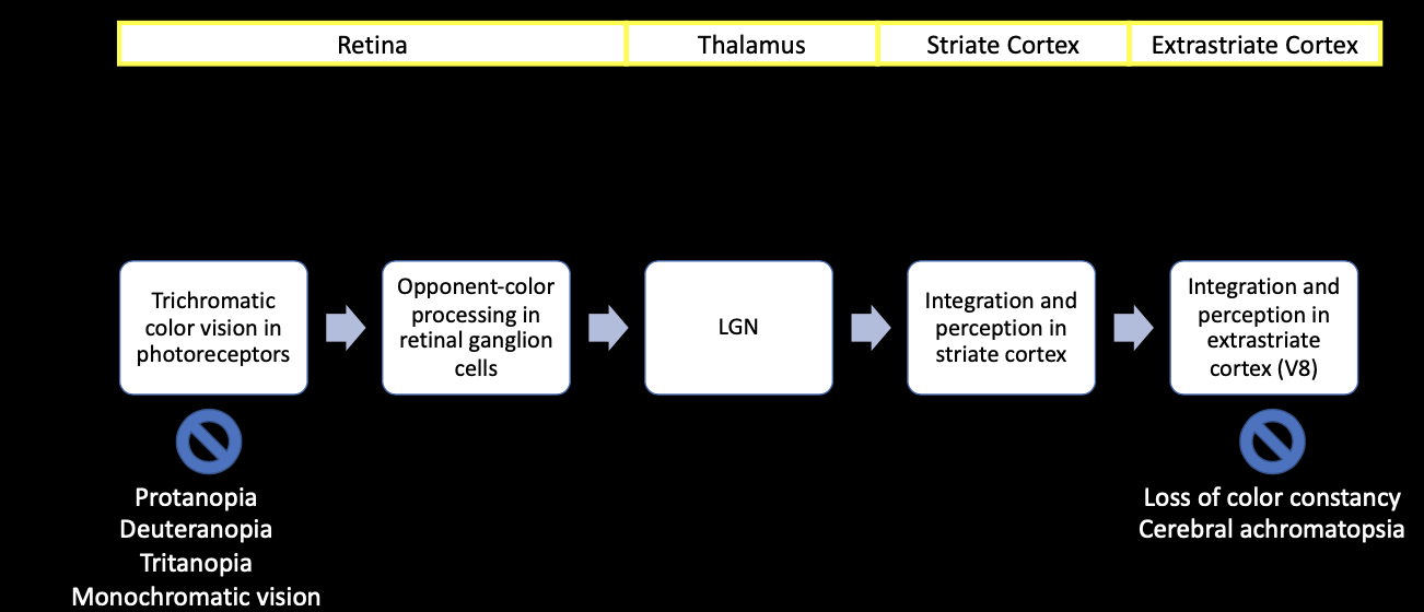
- color perception requires: trichromatic coding in 3 different types of photoreceptors, opponent processing coding in retinal ganglion cells, color processing layers of the striate cortex, further processing in the extra striate cortex
- trichromatic coding in 3 different types of photoreceptors: different photopigments in cones absorb either red green or blue wavelengths, absorption characteristics of photoreceptors determine the amount of light different wavelengths absorb
- protanopia: first color defect, red cones are filled with green opsin so you see in shades of yellow and blue (x linked)
- deuteranopia: second color defect, green codes are filled with red opsin (x linked)
- tritanopia: third color defect, lack blue cones, see in greens and reds
- monochromatic vision: complete lack of cones
- opponent processinf coding in retinal ganglion cells: 2 types of color sensitive ganglion cells (red-green and blue-yellow)
- color procesing layers of the striate cortex
- further processing in the extra striate cortex: cortical processing allows for complex integration of color inputs especially in V8, cortical processing allows for integration of color inputs, maintains color constancy
- color constancy: visual cortex compensates for differences in lighting, the appearance of colors foes not change on cludy days
- cerebral achromatopsia: loss of color vision without loss of visual acuity due to damage to the extrastriate cortex
- after image effect/rebound effect: after prolonges stimulation the opposite color image is seen
Form Perception
- form perception allows for: recognition of shapes, objectsm complex shapes like faces, spatial location
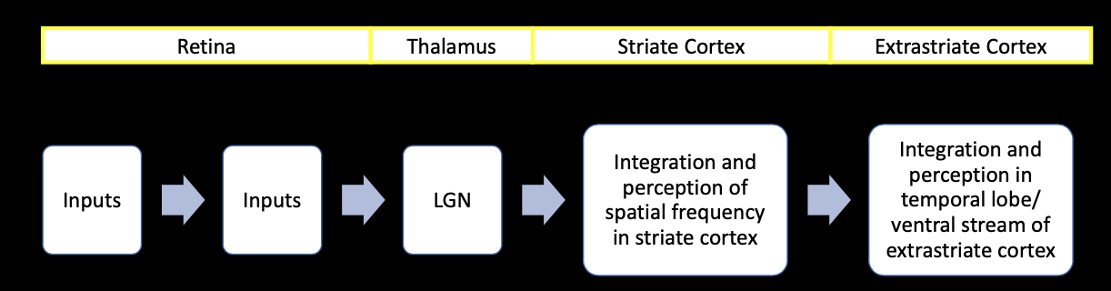
- form perception requirement: striate cortex, extrastriate cortex
- striate cortex:perceprion of form begins with neurons in the striate cortex that are sensitive to spacial frequency
- spatial frequency: variations in light measures in cycles of visual angles
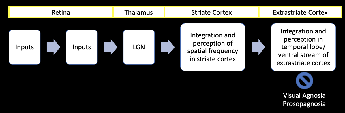
- extrastriate cortex: ventral. system if the visual association cortex terminates in the inferior temporal cortex
- inferior temporal cortex: at the end of the ventral system, responsible for recognition of visual petters, identification of objects, neurons respind to 3D objects, involved in learning
- fusiform face area: responds. specifically to faces
- visual agnosia: cannot identify common objects by sire, caused by damafe ro the regions of the extrastriate cortes that contribute to the ventral system, does not damage vision itself or reading, does not impair the ability to name objects that the patient is holding
- prosopagnosia: cannot recognize faces, FFA is damages or smaller
Perception of Spatial Location
- perception of spatial location contributes to: depth perception, percieveing or remembering location, controlling movements of the eyes and limbs
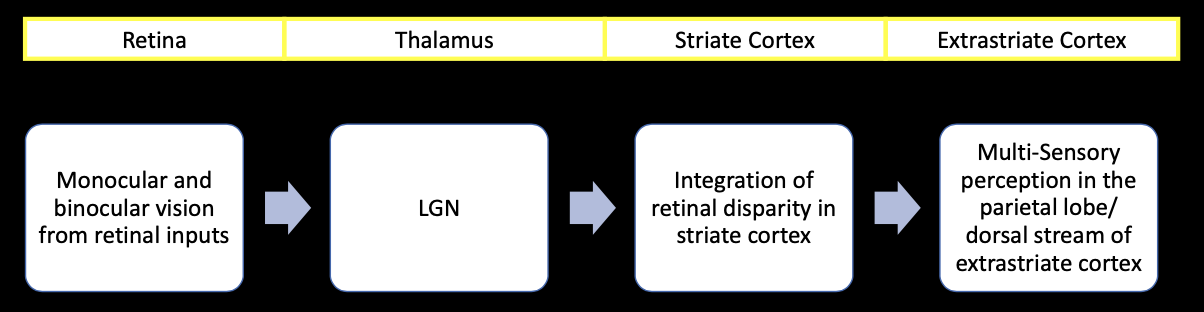
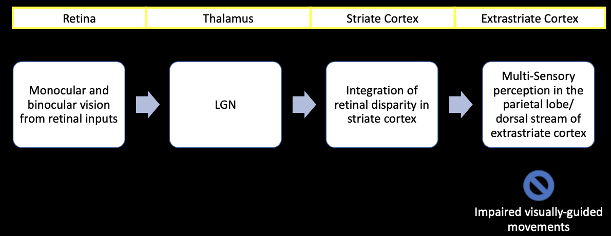
- perception of spatial location requires: retina (monocular and binocular depth perception), striate cortex processes retinal disparity, extrastriate cortex
- retinal disparity: slight differences in retinal imafe due to differences in angles from which eyes view a stimulus
- posterial parietal cortex: integrates somatosensort and spatial information with visualsm auditory, somatosensory, and vestibular information
- intraparietal sulcus: involved in visual attentionm, control of eye movement, visual control of reaching and pointing, visual control of grasping and manipulating hand movements, perception of depth from stereopsis
- damage to dorsal stem: can impair visually guided movement (ability to pick up objects, adjusting hands or fingers before reaching to grasp an object)
Perception of Orientation and Movement
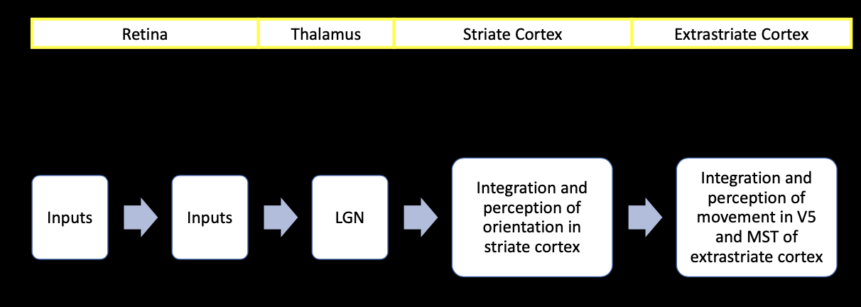
- perception of orientation and movement requires: striate cortex, extrastriate cortex
- Area V5: medial temporal, receives input. from striate cortes, superior colliculus and regions of the extrastriate cortex, afferents are thick and heavily myelinated allowing for rapid AP conduction
- Medial superior temporal: adjacent to V5, receives input from V5, neurons respond to complex patterns of movement (spiral radial circular), analyzes optic flow
- optic. flow: complex motion of points in the visual field, caused by relative movement between observer and the environment
- akinetopsia: inability to perceive movement, moving stimuli are experienced as a series of still images, caused by damage to V5