N110 Midterm 1 notes
Lecture 1- Overview and Neuroanatomy
- Describe historical advances in our understanding of the nervous system, including evidence supporting or refuting major hypotheses
| Camillo Golgi | Santiago Ramon y Cajal | Charles Sherrington |
|---|---|---|
| ==reticular theory (web-like)==Golgi stain b/c the NS responses are instantaneous, nerve cells must form a continuous web | ==Neuron doctrine==perfected the Golgi method; proved that the brain has individual neurons connected together | ==Synapse==junction between 2 nerve cells Studied spinal reflex and how we can override it Ex. hot pan |
- Compare the anatomical features that underlie functional specializations among different kinds of neurons
- form follows function
- cell bodies (DNA storage, protein making, housekeeping)
- Dendrites * i for input; this is where inputs go into the nerve cell
- axons * o for output; input travels down axon, to the synapse, to the next neuron * carries info in form of APs * decisions of cell: fire AP in response to input * release chemicals/neurotransmitters as output, which then serve as the input for the next neuron
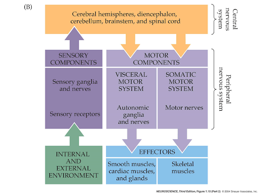
- Describe a simple reflex circuit and how each neuron in the circuit represents a major subdivision in the nervous system.
Lecture 2- Neural Signals are Electrical
- Describe historical advances in our understanding of the nervous system, including evidence supporting or refuting major hypotheses.
- Luigi Galvani- electricity contracts muscles * frog legs
- Alessandro Volta * invented battery to try to disprove Galvani
- Nernst- calculated potential produced by differential ion concentrations across membrane
- 1902: Bernstein’s 2 hypothesis * neurons store energy, a “resting potential” due to selective permeability of membrane (ENERGY STORAGE) * brief increase in permeability to all ions could underlie neural signal (action potential) (ENERGY EXPENDITURE)
- Describe the idea that the nervous system uses “endogenous electricity” and the evidence used by Galvani and Volta for and against this point
- Nervous system uses endogenous electricity
- Galvani's experiments with frog legs supported this idea
- Galvani believed that muscles generate electricity
- Volta's experiments with frog legs contradicted Galvani's findings
- Volta believed that electricity was generated by the contact between different metals
- Debate between Galvani and Volta continued for many years
- Use the Nernst equation to calculate “equilibrium potential” and understand (conceptually) how this depends on the distribution of ions. * If you know the concentrations on either sides of a membrane you can calculate what the potential is when the ions are at equilibrium * E = E° - (RT/nF) ln([X]out/[X]in)
- Describe Bernstein’s two hypotheses, how they are based on Nernst’s ideas, and how they relate to passive and active neural activity (resting potential and action potential), respectively. * Bernstein’s 2 hypotheses about how neurons could generate and store electricity/electrical potential in form of APs * I: how resting potential is created * II: energy expended to generate a neural signal * A semipermeable membrane and a difference in concentrations on either side of the membrane, a resting potential is generated * A release in potential en. created by permeability to just one ion vs. all ions, the equilibrium (revise)
- Explain how ion transporters and ion channels contribute to passive and active neural activity. * K+ highly concentrated inside the cell 400 mM (a ten-fold difference compared to the outside) * Na+ highly concentrated outside the cell 440 mM
Lecture 3 - Membrane Permeability
- Describe historical advances in our understanding of the nervous system, including evidence supporting or refuting major hypotheses. * “Doctor death” famous proponent for assisted suicide if you’re of ==“sound mind”== and a diagnosed terminal illness * This rules out one of the major reasons why people would end their life, by the end stages of neurological illness you are no longer of “sound mind” * Similar to how prisoners are medically executed (==KCl==)- the resting potential in the neurons is neutralized because extracellular environment now saturated with K+ * “Doctor Death” pushed the button for a patient who was paralyzed, and was convicted for murder (ethical dilemma of killing people)
- Describe how Hodgkin and Katz (1949) used the squid giant axon to test ==Bernstein’s first hypothesis== (resting potential).
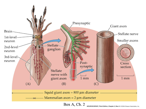
- A squid has a giant ganglion axon it uses for escape behavior- coordinates at the muscles in the squid
BIG AXON=FAST TRANSMISSION
- Big enough to test membrane potentials with wires- you can record transmembrane potential while changing ion concentrations
- Testing whether RMP is due to a difference in one ion (K+), equilibrium potential and transmembrane potential relationship using NERNST equation for what the equilibrium potential should be
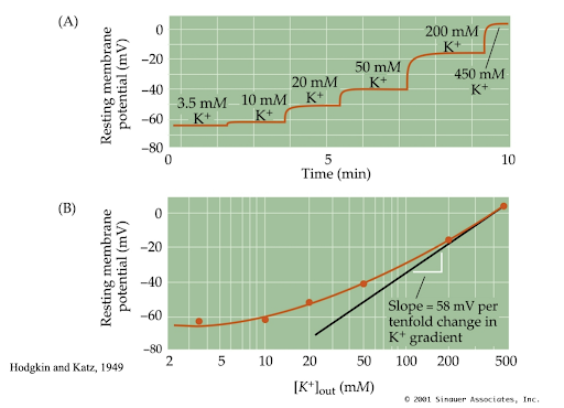
- Putting in 450mM of K+ should lead to a RMP of 0, which it is
- Not perfect, because K+ is not the only ion responsible for RMP (Na+ and Cl-) * Goldman came up with an equation that includes all three ions
- Compare the results of Cole and Curtis (1939) vs Hodgkin and Huxley (1939) when each email tested Bernstein’s second hypothesis (action potential). How did each support, or not support, the hypothesis? * Conductance of the membrane during action potential * Conductance during action potential increases- BUT to transiently release, Bernstein used the conductance of open seawater as the basis for measurement
- Describe how Hodgkin’s insight (hypothesis) on the sequence of permeability changes could explain how the action potential is produced * Action potential able to be recorded in 1939- transmembrane voltage measured, +40 mV and then back down to negative * This was a major discovery, because B predicted an increase only up to 0mV, not all the way up to a positive +40mV value (a value of 0 would indicate the membrane became permeable to all ions, and the resulting difference in potential is zero) * Hodgkin: “RMP one ion dominates permeability K+ at rest, the Na+ dominates during action potential because the membrane is suddenly permeable to Na+” * Sodium permeability is what drives the action potential- the resting potential DOESNT CHANGE, but affects the peak of the action potential (follows Nernst eq.) * Study Voltage Clamps!!! * Voltage clamp is a technique used in electrophysiology to measure the electrical properties of cells * It was first developed by Hodgkin and Huxley in the 1940s to study the ionic basis of the action potential in neurons * The technique involved clamping the membrane potential of a cell at a specific voltage and then measuring the resulting ionic currents that flow across the membrane. * Voltage clamp can be used to study the properties of ion channels and their conductance properties. * the technique has been used extensively to study the properties of voltage-gated ion channels in neurons and other excitable cells
Lecture 4- Action Potential
- Describe historical advances in our understanding of the nervous system including evidence supporting or refuting major hypothesis * 1952- Hodgkin and Huxley published five papers on a quantitative description of ionic basis of Action Potential and underlying membrane permeability
- Describe how the voltage clamp amplifier measures ionic currents flowing across the membrane of the squid axon * Allows for manipulation of voltage * Researchers can set command voltage to whatever they want * Membrane potential is always at 0
- Compare voltage-clamp and conventional voltage recordings to explain how information from voltage-clamp recordings cannot be obtained from conventional recordings. * Conventional voltage recordings measure the membrane potential of a neuron at a given time. In contrast, voltage-clamp recordings measure the current flowing through the membrane of a neuron at a given voltage. * Voltage-clamp recordings allow researchers to control the voltage across the membrane of a neuron and measure the resulting current. This is not possible with conventional voltage recordings. * Voltage-clamp recordings can provide more detailed information about the properties of ion channels in neurons than conventional voltage recordings. * ==Because of different internal solutions that are used in voltage-clamp and current-clamp recordings, only limited information can be obtained from recording the same neuron in both modes==
- Describe how membrane potential, membrane current, and membrane conductance are related to each other. * Membrane potential is the difference in electrical charge between the inside and outside of a cell membrane. ==It is determined by the concentration gradients of ions across the membrane and by membrane permeability to each type of ion.== * Membrane current is the flow of ion across a membrane. ==It is determined by the concentration gradients of ions across the membrane and by the electrical potential difference across the membrane.== * Membrane conductance is a measure of how easily ions can flow across a cell membrane. ==It is determined by the number and type of ion channels in the membrane.== * In summary, membrane potential determines the direction of ion flow across a cell membrane, while membrane current determines the rate of ion flow. Membrane conductance determines how easily ions can flow across a cell membrane1.
- Explain how membrane conductance, but not membrane current, can be used to measure membrane permeability * Membrane conductance is a measure of how easily ions can flow across a cell membrane * Membrane permeability is a measure of how easily molecules can cross the membrane * Membrane conductance and membrane permeability are directly proportional to each other * Membrane conductance is determined by the number and type of ion channels in the membrane * When the number of ion channels in the membrane increases, the membrane conductance also increases * This means that more ions can flow across the membrane, which increases the membrane permeability. * In contrast, membrane current is not directly proportional to membrane permeability because it depends on both the concentration gradient and the electrical potential difference across the membrane,
- Describe how Hodgkin et al. obtained separate descriptions for time- and voltage-dependent changes in Na+ and K+ conductance. * Hodgkin and Huxley used parameters fitted from their voltage clamp technique measurements on the giant axon of the squid. * They were able to describe the time behavior of the intracellular membrane potential and the currents through potassium (K) and sodium (Na) channels with simple first-order ordinary differential equations * Using the voltage clamp amplifier, Hodgkin measured the membrane potential at different concentrations of either [Na+] or [K+]. * The early current depended on Na+ while the late current depended on K+ * The data’s voltage dependence tells us it’s Na+ because at 0mV, the line is close to Na+ eq potential. * In their classical work, Hodgkin and Huxley introduced the concept of voltage-dependent ionic conductance and they even separated the process of conduction from the process of opening and closing the permeability pathway.
Lecture 5- Hodgkin-Huxley Axon
- Describe historical advances in our understanding of the nervous system evidence supporting or refuting major hypotheses. * Hodgkin and Huxley’s fifth published paper was completely computational * They understand about action potentials, and could figure out (something about conductance) * After calculating, they entered the data into a model and it looked just like an action potential * Often called the highlight of 20th century in neuroscience
- Describe how Hodgkin and Huxley obtained descriptions for time- and voltage-dependent changes in Na+ and K+ conductance. * Hodgkin and Huxley obtained descriptions for time- and voltage-dependent changes in Na+ and K+ conductance by performing experiments on the giant axon of the squid. * They measured the changes in Na+ conductances by clamping the membrane potential to various levels. The more the cell is depolarized, the greater is the Na+ conductance. * They were able to construct a detailed mathematical model of the Na+ and K+ conductance changes from their experimental measurements. * The Hodgkin–Huxley model is a mathematical model that describes how action potentials in neurons are initiated and propagated. It is a set of nonlinear differential equations that approximates the electrical characteristics of excitable cells such as neurons and muscle cells.
- Describe the implications of membrane conductance for understanding permeability, i.e., whether ion channels are open or closed. * When ion channels are in a ‘closed’ (non-conducting) state, they are impermeable to ions and do not conduct electrical current * When ion channels are in their open state, they conduct electrical current by allowing specific types of ions to pass through them, and thus, across the plasma membrane of the cell * The opening and closing of the channels are voltage-dependent; thus, the channels are closed at -80 mV but open when the membrane potential is depolarized
- Analyze voltage-activation functions for Na+ and K+ conductance to describe how they can fully explain key features of action potentials. * Both conductance are voltage and time dependent * Sodium conductance activates quickly and then inactivates * Potassium conductance activates slowly and never inactivates * Voltage dependent activation of both ions is almost identical * Sodium enters the cell through positive feedback, depolarizing it * Potassium leaves the cell through negative feedback, repolarizing it * ==Sodium channels close first, and potassium channels close after (at a much slower rate compared to Na+), so even though the action potential is usually closer to the sodium equilibrium at rest (?), for a brief amount of time it is closer to the potassium equilibrium than at rest==
- Describe how features of Na+ and K+ conductance, along with movement of charge down the axon, permit the action potential to propagate. * Same trigger opens channels for both ions, but potassium channel opening is delayed * Membrane potential increases from sodium channels opening, goes back down from potassium channels opening
- Describe how myelination affects action potential propagation. * Sodium channels restricted to nodes of Ranvier * Multiple sclerosis is a demyelinating disease, body produces antibodies against myelin so some nerves lose all myelin, some nerves lose some myelin, causes difficulty in movement
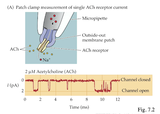
Lecture 6- Ion Channels
- Describe historical advances in our understanding of the nervous system, including evidence supporting or refuting major hypothesis. * History of scientific knowledge on how ions cross membranes: * Pre-1960- unknown mechanism, suggestions included transporters and water-filled pores * 1965-1975- protein channel with water-filled pore and selectivity filter: - TTX (Na+ channel) vs. TEA (K+ channel for AP, not RMP)- rate of ion flux (e.g., 600/ms)- channel radius and selectivity * 1976, Sakmann and Neher- first record of single channel * 1980-present- introduction of molecular technique
- Describe how optogenetic can be used to manipulate neural activity * Optogenetic is a biological technique that allows for the precise control of specific cell types on neuronal circuitries. * It is achieved by expression of light-sensitive ion channels, pumps or enzymes specifically in the target cells * The ultimate goal of optogenetic is the modulation of neuronal circuitries with high temporal and spatial resolution in the behaving animal * In contrast to pharmacological or electrical manipulations, optogenetic allows for the precise control of specific cell types on neuronal circuitries * Controlling neurons with light or chemicals are widely used techniques for manipulating neural activity * Optogenetic affords high temporal control but is invasive as it requires light delivery via optical fibers * Near-infrared manipulation of multiple neuronal populations via optogenetic ahs been demonstrated in recent study
- Summarize evidence supporting the notion of ion channels as the basis of membrane currents * Ion flow across cell membranes was mediated by individual molecular-scale aqueous “channels” across the membrane. * Different channels were thought to have different selectivity among ions—e.g., some selective for potassium, others for sodium ion. * Whether a channel was “open” or “closed” depended on the membrane potential, with the kinetics of response to a change in membrane potential differing among channel types. * The experimentally-determined characteristics of ion flow across squid giant axon membranes could be modeled by cooperative processes, in which a number of subunits of a channel needed all to be in particular voltage-dependent states for the channel to be open. * The first direct evidence for the presence of voltage-sensitive, ion-selective channels in nerve cell membranes came from measurements of the ionic currents flowing through individual ion channels.
- Compare the activity of ion channels and membrane currents to explain the molecular basis of macroscopic currents * Ion channels are proteins that span the cell membrane and allow ions to pass through the membrane. * The activity of ion channels determines the flow of ions across the cell membrane and thus determines the macroscopic current. * Most cells have a range of ion channels within their membrane whose activities under a given condition dictate the macroscopic ionic current flowing across the membrane. * Macroscopic currents can be recorded from a variety of cells using techniques such as two-electrode voltage-clamp, single-electrode switched clamp, and whole-cell patch clamp. * The opening and closing of ion channels is regulated by changes in membrane potential and/or by binding of ligands.
- Compare functional and structural characteristics of different types of ion channels * There are three main types of gated channels: chemically-gated or ligand-gated channels, voltage-gated channels, and mechanically-gated channels. * Ligand-gated ion channels are channels whose permeability is greatly increased when some type of chemical ligand binds to the protein structure. * Voltage-gated ion channels are channels whose permeability is greatly increased when the membrane potential changes. * Mechanically-gated ion channels are channels whose permeability is greatly increased when the cell membrane is physically deformed. * Ion channels can also be classified based on their structural characteristics. For example, some ion channels are tetramers, meaning they consist of four subunits. Other ion channels are monomers or dimers. The subunits of an ion channel can be identical or different.
Lecture 7- Synaptic Transmission I
- Describe historical advances in our understanding of the nervous system, including evidence supporting or refuting major hypothesis. * ~1900, Cajal- Neuron Doctrine; Sherrington- synapse * 1921, Loewi- chemical transmission by “Vangusstoff” (acetylcholine) * 1952, Katz- Quantal hypothesis of release
- Compare the actions of neurotoxins that affect synaptic transmission at the neuromuscular synapse. * Neurotoxins that affect synaptic transmission at the neuromuscular synapse can be classified into different types based on their mechanism of action * Some neurotoxins interfere with voltage-gated ion channels, acetylcholine release, depolarization of the postsynaptic membrane, or generation and spread of muscle action potential * Plant poisons such as curare and bacterial poisons such as botulinum toxin act at single or multiple sites of neuromuscular apparatus. * Clostridial neurotoxins act inside nerves and block neurotransmitter release via their metalloproteolytic activity directed specifically on SNARE proteins * Snake presynaptic neurotoxins with phospholipase A2 activity induce the release of acetylcholine followed by impairment of synaptic functions. * Excitatory latrotoxin-like neurotoxins induce a massive release of neurotransmitter at peripheral and central synapses
- Describe Loewi’s evidence that the nature of synaptic transmission is chemical * Otto Loewi performed an experiment in 1921 that provided the first clear evidence that synaptic transmission was chemical * He electrically stimulated the vagus nerve of an isolated frog heart to decrease the strength and rate of contractions * The bathing solution caused a decrease in the strength and rate of contractions when subsequently applied to a second heart * Loewi called this substance vagusstoff, which was subsequently shown to be acetylcholine, the first neurotransmitter to be identified
- Describe Katz’s study and understand why he compared MEPPs and EPPs obtained in low Ca++ * Bernard Katz studied how nerves talk to muscles * He found out that when a nerve talks to a muscle, it releases chemicals called neurotransmitters * These neurotransmitters cause the muscle to move * Katz discovered that when the concentration of calcium ions is low, the size of the endplate potentials (EPPs) is reduced to about the size of miniature endplate potentials (MEPPs) * Katz compared MEPPs and EPPs obtained in low Ca++ because he wanted to understand how neurotransmitter release works * He found that MEPPs were generated in multiples of a certain amplitude (now known as a “Quantum”) and they all added together to form EPPs.
- Understand the difference between MEPPs and EPPs obtained in low Ca++ * MEPPs are spontaneous depolarizations of the motor endplate that occur in the absence of nerve stimulation. * EPPs are depolarizations of the motor endplate that occur in response to nerve stimulation. * MEPPs are caused by the release of a single quantum of acetylcholine from a single vesicle in the presynaptic terminal. * EPPs are caused by the release of multiple quanta of acetylcholine from multiple vesicles in the presynaptic terminal. * MEPPs are additive, eventually increasing the end-plate potential (EPPs) from about -100mV up to the threshold potential of -60mV. * EPPs are much larger than MEPPs and can reach -40mV to -20mV. * When extracellular Ca2+ concentrations fall too low, the nervous system becomes more excitable as it becomes more permeable to sodium ion. When extracellular Ca2+ concentrations fall to ~50%, spontaneous action potentials occur, causing tetany due to an overactive Peripheral Nervous System.
Lecture 8- Synaptic Transmission II
- Describe historical advances in our understanding of the nervous system, including evidence supporting or refuting major hypotheses. * Blank Space
- Describe the physiological evidence recorded by Katz et al. and how it led to the Quantal Hypothesis * Bernard Katz and Ulf von Euler were awarded the Nobel Prize in Physiology or Medicine in 197 for their work on the release of neurotransmitters * Katz formulated the quantal hypothesis in the 1950s, which states that neurotransmitters are released from presynaptic terminals in discrete ‘quanta’ * Katz and his colleague Horace W.D. Fatt studied the neuromuscular junction of the frog and stimulated motor neurons while recording synaptic potentials (called “end-plate potentials” (EPPs)) in the muscle fiber * They found that EPPs were not continuous but rather occurred in discrete steps or quanta * In 1954, del Castillo and Katz published a paper that showed that the probability of release of a quantum of transmitter was independent of the size of the EPP. This led to the conclusion that each quantum of transmitter released from a presynaptic terminal was sufficient to produce an EPP
- Explain how the use of Poisson statistics to describe the physiological data supported the Quantal Hypothesis
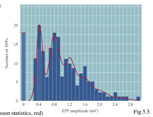 * Poisson statistics is a mathematical tool that can be used to describe the probability distribution of events that occur randomly in time or space.
* The quantal hypothesis states that neurotransmitters are released from presynaptic terminals in discrete ‘quanta’.
* Katz and his colleague Horace W. D. Fatt studied the neuromuscular junction of the frog and found that electrical signals (called “end-plate potentials” (EPPs)) were not continuous but rather occurred in discrete steps or quanta.
* The use of Poisson statistics to describe the physiological data supported the quantal hypothesis because it showed that the probability distribution of the number of quanta released by a presynaptic terminal was consistent with a Poisson distribution.
* Poisson statistics is a mathematical tool that can be used to describe the probability distribution of events that occur randomly in time or space.
* The quantal hypothesis states that neurotransmitters are released from presynaptic terminals in discrete ‘quanta’.
* Katz and his colleague Horace W. D. Fatt studied the neuromuscular junction of the frog and found that electrical signals (called “end-plate potentials” (EPPs)) were not continuous but rather occurred in discrete steps or quanta.
* The use of Poisson statistics to describe the physiological data supported the quantal hypothesis because it showed that the probability distribution of the number of quanta released by a presynaptic terminal was consistent with a Poisson distribution.
- Describe the steps involved in neurotransmitter release and postsynaptic effects.
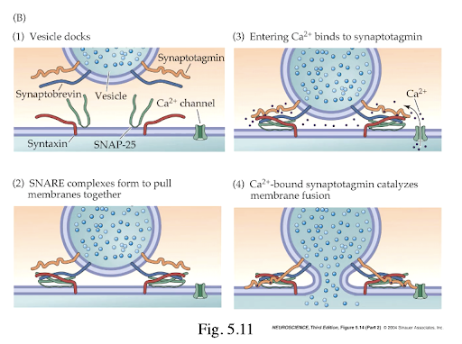 * Synthesis of the neurotransmitter in the neuron’s cell body
* Transport of the neurotransmitter down the axon to the axon terminal where it is stored in vesicles.
* Calcium ions enter the terminal when an action potential reaches the axon terminal, causing the vesicles to migrate towards the cell membrane and fuse together causing the release of the stored neurotransmitter into the synaptic cleft.
* The neurotransmitter travels across the synapse and binds to receptors on target cells (in this case, causing positive ions to flow in).
* The binding of neurotransmitters to receptors on postsynaptic cells can have a variety of effects depending on the type of receptor and the specific neurotransmitter involved.
* Finally, the neurotransmitter is deactivated by either reuptake into the presynaptic neuron or enzymatic degradation
* Synthesis of the neurotransmitter in the neuron’s cell body
* Transport of the neurotransmitter down the axon to the axon terminal where it is stored in vesicles.
* Calcium ions enter the terminal when an action potential reaches the axon terminal, causing the vesicles to migrate towards the cell membrane and fuse together causing the release of the stored neurotransmitter into the synaptic cleft.
* The neurotransmitter travels across the synapse and binds to receptors on target cells (in this case, causing positive ions to flow in).
* The binding of neurotransmitters to receptors on postsynaptic cells can have a variety of effects depending on the type of receptor and the specific neurotransmitter involved.
* Finally, the neurotransmitter is deactivated by either reuptake into the presynaptic neuron or enzymatic degradation
- Compare and contrast postsynaptic potentials and action potentials
 * Action potentials are like electrical signals that travel down the axon of a neuron.
* They are all-or-nothing events that either happen or don’t happen.
* When an action potential reaches the end of an axon, it causes the release of neurotransmitters into the synapse.
* Postsynaptic potentials are changes in the electrical potential of a neuron’s cell membrane that occur when neurotransmitters bind to receptors on the neuron.
* Postsynaptic potentials can be either depolarizing (making the neuron more likely to fire an action potential) or hyperpolarizing (making the neuron less likely to fire an action potential).
* While both action potentials and postsynaptic potentials are involved in communication between neurons, they work in different ways and have different effects on the receiving neuron.
* Action potentials are like electrical signals that travel down the axon of a neuron.
* They are all-or-nothing events that either happen or don’t happen.
* When an action potential reaches the end of an axon, it causes the release of neurotransmitters into the synapse.
* Postsynaptic potentials are changes in the electrical potential of a neuron’s cell membrane that occur when neurotransmitters bind to receptors on the neuron.
* Postsynaptic potentials can be either depolarizing (making the neuron more likely to fire an action potential) or hyperpolarizing (making the neuron less likely to fire an action potential).
* While both action potentials and postsynaptic potentials are involved in communication between neurons, they work in different ways and have different effects on the receiving neuron.
- Describe the nature of ion fluxes underlying synaptic transmission at the neuromuscular junction. * At the neuromuscular junction, an action potential causes the release of calcium ions. * Calcium ions bind to sensor proteins on synaptic vesicles, triggering vesicle fusion with the cell membrane and subsequent neurotransmitter release from the motor neuron into the synaptic cleft. * The neurotransmitter then binds to receptors on the muscle fiber. * This causes ion channels to open and allows positively charged ions like sodium (Na+) and calcium (Ca2+) to enter the muscle fiber. * This influx of positively charged ions depolarizes the muscle fiber and triggers muscle contraction.
Lecture 9- Neurotransmitters & Receptors
- Describe historical advances in our understanding of the nervous system, including evidence supporting or refuting major hypotheses. * 1856: Bernard, curare blocks nerve-muscle excitation * 1910-1960: features of receptors characterized * After 1960: receptors classified as membrane proteins
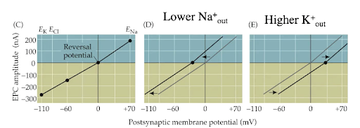
- Explain how EPSPs and IPSPs occur, how they’re different from each other, and how they sum to affect neural activity (action potentials). * Neurotransmitter--→ receptor binds-→ ion channels open/close-→ conductance changes-→ postsynaptic potential changes-→ postsynaptic cells inhibited/excited-→ summation determines action potential occurrence * Both EPSP and IPSP ALWAYS WANT to flow toward Ereversal (equilibrium reversal potential) * EPSP (excitatory post-synaptic potential): A type of potential that triggers an ACTION POTENTIAL. If Ereversal was positive from rest or depolarizes the neuron, then that can be classified as an EPSP * IPSP (inhibitory post-synaptic potential): Type of potential that either hyperpolarizes or depolarizes a neuron.
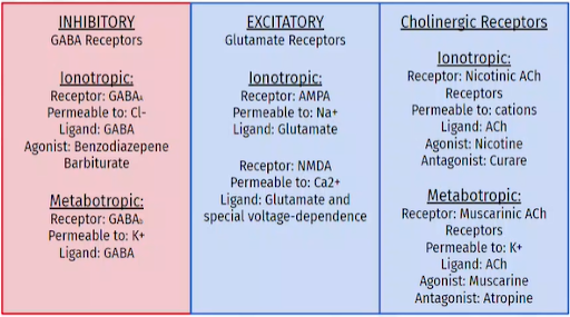
- Compare ionotropic and metabotropic receptors generally, including their characteristic mechanisms and functions * Ionotropic: ligand-gated, fast onset/short duration, mediate fast info processing * Metabotropic: G-protein coupled, slow action/long duration, modulate info processing
- Describe features and functions of ionotropic and metabotropic receptors for acetylcholine and GABA * Nicotinic acetylcholine receptor (ionotropic): agonist is nicotine, antagonist is curare, increases cation permeability * Muscarinic acetylcholine receptor (metabotropic): agonist is muscarine, antagonist is atropine, changes K+ permeability * GABAA receptor (ionotropic): increase Cl- permeability * GABAb receptor (metabotropic): increase K+ permeability
\