Organisation
Cells are the basic building blocks of all living organisms.
A tissue is a group of cells with a similar structure and function.
Organs are aggregations of tissues performing specific functions.
Organs are organised into organ systems, which work together to form organisms.
The Digestive System
the mouth both physically and chemically breaks down food
the oesophagus carries food from the mouth to the stomach
the stomach physically breaks down food by churning it, breaks down proteins and contains hydrochloric acid with a low pH to kill microbes in food so they don’t cause illness
the liver makes bile, which is alkaline to neutralise stomach acid. it also emulsifies fat into small droplets, increasing their surface area, making lipase more effective
the gall bladder stores and secretes bile
the pancreas makes enzymes
the small intestine absorbs nutrients via villi
the large intestine absorbs water from undigested food to produce faeces
enzymes
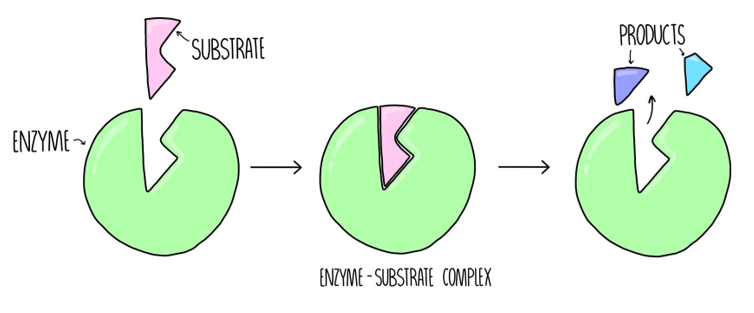
enzymes are biological catalysts, increasing the rate of reactions but not being used up. the way they work can be explained by the lock and key theory
the shape of the active site is specific and complementary to the substrate it can break down.
they collide and become attached, forming an enzyme-substrate complex
the enzyme catalyses the breakdown of the substrate and products are formed and released from the active site
enzymes require an optimum pH and temperature to work. at temperatures/pHs too far above/below the optimum, they will denature and their active site will change shape, meaning that the substrate can no longer fit and it cannot catalyse reactions.
enzyme | what it breaks down | product | where it’s produced | where it works |
|---|---|---|---|---|
carbohydrase | carbohydrates | simple sugars | salivary glands pancreas small intestine | mouth small intestine |
amylase | starch | maltose | salivary glands pancreas | mouth small intestine |
maltase | maltose | glucose | small intestine | small intestine |
protease | proteins | amino acids | stomach pancreas small intestine | stomach small intestine |
lipase | lipids (fats) | fatty acids glycerol | pancreas | small intestine |
Required Practical: food tests
Benedict’s reagent: add it to a food sample and heat in a water bath. should turn anywhere from blue to red depending on if and how much glucose is present
Iodine: add it to a food sample. it will turn from brown to a blue-black if starch is present
Biuret reagent: add it to food sample. will turn from blue to purple if protein is present.
Emulsion/ethanol: add ethanol to the food. shake to combine then pour into water. if the mixture goes cloudy, fats are present.
Sudan III: add to a mixture of food and water. a red layer will form on top if fats are present.
Required Practical: pH and amylase
Method
mark the spotting tile with time in the columns and pH on the rows
add a drop of iodine to each well of the spotting tile
use a water bath to heat a solution of starch, amylase and a buffer which maintain pH. water bath is used to maintain a constant temperature, where it could be a factor affecting rate.
at regular time intervals, something like every 30 seconds, transfer a drop of the solution to a well of the corresponding pH row.
if the iodine turns black, there’s starch present, and the amylase has denatured. so the faster the iodine stops seeing a change in colour, the faster amylase is breaking down starch so the faster the reaction and the more effective the enzyme at that pH
rate = product formed/reactant used / time
The heart
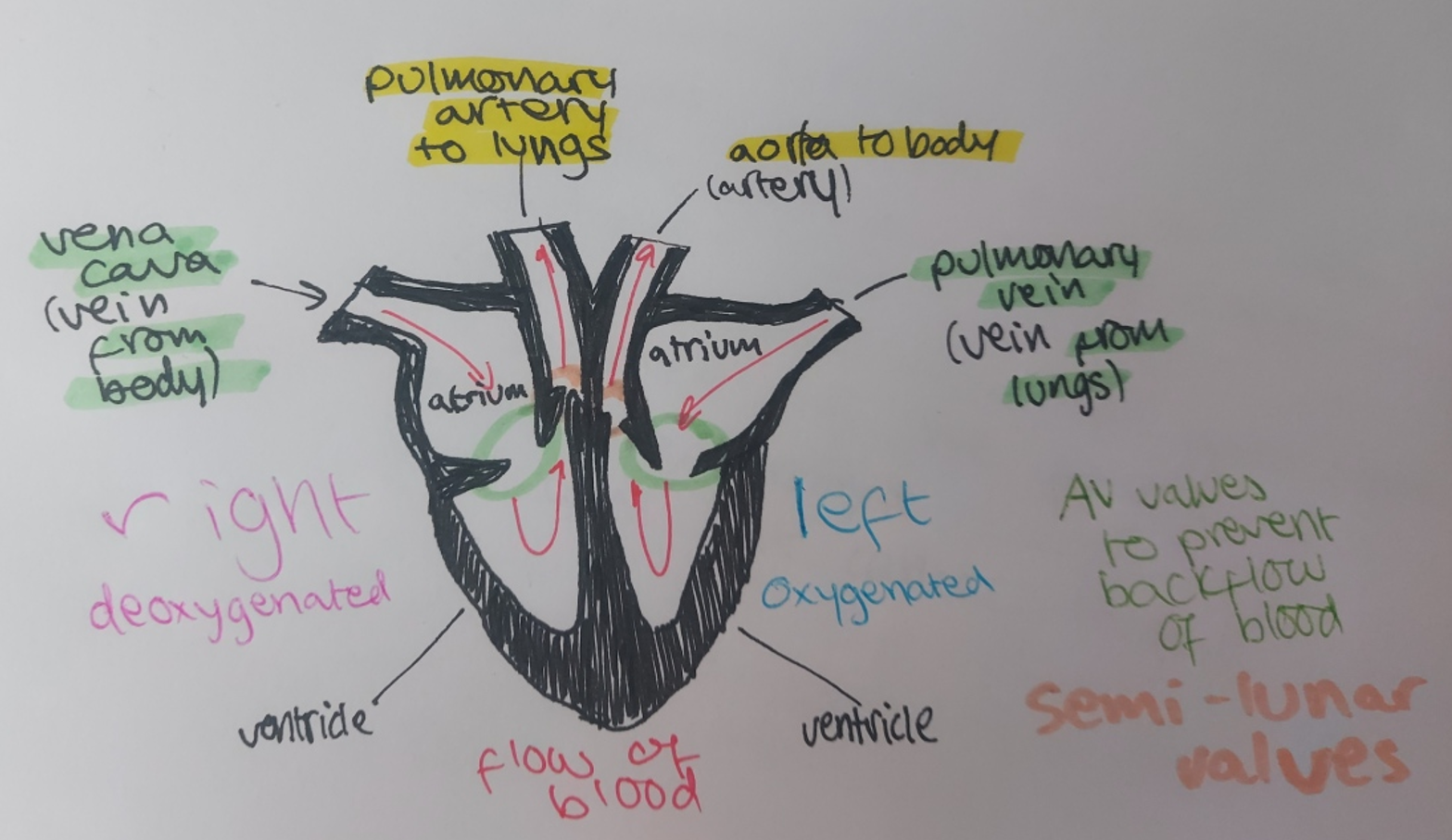
the heart pumps blood around the body in a double circulatory system. this means there are two circuits/routes for blood to flow.
deoxygenated blood
gets pumped in via the vena cava
right atrium contracts to force it into the right ventricle
AV valves close to prevent backflow
ventricle contracts, forcing blood into the pulmonary artery
it then travels to the lungs to get oxygenated
oxygenated blood
gets pumped in via the pulmonary vein
left atrium contracts to force it into the left ventricle
AV valves close to prevent backflow
ventricle contracts, forcing blood into the aorta
it travels around the body delivering oxygen to cells for respiration
blood vessels
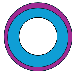
arteries carry blood away from the heart.
they have thick muscular walls to make them strong and thick elastic layer that allows them to stretch to facilitate high blood pressure caused by the heart pumping
they have a narrow lumen (the hole in the blood vessel through which blood flows)
the blood in them is generally oxygenated, save for in the lungs, because this is where blood in them has their oxygen moved to veins to travel back to the heart and be circulated

veins carry blood towards the heart.
they have thin walls due to their large lumen that allows low pressure blood to flow through
they have valves to prevent backflow
the blood in them is generally deoxygenated, save for in the lungs, because this is where blood gets oxygenated and then travels back to the heart
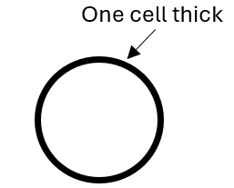
capillaries carry blood close to the cells so substances can move between the two.
they’re one cell thick to make the diffusion pathway as short as possible, and thus the most efficient, and don’t have a muscular or elastic wall to facilitate this.
Blood
blood is a tissue made up of plasma, white blood cells, red blood cells and platelets.
plasma is the liquid that carries the other components of blood. it’s mostly water
white blood cells that protect the body from infection (see infection and response for specifics)
red blood cells that carry oxygen molecules from the lungs to the rest of the body by binding to it with haemoglobin
platelets which are essentially cell fragments that allow blood to clot, which can then dry into a scab that allows new skin growth underneath while keeping microorganisms out
Issues of the Heart
valves: can get damaged over time as they withstand a lot of pressure, so can get stiff and the heart becomes less efficient, or may develop a leak. biological (human and animal) or mechanical equivalents can be transplanted. biological valves work very well but degrade within 15 years while mechanical last for a long time but need to be medicated to prevent blood clots
artificial hearts are used to keep a patient alive while they’re awaiting a transplant
heart rate is controlled by a group of cells in the right atrium. this is known as your pacemaker. if there are irregularities with your heartbeat, an artificial pacemaker can be used.
coronary heart disease
coronary heart disease occurs when fatty material builds up in the coronary arteries that provide blood to the heart itself, and they become blocked. this means less blood flows to the heart, reducing its oxygen supply.
the oxygen supply to the heart needs to be maintained so that aerobic respiration can occur. the heart CANNOT start respiring anaerobically, because this produces lactic acid that causes muscle fatigue, and the heart cannot afford to tire and stop pumping blood, because the rest of you body cells will then stop respiring too.
there are 2 solutions:
stents - wire mesh tubes placed into the coronary arteries to hold them open, but you risk infection and blood clots
statins - drugs that lower cholesterol that leads to coronary heart disease. reduce risks but are only a preventative measure
The lungs
the trachea or windpipe sends air down into the lungs, where it then splits between the two bronchi which further split into bronchioles that lead to the alveoli
when you inhale, your ribcage moves up and out to facilitate an increase in chest volume, which results in low pressure. this causes air to be drawn into the lungs
when you inhale, gas exchange takes place.
alveoli fill with oxygen
blood in surrounding capillaries is deoxygenated as blood has flowed into them from pulmonary artery, and it has a lot of carbon dioxide
oxygen diffuses into the capillaries from the alveoli due to the concentration gradient
carbon dioxide diffuses into the alveoli from the capillaries
blood is now oxygenated and travels towards to heart via the pulmonary vein
alveoli are adapted to be as efficient for this diffusion as possible:
plentiful
small meaning they have a large surface area to volume ratio meaning diffusion occurs quicker
thin to make for a short diffusion pathway
other parts of the lungs are also adapted to make diffusion as efficient as possible:
capillaries are also only one cell thick, making for a short diffusion pathway
capillaries provide a good blood supply, removing oxygenated blood fast to maintain the concentration gradient
the action of breathing in and out maintains the concentration gradient
Health issues
health is a state of physical, mental and social wellbeing.
communicable diseases are infectious and caused by pathogens
non-communicable diseases aren’t passed on from person to person
diseases interact in a multitude of ways:
someone with a weakened immune system is more likely to be infected with a communicable disease because the body is less able to fight off pathogens. they may be immunocompromised due to another disease
infections by viruses in some parts of the body leads to cancers
reduction in physical health can lead to a reduction in mental health
immune responses due to infection can cause allergic reactions
non-communicable diseases can have huge financial and emotional impacts on the individual, their family and the wider community, with masses of people dying due to these diseases and, in some countries, healthcare not being free. it also has a nation-level impact because research into diseases and treatment is expensive. globally, there may be effects when a disease affects the working age population, in regards to productivity.
risk factors are things that increase the likelihood of having a non-communicable disease. they pertain to the person’s lifestyle, now and in the past. a few common ones are:
diet
obesity
smoking
drinking
age
genes
sex
infection
carcinogens/ionising radiation
if one factor increases as another increases, they are correlated. a casual mechanism explains this correlation.
Cancer
cancer is the result of mutations in cells that leads to uncontrolled growth and mitosis.
benign tumours are growths of abnormal cells which are contained in one area, usually within a membrane. They do not invade other parts of the body.
malignant tumour cells are cancers. They invade neighbouring tissues and spread to different parts of the body in the blood where they form secondary tumours.
there are a few specific risk factors for different types of cancer:
smoking
obesity
UV light (skin cancer)
infection
genes
Plant Tissues and Organ systems
meristem tissue differentiates to allow the plant to grow.
the leaf and gas exchange
epidermis tissue (upper and lower): covered with a waxy cuticle to reduce water loss by evaporation and make the plant waterproof
palisade mesophyll: site of photosynthesis. contains lots of chloroplasts and has a long vertical structure, at the top of the leaf, to maximise light received - long structure means that many of them can be lined up at top of the leaf
spongy mesophyll: air space that allows for diffusion of gases during gas exchange
guard cells: swell with water to cause the stomata to open, and lose water to close them
stomata: holes made by guard cells, that enable water loss (transpiration) and gas exchange when they're open. they’ll shut in certain conditions:
when there’s little water: shut to prevent water loss
when it’s night: photosynthesis can’t occur so there’s no point being open to let gas exchange occur and losing water
the leaf is the site of gas exchange in a plant, which enables photosynthesis, that generates the glucose and oxygen needed for respiration, which releases energy and enables the plant to carry out processes.
translocation
the movement of food substances made in the leaves up and down the plant via the phloem.
perforated cell walls (sieve plates) allows water and food (glucose) solution to pass between cells
hollow (bar cytoplasm) to facilitate movement of glucose - companion cells with mitochondria produce energy for the cell
transpiration
this is the loss of water from the leaves and stem as a result of gas exchange, when the stomata open. water is brought up into the plant via the root hair cells when they take up minerals, and is transported to the leaves by the xylem cells.
it’s affected by 4 main factors:
temperature - increases temperature makes water evaporate faster, so rate of transpiration increases
humidity - if it’s more humid there’s more water in the air, it’s more difficult for water to evaporate, so rate of transpiration decreases
light intensity - higher light intensity makes water evaporate faster, so rate of transpiration increases
air flow/wind - carries water away from the leaves, refreshing the concentration gradient and increases the rate of transpiration