MA 4 Chapter Notes
Ch 17 Innate Immunity
17.1 Physical Defenses
Physical Defenses
physical barriers
prevent microbes from reaching tissues that are susceptible to infection
barriers are composed of cells that are tightly joined to prevent invaders from crossing through to deeper tissue
ex. endothelial cells
cell junctions are composed of cell membrane proteins that may connect with the extracellular matrix or with complementary proteins from neighbouring cells
ex. tight junctions, desmosomes (intracellular junctions/ spot welds/bind adjacent animal cells), gap junctions


skin barrier
composed of 3 layers of closely packed cells
epidermis: thin upper layer
cells w/ hella keratin
keratin makes surface tough and resistant to degradation by bacterial enzymes
dead cells remain tightly connected and create dense layers of protein filled cell husks on surface of skin
frequently shed, along with microbes that may be clinging to them
shed skin cells are continually replaced with new cells from below
fatty acids on surface inhibits growth of some microbes and highly resistant to breakdown by bacterial enzymes
dermis: thicker, with hair follicles, sweat glands, nerves, and blood vessels
hypodermis: fatty tissue layer with blood and lymph vessels
mucous membrane
line the nose, mouth, lungs, and urinary and digestive tracts
consist of a layer of epithelial cells bound by tight junctions
secrete a moist, sticky substance called mucus
covers and protects more fragile cell layers and traps debris and particulate matter, including microbes
secretions contain antimicrobial peptides
mechanical actions flush mucus out of body
removal system = mucociliary escalator
mucociliary blanket = debris caught in mucus lining RT
ciliated epithelial cells = EC lining the upper RT
has cilia
Endothelia
epithelial cells that line the urogenital tract, blood vessels, lymphatic vessels, and other tissues
endothelia of blood-brain barrier protect from the central nervous system (brain/spinal cord)
microbial infections of CNS lead to serious/fatal inflammation
cell junctions in blood vessels of CNS are tightest/toughest in body
Mechanical Defenses
shedding of skin cells, expulsion of mucus (mucociliary escalator), excretion of feces through intestinal peristalsis
flushing of urine and tears—> carry microbes away from body
urinary tract: flushing of urine —> clearing of kidneys, ureters, urinary bladder
urine passing out of body washes out transient MOs
eyes: physical and mechanical defense mechanisms for preventing infections
blinking
eyelashes and eyelids
Physical Defenses of Nonspecific Innate Immunity
Defense | Examples | Function |
Cellular Barriers | skin, mucous membrane, endothelial cells | deny entry to pathogens |
Mechanical Defenses | shedding of skin cells, mucociliary sweeping, peristalsis, flushing action of urine and tears | remove pathogens from potential sites of infection |
Microbiome | resident bacteria of the skin, upper respiratory tract, gastrointestinal tract, and genitourinary tract | Compete with pathogens for cellular binding sites and nutrients |
17.2 Chemical Defenses
Chemical and Enzymatic Mediators Found in Body Fluids
endogenous mediator:
naturally occuring substance produced by body that acts as a chemical messenger or link to trigger/regulate/stop a specific biological process
endogenous: originating or produced internally (within cell, organism, or tissue)
mediator: a substance (protein, peptide, or lipid) that acts as a go-between to transmit signals or influence the activity of other cells
i.e. inflammation, pain, immune responses
common examples include: histamines, specialized pro-resolving mediators (SPMs), prostaglandins (mediate processes like pain, fever, and inflammatory response to injury)
exogenous mediator:
external agent introduced to facilitate a chemical, biological, or electrical reaction
in immunology/inflammation: describe external triggers that enter the body and induce an immune response
*exogenous administration of healing agents—> (like SPMs) used therapeutically to promote tissue healing from the outside
in bioelectrochemistry/microbial fuel cells: external chemicals added to bacterial systems to act as “electron shuttles”
fluids produced by skin can be both endogenous and exogenous mediators
sebaceous glands secrete sebum —> released in the skin surface through hair follicles
endogenous mediator
seal off the pore via hair follicle
Cutibacterium acnes/malassezia use lipasre enzymes to degrade sebum
produces oleic acid—> creates mildly acidic env inhospitable for pathogenic microbes
oleic acid: exogenously produced mediator—> because produced by resident microbes and not directly by body cells
environmental factors that affect microbiota have a direct impact on the production of chemical mediators
low humidity/decreased sebum production could make the skin less habitable for microbes that produce oleic acid
making skin more susceptible to pathogens normally inhibited by skin’s pH
digestive tract produces hella chemical mediators that inhibit or kill microbes
Antimicrobial Peptides (AMPs)
special class of nonspecific cell-derived mediators with broad spectrum antimicrobial properties
most AMPs are produced in response to invading pathogen
may induce cell damage in MOs
inflicting damage to membranes
destroying DNA/RNA
interfering with cell-wall synthesis
depending on AM mechanism, AMPs may only inhibit certain groups of microbes (gram-positive/negative) or may be more effective against bacteria, fungi, protozoa, and viruses
mostly found on skin but can be found in other regions of the body
family “defensins” —> prod. by epithelial cells as macrophages/neutrophils
may be secreted/act inside host cells
combat MOs by damaging plasma membranes
AMPs called bacteriocins—> prod. exogenously by certain members of resident microbiota within gastrointestinal tract
bacteriocins—> genes carried on plasmids and can be passed between different species via lateral/horizontal gene transfer
Characteristics of Selected AMPs
AMP | Secreted By | Body Site | Pathogens Inhibited | Mode of Disruption |
bacteriocins | resident microbiota | GI tract | bacteria | disrupt membrane |
cathelicidin | epithelial cells, macrophages, other cell types | skin | bacteria and fungi | disrupts membrane |
defensins | epithelial cells, macrophages, neutrophils | throughout body | fingu, bacteria, many viruses | disrupt membrane |
dermcidin | sweat glands | skin | bacteria and fungi | disrupts membrane integrity and ion channels |
histatins | salivary glands | oral cavity | fungi | disrupt intracellular function |
Plasma Protein Mediators
nonspecific innate immune factors are found in plasma (fluid portion of blood)
plasma contains electrolytes, sugars, lipids, proteins; also proteins involved in clotting blood; also acute-phase proteins, complement protein, and cytokines
acute phase proteins
produced in liver and secreted into blood in response to inflammatory molecules from the immune system
ex. C-reactive protein, serum amyloid A, ferritin, transferrin, fibrinogen, and mannose-binding lectin
Functions of Some Acute-Phase Proteins
Opsonization, preparing them for ingestion by phagocytes | C-reactive protein |
Serum amyloid A | |
Bind and sequester iron, thereby inhibiting the growth of pathogens | ferritin |
transferrin | |
Involved in formation of blood clots that trap bacterial pathogens | Fibrinogen |
Mannose-binding lectin | Activates complement cascade |
Complement System
group of plasma protein mediators that can act as an innate nonspecific defense
composed of more than 30 proteins; normally precursor proteins in blood
can become stimulated via presence of MOs
complement proteins = innate specific immunity—> always present in blood/tissue fluids (allows for them to be activated quickly)
Complement Activation
process by which circulating complement precursors become functional
can be triggered by:
alternative pathway
initiated by spontaneous activation of C3
hydrolysis of C3 produces C3a and C3b
without presence of invader microbes—> C3b degraded in a hydrolysis reaction using water in the blood
with presence of invader microbes—>C3b attaches to the surface of these microbes and will recruit other CP in a cascade
classical pathway:
must be initiated via specific antibody—> binds to the pathogen to form an antibody-antigen complex
this step activates the first protein in the complement cascade (C1 complex)
C1 complex is a multipart protein complex; each component participates in the full activation —> remaining classical pathway complement proteins are recruited and activated in a cascading sequence
lectin activation pathway:
similar to classical pathway but is triggered by the binding of mannose-binding lectin to carbohydrates on microbial surface
lectins—> prod. by liver cells
common response to inflammatory signals received by body during infection

all pathways produce the same protective outcomes:
opsonization: coating of a pathogen by a chemical substance (opsonin)
opsonins from CC (C1q, C3b, C4b)
*also imp. mannose-binding proteins and antibodies
complement fragments C3a/C5a are anaphylatoxins (inflammatory functions!)
anaphylatoxins: activate mast cells—>causing degranulation and release of inflammatory chemical signals
including mediators that cause vasodilation and increased vascular permeability
C5a =potent chemoattractants for neutrophils and other white blood cells
membrane attack complex: C6, C7, C8 and C9
enables C9 polymerize into pores in the membranes of gram-negative bacteria
pores facilitate water, ions,and other molecules to move in and out of targeted cells
*MAC cannot penetrate peptidoglycan in gram-positive bacteria
because of this lack of threat to g+ bacterial pathogens, complement-mediated opsonization is more important for clearance.
cytokines:
soluble proteins that acts as communication signals between cells
in nonspecific innate immune response; cytokines may be released to stimulate production of chemical mediators or other cell fns
proliferation, cell differentiation, inhibition of cell division, apoptosis, and chemotaxis
when bound to target receptor: effect can vary widely depending on type of cytokine/type of cell or receptor to which it has bound
Autocrine | same cell secretes and receives cytokine signal | 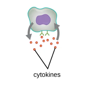 |
Paracrine | cytokine signal secreted to a nearby cell |  |
Endocrine | cytokine signal secreted to circulatory system; travels to different cells | 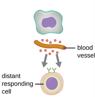 |
Important Classes of Cytokines
Interleukins |
|
Chemokines |
|
Interferons |
|

Inflammation-Eliciting Mediators
chemical mediators previously mentioned contribute in some way to inflammation and fever—>nonspecific immune responses
cytokines stimulate prod. of acute-phase proteins (C-reactive/mannose-binding lectin) in liver
these proteins act as opsonins—> activate CC through lectin pathway
some cytokines also bind mast cells and basophils—>release histamine
histamine: proinflammatory compound
receptors found on variety of cells and mediate proinflammatory events
mast cells may also release leukotrienes
leukotrienes: lipid-based proinflammatory mediator prod. from metabolism of AA in cell membrane of leukocytes and tissue cells
effects are more potent and long lasting than histamines
leukotrienes + histamines = coughing, vomiting, diarrhea —> expel pathogens from body
cytokines can stim. prod. of prostaglandins
prostaglandins: chem. mediator that promote inflammatory effects of kinins and histamines
also set body temp. higher—>fever
promotes active white blood cells and inhibits the growth of pathogenic microbes
bradykinin: contributes to edema; when fluids and leukocytes leak out of bloodstream into tissues
Defense | Examples | Function |
Chemicals and enzymes in body fluids | sebum from sebaceous glands | Provides oil barrier protecting hair follicle pores from pathogens |
oleic acid from sebum and skin microbiota | lowers pH to inhibit pathogens | |
lysozyme in secretions | kills bacteria by attacking cell wall | |
acid in stomach, urine, and vagina | inhibits or kills bacteria | |
digestive enzymes and bile | kill bacteria | |
lactoferrin and transferrin | bind and sequester iron, inhibiting bacterial growth | |
surfactant in lungs | kills bacteria | |
Antimicrobial peptides | defensins, bacteriocins, dermcidin, cathelicidin, histatins | kill bacteria by attacking membranes or interfering with cell functions |
Plasma protein mediators | acute-phase proteins (C-reactive protein, serum amyloid A, ferritin, fibrinogen, transferrin, and mannose-binding lectin) | inhibit the growth of bacteria and assist in the trapping and killing of bacteria |
complements C3b and C4b | opsonization of pathogens to aid phagocytosis | |
complement C5a | chemoattractant for phagocytes | |
complements C3a and C5a | proinflammatory anaphylatoxins | |
Cytokines | interleukins | stimulate and modulate most functions of immune system |
chemokines | recruit white blood cells to infected area | |
interferons | alert cells to viral infection, induce apoptosis of virus-infected cells, induce anticrial defenses in infected and nearby uninfected cells, stimulate immune cells to attack virus-infected cells | |
Inflammation-eliciting mediators | histamine | smooth muscle contraction, increased secretion and mucus production |
leukotrienes | promote inflammation; stronger and longer lasting than histamine | |
prostaglandins | promote inflammation and fever | |
bradykinin | increases vasodilation and vascular permeability, leading to edema |
17.3 Cellular Defenses
Hematopoiesis
formed elements of blood come from HEMATOPOIETIC STEM CELLS (HSCs)
from bone marrow
as HSC makes copies of themselves in bone marrow, individual cells receive different cues from body on how to develop and mature
HSCs diferentiate into blood types that will circulate in peripheral blood
process of differentiation: hematopoiesis

nonfluid portion of blood; formed elements: all formed from same stem cells found in bone marrow
3 major categories:
erythocytes (red blood cells) |
|
thrombocytes (platelets) |
|
leukocytes (white blood cells) |
|
GRANULOCYTES:
can be distinguished by appearance of nuclei and contents of granules (confer traits, functions, staining properties)
neutrophils (polymorphonuclear neutrophils/PMNs):
nucleus with 3-5 lobes and small, numerous, lilac coloured granules
each lobe is connected
eosinophils: fewer lobes in nucleus (2-3) and larger granules that stain reddish/orange
basophils
2 red lobed nucleus and large granules that stain dark blue/purple

Neutrophils (PMNs) |
|
Eosinophils |
|
Basophils |
|
Mast Cells |
|
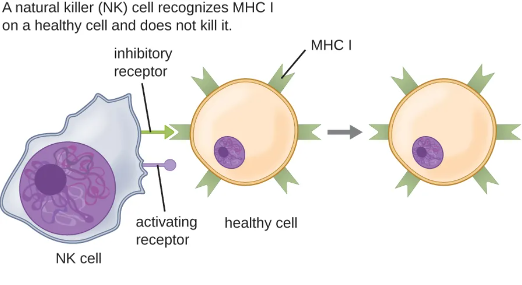
Natural Killer Cells
mononuclear lymphocytes—> nonspecific mechanisms to recognize and destroy cells that are abnormal in some way
abnormal cells: cancer cells/infected cells