Chromosomes and Chromosome Numbers
The Organization of DNA Into Chromosomes
Chromatin
Chromatin: term that describes DNA in its usual unorganized state.
DNA Organization
In the G2 portion of interphase, the replicated DNA will be organized into chromosomes.
The cell constructs chains of protein molecules known as histones.
Each replicated strand of DNA is then coiled around a histone backbone.
This helps the DNA strand maintain its shape and adds strength.
This unit of genetic material is now known as a chromatid.
Because of replication, each chromatid has an identical twin.
These are referred to as sister chromatids.
The sister chromatids are brought together and joined at a location near their center known as a centromere.
Once joined, the overall structure will be known as a chromosome.
Chromosome Number
Each species of organism has certain number of chromosomes that are associated with its cells.
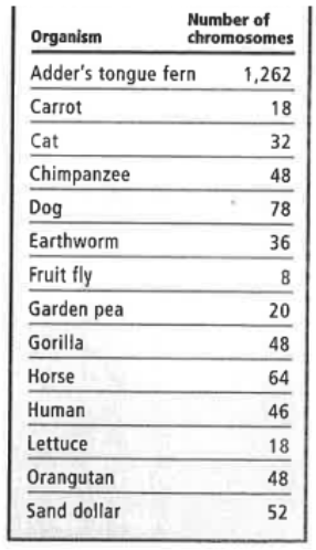
Chromosome number is not a function of size, complexity, sophistication, etc.
In organisms that are created by means of sexual reproduction, chromosomes occur in pairs. Each parent contributes one copy of each form of chromosome.
Chromosomes can either be classified as sex chromosomes or autosomes.
The autosomes are assigned numbers according to descending size order (Largest to smallest).
The single pair of sex chromosomes will always be designated as the last numbered pair.
The two copies of each autosomal pair are described as homologous chromosomes.
This means that they contain genetic information about the same traits, but do not necessarily call for the same exact form or allele for each trait.
Scientists are able to arrange and photograph the chromosomes of a cell. This process is known as karyotyping.
Chromosome Structure & Appearance Within Karyotyping
Each chromosome, as you know, is made up of two sister chromatids that are joined at their centromere.
The centromere constriction forms distinct arms within the chromosome.
In most cases, the centromere is off-center and will produce a pair of shorter arms and a pair of longer arms.
Shorter arms: p arms (p = “petit”)
Longer arms: q arms
G Banding (Giemsa banding)
Giemsa is a stain that can be applied to chromosomes during metaphase of cell division to produce karyotyping images that reveal patterns of dark and light bands.
These bands help to further identify a given chromosome and its homolog. Changes in these bands can also help to reveal disorders.
Light bands (Negative bands): Euchromatin G-C rich areas. More active.
Approx. 92% of the active genes within the genome are found within euchromatin.
Dark bands (Positive bands): Heterochromatin A-T rich areas Less active.

Ploidy Level
Somatic Cells: ordinary body cells. These cells will generally have homologous pairs of chromosomes and are said to be diploid. Diploid is commonly abbreviated using the symbol 2n.
Gametes or Germ cells: these are the sex cells (sperm, eggs, etc.) These cells have only one copy of each type of chromosome and are said to be haploid. Haploid is commonly abbreviated using the symbol 1n or n.
In certain cases, cells may have extra chromosomes. This condition may be referred to as polyploidyism.
In animals, this is always a negative condition.
In plants, polyploidism is more common and can sometimes prove to be beneficial.