Intro to Protein Secretion in Bacteria
Protein Secretion in Bacteria
Overview of Secretion and Translocation
Protein secretion pathways overview.
Basic machineries: Sec and TAT translocation.
Lecture series structure:
Lecture 1: Overview of secretion and basic machineries (Sec and TAT).
Lecture 2: Type secretion machineries.
Lecture 3: Approaches, methods (in vitro, in vivo), and structural studies.
Learning Outcomes
Describe key features of bacterial secretion systems.
Understand basic components and mechanism of translocation by Sec and TAT systems.
Describe similarities and differences between Types 1-6 secretion systems (Type 7 covered later).
Describe how to investigate protein secretion in a lab.
The Importance of Protein Localization
Proteins must be localized to the correct compartment to function.
Central dogma: DNA → RNA → protein is incomplete without considering protein localization.
Polypeptides made by ribosomes in the cytoplasm, need to fold into functional proteins.
A subset of proteins needs to be localized to the correct compartment to function.
Polypeptides are produced in the cytoplasm.
Localisation can occur before or after the polypeptides are folded into a functional protein.
Reasons for Protein Localization Outside the Cytoplasm in Bacteria
Cell shape and stability - governed by cell wall which is outside cytoplasm.
Resistance to environmental stresses- outer membrane.
Environmental sensing.
Nutrient acquisition.
Energy metabolism.
Niche colonization.
Motility.
Adherence/colonization of a surface.
Direct competition with other bacteria.
Secretion Machineries and Protein Relocation
Secretion machineries relocate newly synthesized proteins.
Gram-positive vs. Gram-negative bacteria:
Gram-positive: Cytoplasm, cytoplasmic membrane, extracellular milieu.
Gram-negative: Cytoplasm, cytoplasmic membrane, periplasmic space, outer membrane, extracellular milieu, target cell.
Membranes and Secretion
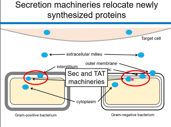
Crossing each membrane requires a different machinery or channel. The membranes from inner to outer are as follows: cytoplasmic membrane, outer membrane and then the membrane of target cell. To move proteins between membranes you need secretion systems.
The two main membranes that we are looking at are Sec and TAT machineries which transporting proteins into or out of the cytoplasm. These can be found in most organisms including Gram negative and positive bacteria.
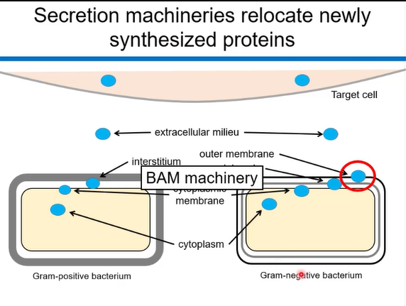
BAM machineries can be found in gram negative bacteria and are responsible for the assembly of β-barrel proteins in the outer membrane, playing a crucial role in the protein secretion pathway.
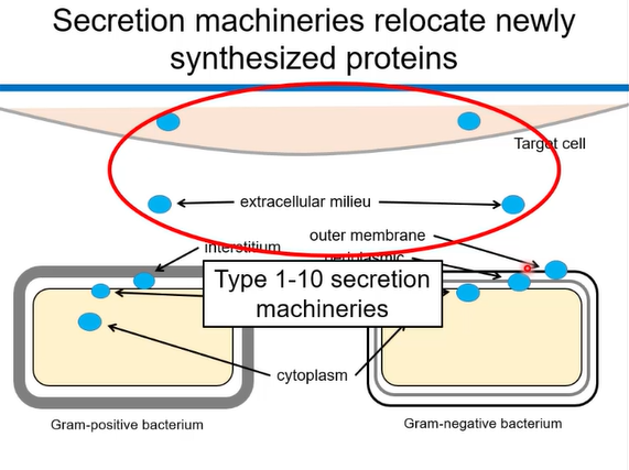
Type 1-10 secretion machines: involved in transporting proteins out cell into the extracellular milieu or directly into the target cell.
Key components of the Secretion Machineries
A means of recognizing substrate proteins.
A protein-conducting channel.
A mechanism that drives translocation of the protein through the channel.
The Sec Pathway
Recognition of substrate proteins.
The Signal Hypothesis (Blobel and Dobberstein):
Secreted proteins are synthesized as longer precursors that are processed upon translocation.
The processed portion contains a signal (“molecular postcode”) which targets it for secretion.
Sec Signal
N-region, H-region, C-region.
General structure:
Minimum length for integral membrane protein:
The signal peptide, typically found at the N-terminus of preproteins (called this before cleaving), is a short amino acid sequence that contains a positively charged N-terminus, a hydrophobic central domain, and a polar C-terminus. This sequence acts as a signal for the Sec pathway to recognize and transport the protein. The signal peptide is connected to the protein sequence and has a recognition site- AXA, that can be cleaved/cut off by signal peptidase (SPase I). Once it has been cleaved off the sequence is now called matured protein.
How are Sec substrates recognised : Sec Substrate Recognition
Two recognition pathways:
SecA: recognise substrates
SRP: recognise substrates that are destined for the membrane, guiding them to the secretion machinery efficiently.
Sec Channel: SecYEG
SecYEG, a protein-conducting channel in bacterial inner membranes, facilitates protein translocation by opening and closing its lateral gates. The channel is composed of SecY, SecE, and SecG subunits, with SecY forming the pore through which proteins pass. The process involves SecA, an ATPase, which interacts with SecYEG to drive the translocation of preproteins
SecYEG is a conserved protein complex forming a channel for unfolded proteins during secretion.
It features an hourglass shape with a central constriction lined by hydrophobic amino acids, allowing only unfolded proteins to pass.
A short α-helix plugs the periplasmic cavity, and a gate enables lateral partitioning of integral membrane proteins into the cytoplasmic membrane.
Mechanism of Translocation Through SecYEG
Here's a step-by-step breakdown:
Signal Sequence Recognition: Proteins destined for translocation possess an N-terminal signal sequence that is recognized by the Sec machinery.
Chaperone Assistance (Post-translational): For post-translational translocation, the unfolded preprotein is bound by SecB, a molecular chaperone, which keeps it in an unfolded state and directs it to SecA.
SecA Interaction: SecA, an ATPase, binds to the preprotein, forming a complex.
SecYEG Interaction: The SecA-preprotein complex interacts with SecYEG, the translocase.
ATP Hydrolysis and Translocation: SecA hydrolyzes ATP, which provides the energy for the stepwise translocation of the preprotein through the SecY channel.
Membrane Insertion/Secretion: As the preprotein moves through the channel, it either gets inserted into the membrane or released into the periplasm
Holotranslocon Complex
Several auxiliary components assist translocation:
YidC
SecD/SecF
YajC
These components form a complex with SecYEG.
Molecular mechanisms are debated.
Twin Arginine Translocation (TAT) Pathway
TAT Signal Sequence
The Twin-Arginine Translocation (Tat) pathway transports folded proteins across biological membranes. It's a unique pathway that relies on a conserved "twin-arginine" motif in the N-terminal signal peptide of the substrate protein. The process involves several key steps: signal peptide recognition, membrane insertion, and translocation across the membrane
TAT Substrate Recognition
In this recognition process, specific chaperones bind to unfolded proteins and facilitate their targeting to the TAT translocase for secretion across the inner membrane.
TatBC recognizes folded substrates at the membrane.
Recognition triggers recruitment of multiple TatA proteins.
TatA forms a channel.
Folded proteins are transported through the channel.
Mechanism of Translocation:
Here's a step-by-step breakdown of the Tat pathway:
1. Signal Peptide Recognition and Binding:
The folded Tat substrate protein, containing the twin-arginine motif, is recognized and bound by the TatBC receptor complex, primarily by TatC. The twin-arginine motif (RR) is crucial for this interaction.
2. Deep Insertion of the Signal Peptide:
The signal peptide, along with the rest of the substrate protein, is deeply inserted into a groove on TatC, likely facilitated by the protein-motive force (PMF) across the membrane.
3. TatA Recruitment and Oligomerization:
TatA, another component of the Tat translocase, is recruited to the TatBC complex, potentially forming a pore-like structure. This recruitment and oligomerization are also influenced by the PMF.
4. Protein Translocation:
Once the Tat translocase is formed, the folded substrate protein is translocated across the membrane. This process is also driven by the PMF.
5. Signal Peptide Cleavage (if applicable):
In some cases, the signal peptide is cleaved after translocation, releasing the mature protein in the destination compartment.
In essence, the Tat pathway is a specialized system for delivering folded proteins across membranes, relying on the interaction of the signal peptide with the Tat translocase and the energy provided by the proton-motive force.
TAT Driving Force
Driving force unclear= In many instances, factors such as membrane potential, pH gradients, and ATP availability play crucial roles in regulating the Tat pathway's performance, highlighting the complexity of protein transport mechanisms.