7.2: The Skull
Learning Outcomes
Describe the location, structural features, and functions of each of the cranial bones, and the main sutures that unite them
Describe the location, structural features, and functions of each of the facial bones and the hyoid bone
Explain the structural features and functions of the orbit, nasal cavity, and paranasal sinuses
Compare and contrast the skull of a fetus or infant to that of an adult
Overview of Skull structure
2 Groups of bones: Facial and cranial
8 Cranial bones:
Four Single bones
frontal
occipital
ethmoid
sphenoid
two paired bones:
temporal
parietal
14 facial bones
Paired
maxillary
zygomatic
nasal
lacrimal
paltine
inferior nasal conchal bones
Unpaired
Mandible
vomer
Besides the mandible, all skull bones are united through sutures
8 cranial bones form the cranial cavity, which is divided into:
cranial vault or calvaria (superior)
cranial base (inferior)
divided into anterior, middle and posterior cranial fossae
Smaller cavities
The orbits contain the eyeballs
Nasal cavity holds the sensory receptors for smell
oral cavity surrounds the mouth
Other cavities house sensory organs for hearing and balance
Many bones contain air filled sacs called sinuses
four bones around the nasal cavity have large sinuses called paranasal sinuses
Cranial Bones
Pneumonic:
PEST OF 6
Parietal Bones
The two bones meet at the sagittal suture to form the lateral wall
Meet the frontal bone at the coronal suture
Each bone meets a temporal bone at the squamous suture
Both meet the occipital bone at the lambdoid suture
Ethmoid Bone
Is anterior, between the eyes, and posterior the nasal bones
the cribriform plate forms the roof of the nasal cavity, which have holes in it called cribriform (olfactory) foramina that have nerves that detect smell
The Crista galli is superior to the plate, where membranes from the brain attach
Inferior to the crista galli is the perpendicular plate, forming part of the nasal septum
Sphenoid bone
articulates with every cranial bone
anterior view looks like a bat, superior view looks like a stingray
4 main components:
Central Body:
Hollow containing sphenoidal sinuses
anterior of central body are 2 optic foramina, that lead to 2 optic canals
On superior surface, has saddle-shaped depression called sella turcica, which houses the pituitary gland
Greater wings:
extend laterally from the body
wings of the bat
with temporal bone, form part of the middle cranial fossae
with frontal bone, form part of the posterior walls orbit
3 foramina for blood vessels and nerves:
anterior foramen rotundum
middle foramen ovale
posterior foramen spinosum
Lesser wings:
Wings of stingray
project from the superior surface of the body
in the anterior can be found superior orbital fissures, a slit between the lesser and greater wings, which contain nerves for eye movement

Temporal Bones
forms lateral region
4 regions:
Squamous:
broad flat surface
has the zygomatic process, which forms part of the zygomatic arch and mandibular fossa (where the mandible attaches)
Tympanic:
has external acoustic meatus
inferior to the external acoustic meatus is the styloid process
Mastoid:
Is thick and located posterior and lateral to the styloid process
filled with tiny sinuses called mastoid are cells
Petrous:
internal surface of the temporal bone, where it forms part of the middle cranial fossa
contains 4 important features:
internal acoustic meatus
a canal leading to the middle ear
jugular foramen
posterior opening where temporal bone meets the occipital bone, through which the internal jugular vein and nerves pass through
carotid canal
internal carotid artery passes and enters the cranial cavity
foramen lacerum
Allows passage of small blood vessels and nerves

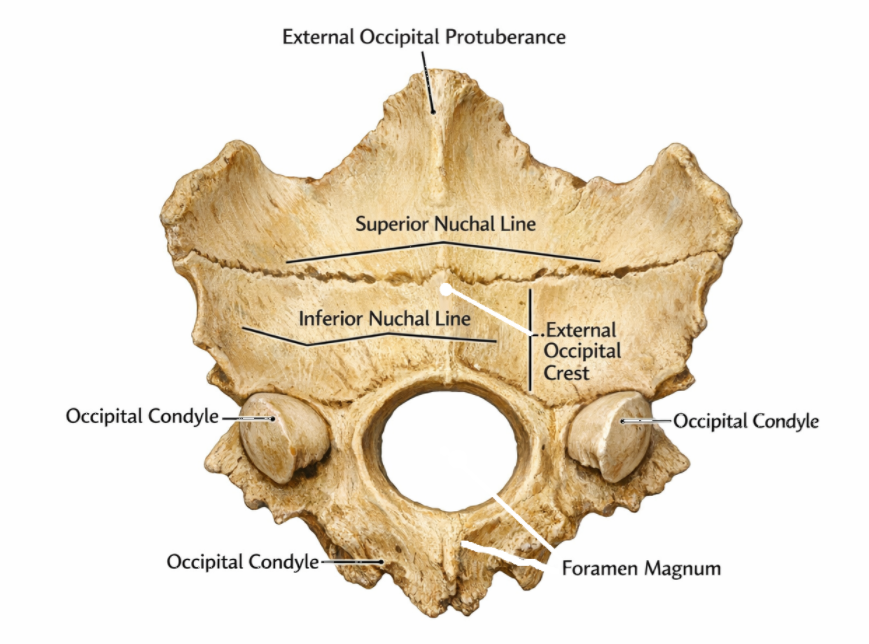
Occipital Bone
On anterior and inferior surface is the foramen magnum, where the spinal cord passes through
on the sides of the foramen magnum are the occipital condyles, which articulate with the first cervical vertebra
The posterior surface has two parallel ridges the superior and inferior nuchal lines
The external occipital protuberance runs through the superior nuchal line and continues as external occipital crest
Frontal bone
forms the frontal squama
inside is the frontal sinuses, which are part of the paranasal sinuses
Has 3 features:
supraorbital margins
sharp ridges forming the superior and superomedial boundaries of the orbit
supraorbital foramen
middle of the supraorbital margin
allows blood vessels and nerves to forehead
glabella
smooth region between the 2 supraorbital margins
Facial bones
Pneumonic:
Virgil Is Now Making My Pet Zebra Laugh
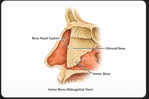
Vomer bone
Deepest facial bone
forms the inferior and posterior of the nasal septum
inferior nasal conchae
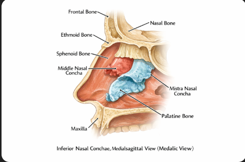
Located in the lateral walls of the nasal cavity, inferior to the middle nasal conchae of the ethmoid bone
Nasal bones
form the bridge of the nose
articulate with the hyaline cartilage that forms the nose
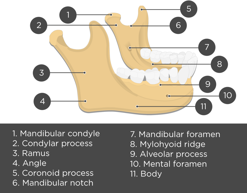
Mandible
only moveable bone in the skull
consists of the mandibular body, and the mandibular rami (ramus = branch)
mandibular body forms the chin and meets the rami at the mandibular angles
on the inside of the body, on each side, is a small mental foramen
the bottom teeth are housed in deep sockets, dental alveoli, within a ridge called the alveolar process
each ramus is topped with a u-shaped mandibular notch, inferior to which is the mandibular foramen
Anterior side of the mandibular notch is the coronoid process, which serves as an attachment point for muscles of mastication
Posterior side has the condylar process, at the top of which is the mandibular condyle, which articulates the mandibular fossa, to form the temporomandibular joint
Maxilla
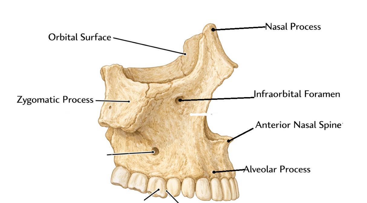
the maxillae fusion creates a bony midline protrusion called the anterior nasal spine
medial the alveolar processes, they form 2 horizontal palatine processes, meeting at the intermaxillary suture
the 2 processes form the anterior of the palate
The anterior and superior surfaces of the maxillary bone form part of the inferomedial walls of the orbit
immediately below the inferior margin, arteries and nerves pass through the inferior orbital fissure and the infraorbital foramen, around which you find large maxillary sinuses
laterally articulates with with zygomatic bones via zygomatic process
Palatine bones
2 L-shaped bones
posterior nasal cavity, between the maxillae and pterygoid process of the sphenoid
each bone is made of the perpendicular plate and the horizontal plate
Horizontal forms the posterior part of the hard palate
perpendicular are vertical projections that form part of the lateral walls of the nasal cavity
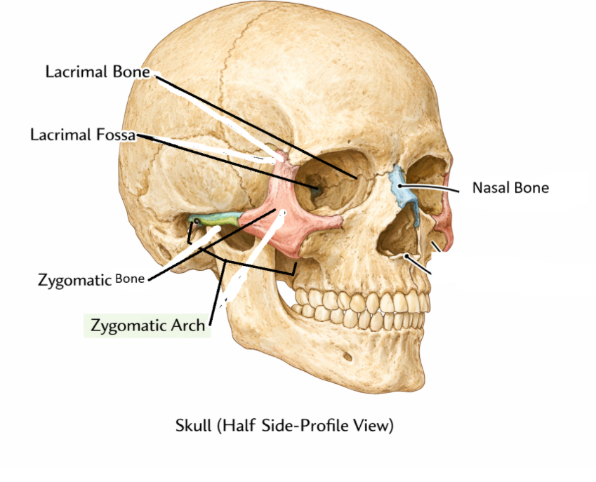
zygomatic bones
Along the zygomatic process of the temporal and maxillae
Form majority of cheekbone or zygomatic arch
form lateral wall and part of inferior border of the orbit
lacrimal bones
Smallest and most delicate facial bones
deep in the medial wall of the orbit
contain a depression called the lacrimal fossa, where tears drain
Cavities of the Skull
Orbit
Houses eyeball, and lacrimal gland (produces tears)
formed by 7 bones
Frontal forms the superior and posterosuperior walls
maxilla forms the posteroinferior wall (partly along with the palatine bone)
zygomatic bone forms the anterolateral wall
sphenoid forms posterior wall
ethmoid, lacrimal, and palatine bones form the medial wall
Nasal Cavity
Posterior wall:
formed by the sphenoid body and pterygoid process
lateral walls:
made of ethmoid, perpendicular plate of palatine bone, inferior nasal conchae, and maxilla
roof and floor:
cribriform plate of ethmoid bone forms roof
floor is hard palate, made of palatine bones and palatine process of maxillae
anterior wall:
nasal bones and maxillae form it
cartilage and connective tissue of nose attach to the margins of the bones
nasal septum:
nasal cavity is divided into 2, each being a nasal fossa, divided by nasal septum
anteriorly made of hyaline cartilage
posteriorly made of perpendicular plate of ethmoid bone and vomer
Paranasal Sinuses
Include sinuses within frontal, sphenoid, ethmoid, and maxillary bones
lined with mucous membranes
lighten the skull and aid in voice resonance
Sinuses can be blocked from excessive mucous, causing sinus headaches, which the pain relates to the areas of the sinuses
above the eyes: frontal sinuses
between the eyes: ethmoidal sinuses
behind the eyes: sphenoidal sinuses
below the eyes: maxillary sinuses
Oral Cavity
roof is formed by hard palate, the same of the floor of the nasal cavity
anterior and lateral walls are maxillae and mandible
floor and posterior wall are soft tissue
Fetal Skull
Soft spots are fontanels, resulting from ossification not being completed, and the skull not fully solidifying
Fontanel spots
anterior fontanel
where the coronal and sagittal sutures meet
between the frontal and parietal bones
posterior fontanel
between the parietal and occipital bones at the apex of the lambdoid suture
sphenoid fonanel
temples, where the sphenoid bones meet other cranial bones
mastoid fontanels
junction of the lambdoid and squamous sutures
where the parietal, temporal and occipital bones meet
Sutures between bones are not fully fused
Hyoid Bone
small C-shaped bone (not an actual bone) in the superior neck
suspended by muscles and ligaments that attach to the styloid process and the larynx
important as it supports many bones for swallowing and speech