9a: Parallel pathways in the brain
Multiple Processing Pathways and Stages
Characteristics of the Visual System:
Multiple Pathways:
Tuned to various aspects of visual stimuli.
Specialized for different types of information (e.g., form vs. motion).
Multiple Processing Stages (within pathways):
Numerous stages exist; each extracts different information types.
Each stage transforms visual input to process various visual aspects.
Initial stages are hierarchical (especially V1) but less so beyond V1.
stages not so hierarchical after v1
each stage transforms visual info in some way
Types of Parallel Pathways
Key Areas of Focus:
Cortical vs. Sub-cortical Pathways
Superior Colliculus (sub cortical): A brain structure involved in visual processing.
Magnocellular & Parvocellular Distinctions (cortical):
Important from the retina onward.
Multiple Cortical Pathways:
Dorsal and Ventral pathways are critical for visual processing.
Other Parallel Pathways:
Include multiple motion pathways.
Superior Colliculus (a structure in the brain) in Primates
Location and Phylogenetics (evolution):
Positioned at the top of the brainstem; phylogenetically older than the cortex.
In many lower animals (e.g., frogs and fish), the superior colliculus (SC) serves as the primary visual projection site.
Receptive Field Characteristics in Primates:
Poorly defined On and Off regions that respond to a wide range of stimuli.
Multisensory properties: Some neurons also receive auditory input.
general-purpose detectors
neurons only responds to auditory input if both signals come from the same space/location
This suggests the SC's likely role is to determine 'where' an object is, not 'what' it is, aiding in the control of eye movements and visual orienting responses.
Control of Eye Movements
Neurons in the SC exhibit a burst of activity prior to eye movements, specifically when the eyes fixate on a light in peripheral vision.
There is no activity noted when eye movements occur in the dark, indicating that the SC is responsive to visual stimuli.
SC, superior colliculus is stimulus driven
reacts to external visual cues involuntarily (glimpse of light in peripheral vision)
Function of the Superior Colliculus
The SC detects objects outside the fixation point, guiding orienting movements of the eye and head.
Notes that detailed analysis of objects is the function of cortical pathways, not the SC.
SC just brings the fixation point to the object but does not analyse it.
Subcortical Pathways in Frogs
Research on SC cell tuning properties in frog visual systems supports the feature-detector approach.
“Feature Detectors” in Frogs
Letvin (1961) Identify 5 Types of Feature Detectors:
On Detectors:
Respond to an increase in light levels.
Off Detectors:
Respond to a decrease in light levels.
On-Off Detectors:
Respond to both increases and decreases in light levels.
Line/Edge Detectors:
Respond to moving lines/edges of particular orientations.
Bug Detectors:
Detect small, dark, moving spots; can trigger a capture response (e.g., tongue snapping to catch bugs).
Cortical Pathways
Magnocellular (M) & Parvocellular (P) Pathways:
Begin in the retina and project to the cortex via the LGN.
Named after the cell types located in the lateral geniculate nucleus (LGN).
M & P cell differentiation is crucial in understanding visual processing.
Magnocellular & Parvocellular Pathways
Key Distinctions in Primates' Visual Systems:
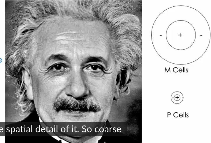
Magnocellular Cells:
Larger cells abbreviated as M cells.
Responsible for motion detection and fast processing.
Parvocellular Cells:
Smaller cells abbreviated as P cells.
Key for fine detail and color detection.
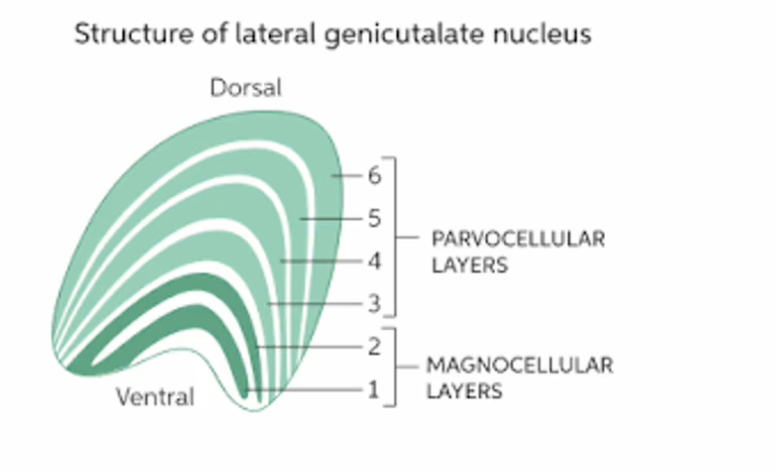
Layer Organization in Lateral Geniculate Nucleus:
2 layers for M cells (one for each eye) and 4 layers for P cells.
possibly due to separate on and off layers?
Main M & P Cell Differences
M Cells Characteristics (Approx. 15% of total cells):
Large receptive field size.
Fast axonal conduction speed.
More transient responses.
Insensitive to colour differences.
High temporal-frequency sensitivity; better at detecting flickering or rapidly moving stimuli.
High contrast sensitivity but saturation occurs.
P Cells Characteristics (Approx. 80% of total cells):
Smaller receptive field size.
Better spatial acuity; can perceive fine details.
Slower axonal conduction speed.
More sustained responses.
Sensitive to color differences.
Low temporal-frequency sensitivity and contrast sensitivity.
Contrast Sensitivity
Defined as how rapidly the cell's response increases as contrast increases.
rapid cell response = very high contrast
Contrast:
The distinction in luminance between a stimulus/object and the background.
Functional Implications:
M cells might saturate at high contrast, limiting their effectiveness in distinguishing between different contrast levels, while some argue response slopes remain constant at high contrasts.
M cells respond strongly at low contrast
response rate flattens out (saturates) at high contrast (m cells maxed out?)
devateable
Large vs. Small Receptive Fields
Information Extraction from Stimuli:
M cells (large RF): Capture broad stimulus information (e.g., overall movement).
P cells (small RF): Focus on fine details (e.g., facial features).
Transmission Speeds:
M cells relay information to V1 faster than P cells due to differences in receptive field size and response characteristic.
Coarse to Fine Scale Processing
Information Gradient:
M cells extract coarse information, reaching V1 quicker, while P cells provide detailed information later.
The brain conducts a coarse-to-fine analysis, establishing a broad outline of an image and gradually filling in details.
brain analyses image from blots then details.
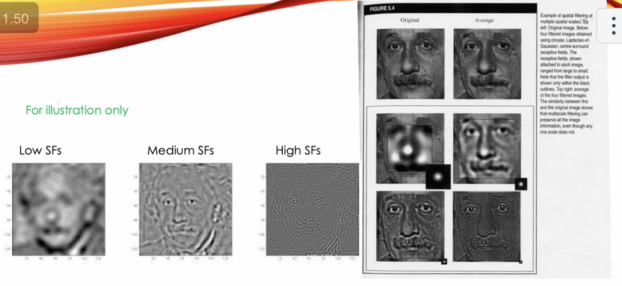
Understanding Spatial Filtering
Illustration of Spatial Filtering:
Multi-scale filtering can preserve image information while emphasizing varied spatial scales (low to high spatial frequencies).
Transient vs. Sustained Responses
Likely functions based on cell response characteristics:
M Transient Cells:
Focus primarily on motion detection; detect rapid changes.
P Sustained Cells:
Essential for processing stable, detailed aspects of visual tasks.
Types of Information Processed by Visual System
Two main types:
Form (object) Information:
Involves identifying what an object is.
Motion Information:
Pertains to how objects move.
This process involves sustained responses for objects at rest and transient responses for moving objects.
M and P Cell Functions
There are distinct roles associated with M and P pathways:
P Cells: Responsible for fine spatial vision.
M Cells: Specialized for motion detection.
This separation has led to the premise that certain visual tasks (like motion perception) may be color-blind due to reliance on M pathways.
No Simple Segregation in Function
An overlap exists between M and P cells, extending visual system sensitivity.
M cells respond to rapid temporal (time) changes, while P cells respond to rapid spatial changes.
Emphasises the concept of coarse-to-fine processing as a fundamental aspect of visual perception.
Linking Pathologies to M or P Dysfunction
Dysfunctions in either M or P systems could lead to specific visual impairments.
Dyslexia Connection:
P system’s role in spatial detail perception underlines potential challenges encountered in reading.
Questions for Further Consideration:
Would reading be affected with P system impairments?
How does M system impairment affect reading mechanics?
Dyslexia
Crucial for perceiving fine spatial detail, an impaired P system could affect reading as it demands precise eye movements and spatial attention control.
M system impairment: impairs visual timing, motion detecion and attentional shifting.
can’t do rapid eye movements to jump between words - disrupts reading flow
difficulty with tracking moving test or adjusting to new lines
word/letter order perception timing order
P system impairment: lacks support of spatial detail, colour and sustained visual imput
can’t do precise fixation on individual letters and words
can’t discriminate words and letters
Blink and Saccadic Suppression
Eye movements and blinking can obscure visual information but are necessary to prevent distractions.
Effects on M Cell Activity:
Both blinking (brief brightness reduction) and saccades (producing blurred images) can reduce M cell activity, highlighting their involvement in spatial attention processes.