Understanding body motion
Bones and Muscles

One of the characteristics of living things is movement. The human body is capable of a wide range of simple and complex movements. This includes actions like running and jumping or talking and chewing. Movement needs coordination between your bones and skeletal muscles. In this Lesson, you will learn the bones and skeletal muscles that interact to allow movement.
In future Lessons, you will also learn about the muscles that work behind the scenes to run the processes of your organs. We also have movements that occur in places like our heart, blood vessels, bladder, and intestines.
Below are some standard terms you might see as muscles and bones are discussed. The way bones and muscles are connected allows for specific ranges of motion (ROM) to occur. Explore the terms and try them out to test your own range of motion.
Abduction

Abduction is the movement of a limb away from the midline of the body.
Adduction

Adduction is the movement of a limb towards the midline of the body.
Supination

Supination describes the movement of a hand, arm, or foot into a face-up position.
Externally rotated so your palm is facing up
Pronation

Pronation describes the movement of a hand, arm or foot into a face-down position.
Internally rotated so your palm is facing down
Flexion

Flexion decreases the angle between two limbs of a joint.
For instance, bending your arm at the elbow is flexion.
Extension

Extension increases the angle between two limbs of a joint.
For instance, straightening your arm is extension.
Axial Bones and Muscles
Focus Question
What are the bones and muscles of the axial skeleton?
The axial skeleton contains the cartilage and bones that support and protect the organs of the head, neck, and trunk.
Axial Skeleton
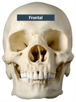
Skull - The skull is made up of the cranium (brain case) and facial bones.
Hyoid Bone -
Located in the neck between the larynx and lower jaw, the hyoid bone supports the tongue and the muscles that move the tongue. It is not attached to any other bones but is held in position by muscles and ligaments.

Vertebral Column -
Also called the spinal column, it forms the central axis of the skeleton.
The vertebral column is made up of many vertebrae bones that are separated by discs that consist of fibrous cartilage and a gelatin-like core.
Vertebrae and many facial bones are examples of irregular bones. Irregular bones are bones with a variety of shapes.

Sacrum -
Five vertebrae fuse together near the distal end of the vertebral column to form the sacrum, part of the pelvis. A small tailbone, formed by four fused vertebrae, is attached to the end of the sacrum. This tailbone is called the coccyx [kok-siks].

Rib Cage -
Also called the thoracic cage, the rib cage is made up of twelve pairs of ribs connected posteriorly to the thoracic vertebrae. Most of the ribs are also attached anteriorly to the sternum, or breastbone. The ribs are flat bones: plate-like bones with broad surfaces. Some bones in the skull are also flat bones.

The axial skeleton:
skull
rib cage
spinal column

Just like bones, muscles can be categorized based on their location. Axial muscles are any of the skeletal muscles of the trunk or head.
Axial Muscles
Trapezius -
The trapezius is a large superficial muscle that extends from the cranium to the lower thoracic vertebrae. It is one of several muscles involved in the movement of the scapula. You use your trapezius to shrug your shoulders.
Actions: Rotation, retraction, elevation, and depression movements of the scapula and support of the arm

Pectoralis major -
The pectoralis major is a fan-shaped muscle on the chest, making up the bulk of the male chest and found under the breast in a female.
Actions: Flexes, extends, adducts, and rotates the arm at the shoulder

Pectoralis minor -
The pectoralis minor is a thin, triangular muscle found deep to, or underneath, the pectoralis major.
Actions: Draws the scapula forward and downward

Latissimus dorsi -
The latissimus dorsi , commonly known as "lats," are large, flat muscles posterior to the arm and partly covered by the trapezius. This is the broadest muscle in the back.
Actions: Adducts, medially rotates, and extends the arm at the shoulder

External oblique -
The external oblique is a broad and thin muscle found on the lateral and anterior parts of the abdomen. It is called the "external" oblique because is the most superficial, or outermost, of the three flat muscles of the lateral anterior abdomen.
Actions: Flexes the vertebral column by drawing the thorax inward, rotates and laterally flexes the vertebral column (torso), and compresses the abdomen

Rectus abdominis -
The rectus abdominis muscle, commonly called the "abs," is a paired muscle that runs vertically on the anterior wall of the human abdomen. These two parallel muscles are separated by a midline band of connective tissue.
Actions: Flexes the vertebral column (and torso), compresses the abdomen, and assists in breathing

Muscle names often describe one or more properties of the muscles, indicating location, action, attachment, shape, or size. For example, the pectoralis major and pectoralis minor are two chest muscles where the name has been influenced by the compared size between one and the other. This can be useful when you’re learning their names and properties.


Appendicular Bones and Muscles
Focus Question
What are the bones and muscles of the appendicular skeleton?
The appendicular skeleton consists of the bones in the upper and lower limbs, as well as the bones that anchor those limbs to the axial skeleton.
Appendicular Skeleton
Lower Limbs - Appendicular Bones and Muscles
Focus Question
What are the bones and muscles of the appendicular skeleton?
The appendicular skeleton consists of the bones in the upper and lower limbs, as well as the bones that anchor those limbs to the axial skeleton.
Appendicular Skeleton
Lower Limbs
Pectoral Girdle
Upper Limbs
Pelvic Girdle

Each lower limb is made up of:
Femur: Thigh bone. The femur is an example of a long bone. This is an elongated bone with expanded ends. The bones in the forearms and thighs are long bones.
Tibia: Shin bone
Fibula: Slender leg bone next to the tibia
Patella: Kneecap, covering the area where the femur and tibia connect
Tarsals: Seven ankle bones
Metatarsals: Five bones in the inner part of the foot
Phalanges: Fourteen bones of the toes (same name as the fourteen bones of the fingers)
Appendicular muscles are any of the muscles of the upper or lower limbs. They control the movement of limbs and also stabilize and control the movements of pectoral and pelvic girdles.
Appendicular Muscles
Deltoid
Triceps brachii
Biceps brachii
Gluteus maximus
Hamstrings
Quadriceps femoris
Gastrocnemius


Pectoral Girdle - Appendicular Bones and Muscles
Focus Question
What are the bones and muscles of the appendicular skeleton?
The appendicular skeleton consists of the bones in the upper and lower limbs, as well as the bones that anchor those limbs to the axial skeleton.
Appendicular Skeleton
Lower Limbs
Pectoral Girdle
Upper Limbs
Pelvic Girdle
The pectoral girdle is made up of the scapula (shoulder blade) and the clavicle (collar bone). These bones are found on both sides of the body, connecting the bones of the upper limbs to the axial skeleton and aiding in limb movement.

Appendicular muscles are any of the muscles of the upper or lower limbs. They control the movement of limbs and also stabilize and control the movements of pectoral and pelvic girdles.
Appendicular Muscles
Deltoid
Triceps brachii
Biceps brachii
Gluteus maximus
Hamstrings
Quadriceps femoris
Gastrocnemius
Upper Limbs - Appendicular Bones and Muscles
Focus Question
What are the bones and muscles of the appendicular skeleton?
The appendicular skeleton consists of the bones in the upper and lower limbs, as well as the bones that anchor those limbs to the axial skeleton.
Appendicular Skeleton
Lower Limbs
Pectoral Girdle
Upper Limbs
Pelvic Girdle

The upper limbs (arms and hands) are composed of:
Humerus: Upper arm bone
Radius and Ulna: Two forearm bones
Carpals: Eight wrist bones. The carpals are short bones. These are cube-shaped bones with roughly equal lengths and widths. The bones in the wrists and ankles are short bones.
Metacarpals: Five bones of the palm
Phalanges: Fourteen finger bones
Appendicular muscles are any of the muscles of the upper or lower limbs. They control the movement of limbs and also stabilize and control the movements of pectoral and pelvic girdles.
Appendicular Muscles
Deltoid
Triceps brachii
Biceps brachii
Gluteus maximus
Hamstrings
Quadriceps femoris
Gastrocnemius
Pelvic Girdle - Appendicular Bones and Muscles
Focus Question
What are the bones and muscles of the appendicular skeleton?
The appendicular skeleton consists of the bones in the upper and lower limbs, as well as the bones that anchor those limbs to the axial skeleton.
Appendicular Skeleton
Lower Limbs
Pectoral Girdle
Upper Limbs
Pelvic Girdle
The pelvic girdle is composed of the left and right os coxae. They are attached to the sacrum posteriorly and to each other anteriorly. The left and right os coxae connect the lower limbs to the axial skeleton. The left and right os coxae, sacrum, and coccyx form the pelvis, which protects the lower abdomen and reproductive organs.

Appendicular muscles are any of the muscles of the upper or lower limbs. They control the movement of limbs and also stabilize and control the movements of pectoral and pelvic girdles.
Appendicular Muscles
Deltoid
Triceps brachii
Biceps brachii
Gluteus maximus
Hamstrings
Quadriceps femoris
Gastrocnemius
Appendicular Skeleton:
Lower limbs -
The deltoid muscle forms the rounded shape of the shoulder. It earned this name because of its triangular shape, similar to Greek letter Delta (triangle).
Actions: Abduction, flexion, and extension of the shoulder

Pectoral girdle -
Upper limbs
Pelvic girdle
These bones are further categorized based on their location in the body and size.

Appendicular muscles are any of the muscles of the upper or lower limbs. They control the movement of limbs and also stabilize and control the movements of pectoral and pelvic girdles.
Appendicular Muscles
Deltoid -
The deltoid muscle forms the rounded shape of the shoulder. It earned this name because of its triangular shape, similar to Greek letter Delta (triangle).
Actions: Abduction, flexion, and extension of the shoulder
Triceps brachii -
The triceps brachii muscle, commonly called the "triceps" is the large muscle on the back of the arm. It is sometimes called a three-headed muscle because there are three bundles of muscles, each with different origins, joining together at the elbow.
Actions: Extends the forearm and straightens the elbow

Biceps brachii -
The biceps brachii, often called "biceps," is a two-headed muscle (bi- means "two"). It lies on the upper arm between the shoulder and the elbow.
Actions: Flexes the elbow and suppinates (rotates) the forearm

Gluteus maximus -
The gluteus maximus is a narrow and thick muscle that makes up a large portion of the shape and appearance of the buttocks. Together with the gluteus medius and gluteus minimus, they form the "glutes."
Actions: External rotation and extension of the hip joint

Hamstrings -
The hamstrings are a large muscle group that occupies the back of the thigh. It is made up of three muscles:
semimembranosus
semitendinosus
biceps femoris
Actions: Flexes the knee joint, rotates the knee joint laterally, and extends the thigh

Quadriceps femoris -
The quadriceps femoris is a large muscle group that occupies the front and sides of the thigh. As the prefix quad- indicates, it is made up of four muscles:
rectus femoris
vastus lateralis
vastus medialis
vastus intermedius
Actions: Knee extension and hip flexion

Gastrocnemius -
The gastrocnemius [/ˌɡæstrɒkˈniːmiəs/] muscle on the back of the leg forms part of the calf. It is a powerful muscle that, along with the soleus muscle, forms the calf.
Actions: Plantar flexion of the foot (flexing the foot toe-down, greater than 90 degrees), flexion of the knee

Adjacent Muscles
Focus Question
What muscles and bones work together to allow movement?
Bones and their adjacent muscles allow for movement of the body. Explore some adjacent bones and muscle examples below. The first muscle listed is the main muscle, followed by the adjacent muscles, and the connected bones.
Shoulder Movement
Leg Movement -


Muscle
Quadriceps femoris
Adjacent Muscles
Hamstring, Gluteus maximus
Connected Bones
Femur, Patella
Ankle Movement -


Muscle
Gastrocnemius
Adjacent Muscles
Hamstring, Soleus
Connected Bones
Fibula, Tibia
Abdominal Movement -


Muscle
External oblique
Adjacent Muscles
Latissimus dorsi, Rectus abdominis
Connected Bones
Ribs, Pubic bone


Muscle | Deltoid |
Adjacent Muscles | Biceps brachii, Triceps brachii |
Connected Bones | Clavicle, Humerus |
Support for Motion
Focus Question
What other structures help with movement of the body?
The bones and muscles work together to allow for every movement. Other structures help tie it all together so that movement can happen and the correct range of motion occurs.
Joints are the locations that occur between the major bones. Movement occurs where joints meet. Range of motion is impacted by the flexibility of joints. Explore a few of the types of joints below. You should notice joints are often named based on how they work in the body.
Ball/Socket
Gliding - Support for Motion
Focus Question
What other structures help with movement of the body?
The bones and muscles work together to allow for every movement. Other structures help tie it all together so that movement can happen and the correct range of motion occurs.
Joints are the locations that occur between the major bones. Movement occurs where joints meet. Range of motion is impacted by the flexibility of joints. Explore a few of the types of joints below. You should notice joints are often named based on how they work in the body.
Ball/Socket
Gliding
Hinge
Pivot
Saddle
Gliding is when two bones with smooth surfaces slide over one another to produce restricted movement.
Examples: ankle, wrist, and spine


Specific joints work with groups of bones to support movement. In the images below, you can see groups of bones that work with the knee, shoulder, and hip joints.
Knee

The knee joint consists of the femur, patella, tibia, and fibula.
Shoulder

The shoulder joint consists of the clavicle, scapula, and humerus.
Hip

The hip joint consists of the hip bone, sacrum, and femur.
Tendons and Ligaments
Joints are connected to tendons and ligaments. Soft tissues of the body offer connection and support for joints and muscles.
Two that help connect the muscles to the joints are tendons and ligaments.
Let's delve into these structures by examining a knee joint.
Hinge - Support for Motion
Focus Question
What other structures help with movement of the body?
The bones and muscles work together to allow for every movement. Other structures help tie it all together so that movement can happen and the correct range of motion occurs.
Joints are the locations that occur between the major bones. Movement occurs where joints meet. Range of motion is impacted by the flexibility of joints. Explore a few of the types of joints below. You should notice joints are often named based on how they work in the body.
Ball/Socket
Gliding
Hinge
Pivot
Saddle
Hinge is when two bones are molded together to allow movement in one direction or plane.
Examples: knee and elbow


Specific joints work with groups of bones to support movement. In the images below, you can see groups of bones that work with the knee, shoulder, and hip joints.
Knee
The knee joint consists of the femur, patella, tibia, and fibula.
Shoulder
The shoulder joint consists of the clavicle, scapula, and humerus.
Hip
The hip joint consists of the hip bone, sacrum, and femur.
Tendons and Ligaments
Joints are connected to tendons and ligaments. Soft tissues of the body offer connection and support for joints and muscles.
Two that help connect the muscles to the joints are tendons and ligaments.
Let's delve into these structures by examining a knee joint.
Pivot - Support for Motion
Focus Question
What other structures help with movement of the body?
The bones and muscles work together to allow for every movement. Other structures help tie it all together so that movement can happen and the correct range of motion occurs.
Joints are the locations that occur between the major bones. Movement occurs where joints meet. Range of motion is impacted by the flexibility of joints. Explore a few of the types of joints below. You should notice joints are often named based on how they work in the body.
Ball/Socket
Gliding
Hinge
Pivot
Saddle
Pivot is a cylinder-shaped bone that rotates inside another bone.
Examples: neck and forearm


Specific joints work with groups of bones to support movement. In the images below, you can see groups of bones that work with the knee, shoulder, and hip joints.
Knee
The knee joint consists of the femur, patella, tibia, and fibula.
Shoulder
The shoulder joint consists of the clavicle, scapula, and humerus.
Hip
The hip joint consists of the hip bone, sacrum, and femur.
Tendons and Ligaments
Joints are connected to tendons and ligaments. Soft tissues of the body offer connection and support for joints and muscles.
Two that help connect the muscles to the joints are tendons and ligaments.
Let's delve into these structures by examining a knee joint.
Saddle - Support for Motion
Focus Question
What other structures help with movement of the body?
The bones and muscles work together to allow for every movement. Other structures help tie it all together so that movement can happen and the correct range of motion occurs.
Joints are the locations that occur between the major bones. Movement occurs where joints meet. Range of motion is impacted by the flexibility of joints. Explore a few of the types of joints below. You should notice joints are often named based on how they work in the body.
Ball/Socket
Gliding
Hinge
Pivot
Saddle
Saddle is a joint with a saddle-shaped surface that is convex in one direction and concave in another.
Example: only the thumb


Specific joints work with groups of bones to support movement. In the images below, you can see groups of bones that work with the knee, shoulder, and hip joints.
Knee
The knee joint consists of the femur, patella, tibia, and fibula.
Shoulder
The shoulder joint consists of the clavicle, scapula, and humerus.
Hip
The hip joint consists of the hip bone, sacrum, and femur.
Tendons and Ligaments
Joints are connected to tendons and ligaments. Soft tissues of the body offer connection and support for joints and muscles.
Two that help connect the muscles to the joints are tendons and ligaments.
Let's delve into these structures by examining a knee joint.
Ball and socket is when the surface of one rounded bone fits into the depression of another bone.
Examples: hip and shoulder


Specific joints work with groups of bones to support movement. In the images below, you can see groups of bones that work with the knee, shoulder, and hip joints.
Knee
The knee joint consists of the femur, patella, tibia, and fibula.
Shoulder
The shoulder joint consists of the clavicle, scapula, and humerus.
Hip
The hip joint consists of the hip bone, sacrum, and femur.
Tendons and Ligaments
Joints are connected to tendons and ligaments. Soft tissues of the body offer connection and support for joints and muscles.
Two that help connect the muscles to the joints are tendons and ligaments.
Let's delve into these structures by examining a knee joint.
Saddle
Saddle is a joint with a saddle-shaped surface that is convex in one direction and concave in another.
Example: only the thumb
Specific joints work with groups of bones to support movement. In the images below, you can see groups of bones that work with the knee, shoulder, and hip joints.
Knee
The knee joint consists of the femur, patella, tibia, and fibula.
Shoulder
The shoulder joint consists of the clavicle, scapula, and humerus.
Hip
The hip joint consists of the hip bone, sacrum, and femur.
Tendons and Ligaments
Joints are connected to tendons and ligaments. Soft tissues of the body offer connection and support for joints and muscles.
Two that help connect the muscles to the joints are tendons and ligaments.
Let's delve into these structures by examining a knee joint.

Tendons
A tendon is a connective tissue that attaches muscle to bone.

Joints are connected to tendons and ligaments. Soft tissues of the body offer connection and support for joints and muscles.
Two that help connect the muscles to the joints are tendons and ligaments.
Let's delve into these structures by examining a knee joint.