BIOL 1102 Study Guide part 1
Study Guide for Midterm 3
Electrical Signaling
- Rules for ion movement
- Higher to lower concentration
- Away from like chargers, towards the opposite charge
- Depends on the membrane permeability
- Membranes Can be depolarized or hyperpolarized by the opening and closing of different ion channels
- Resting potential= only K+ channels are open
- Depolarized= voltage gated Na+ channels open
- Hyperpolarized= chemically gated K+ channel open
- Production of Action Potentials
- Most neurons depolarization is graded only up to a certain membrane voltage called the threshold potential
- Stimulus strong enough to produce a depolarization that depolarization that reaches the threshold potential triggers a different response= action potential
- Action potential is a all or none thing= once it is triggered it has a magnitude that is independent of the strength of the triggering stimulus
- Action potentials are signals that carry information from axons
- Most are very brief which allows for the production of high frequency signals
- Neurons generally encode information in action potential frequency and not amplitude
- Frequency= how many occur per unit of time
- amplitude= amount of voltage change per action potential
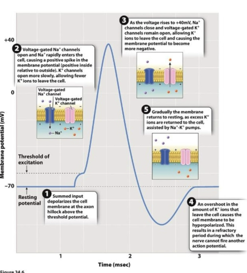
- Refractory period= a state in which the no signal can be sent because the ions are deactivated
- Myelination by Glia Speeds Signal Transmissions
- The electrical charge jumps from space to space in the Nodes of Ranvier.
- Layers of myelin insulate the axon. As a result, action potentials “jump” from node to node increasing the speed of conduction
- At nodes of Ranvier there is a buildup of + charges and inside and - charges outside the axon.
- Known as salutary action potentials.
- Neurons communicate with other cells
- Synapse= two neurons get together in order to transmit info from the presynaptic cell to the postsynaptic neuron
- Given condition, should we go under an action potential or not
- Events in a Chemical synapse
- Blue arrow= showing that the presynaptic cell just had an action potential
- When calcium voltage channels reach a certain voltage they open up and make vesicles that contain neurotransmitters and bind to the pre synaptic membrane at the axon terminal
- On the postsynaptic membrane there are chemically gated channels that bid into the chemically gated channels. Allows sodium to come in and can cause depolarization.
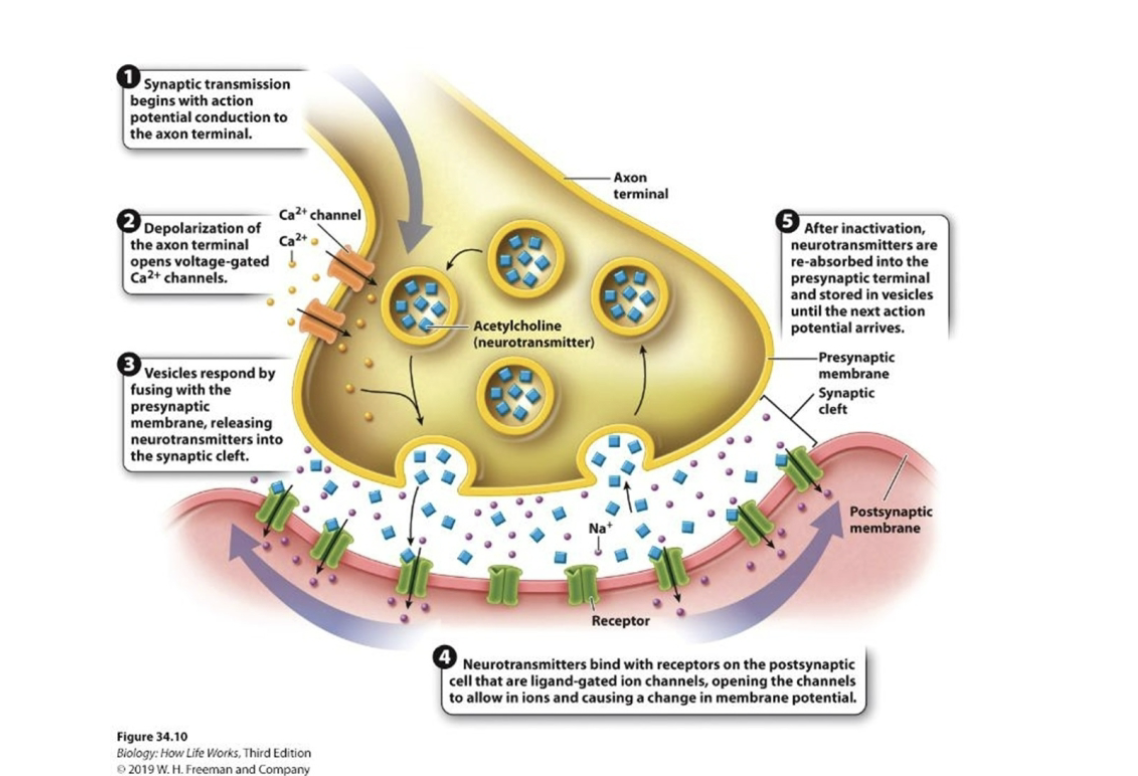
- Neurotransmitters can be repacked into vesicles and brought back to the presynaptic cell, degraded naturally or degraded by enzymes
- Synapses can be;
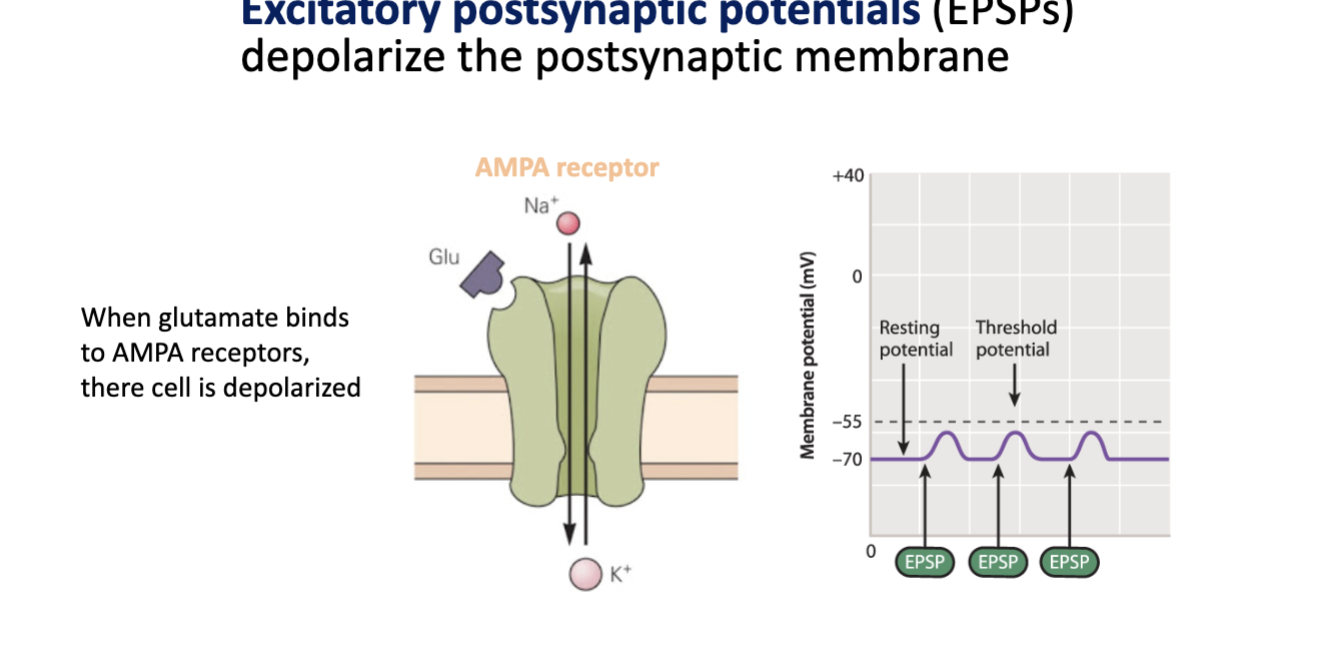
- excitatory= result in depolarization (open sodium channels)
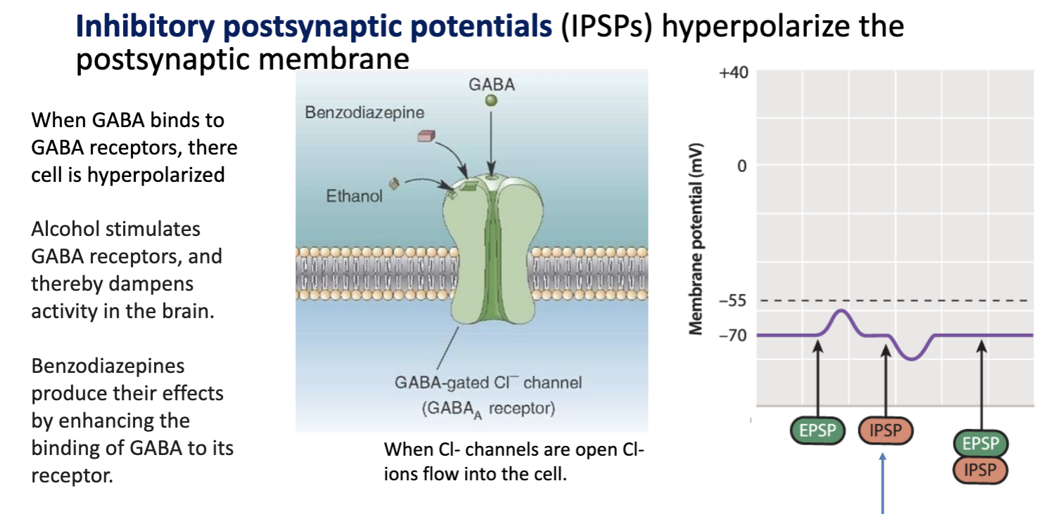
- inhibitory= result in hyperpolarization (open chloride channels)
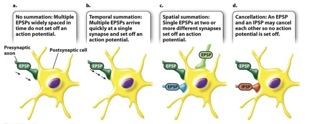
- Postsynaptic cells sums excitatory and inhibitory inputs
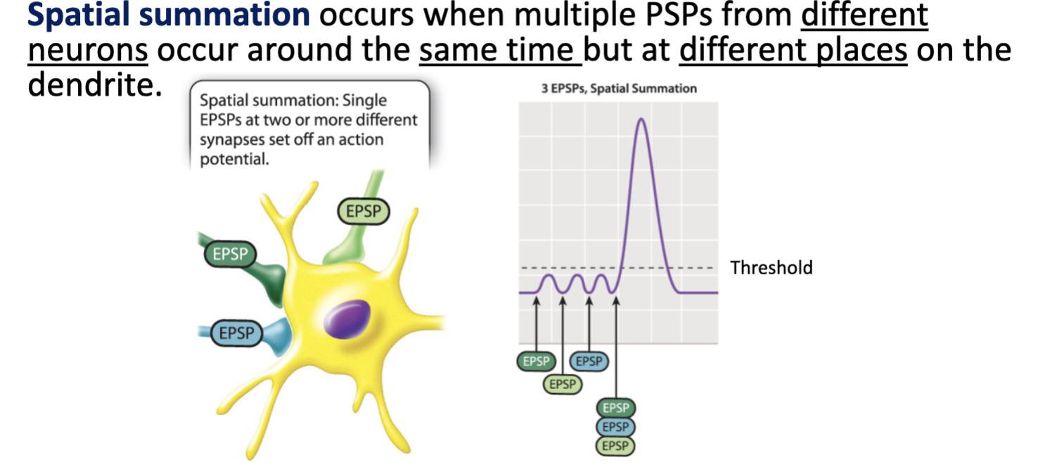
- Spatial summation
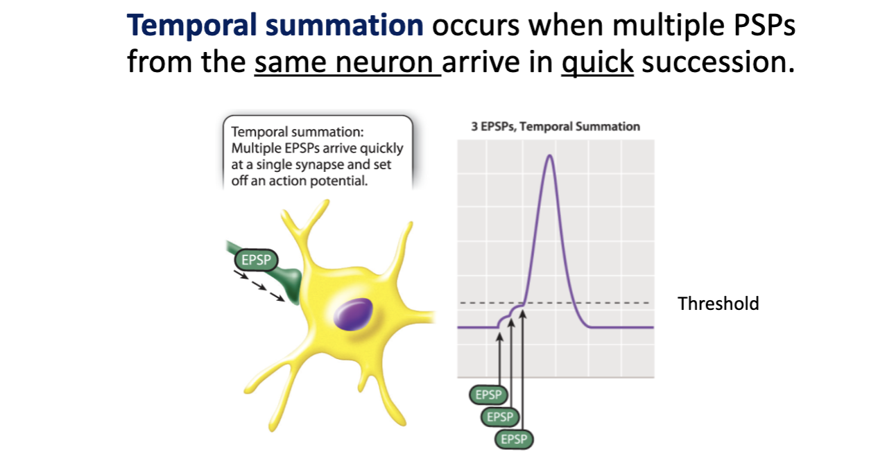
- Temporal summation
- Inputs are integrated at the axon hillock= takes all of the inputs together and decide if an action potential is going to happen
- Small changes (gradient potential→ excitatory and inhibitory)
- Gradient change potential= graded change in postsynaptic cell= moderate change in the membrane potential of the postsynaptic cell. However if the depolarization passes the threshold an action potential can still occur.
- Excitatory post synaptic potentials (EPSPs)= depolarize the post synaptic neuron. Binding of neurotransmitters to the postsynaptic receptors opens gated channels that allow Na+ to diffuse into the cells.
- Inhibitory postsynaptic potentials (IPSPs) = hyperpolarize the postsynaptic neuron= binding of neurotransmitter to postsynaptic receptors opens gated channels that allow K+ to diffuse out of the cell or Cl- to diffuse into the cell
Summed EPSPs and IPsps
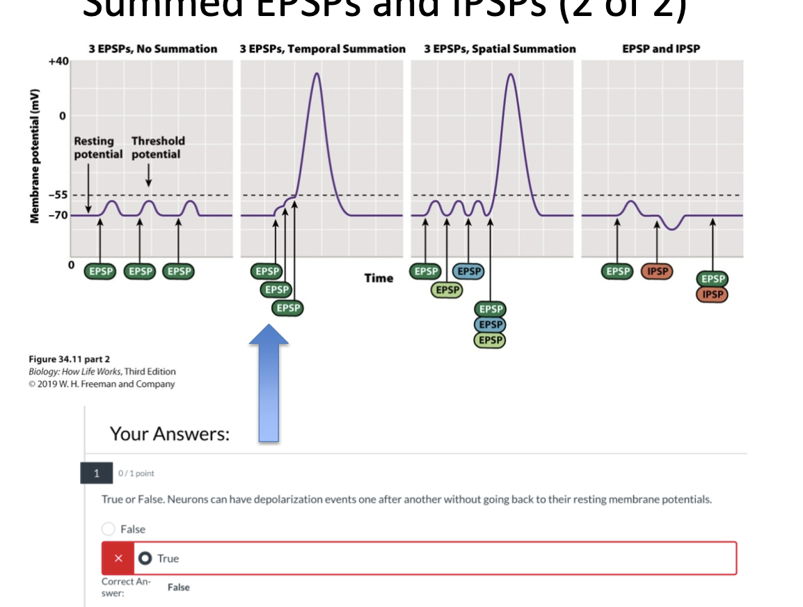
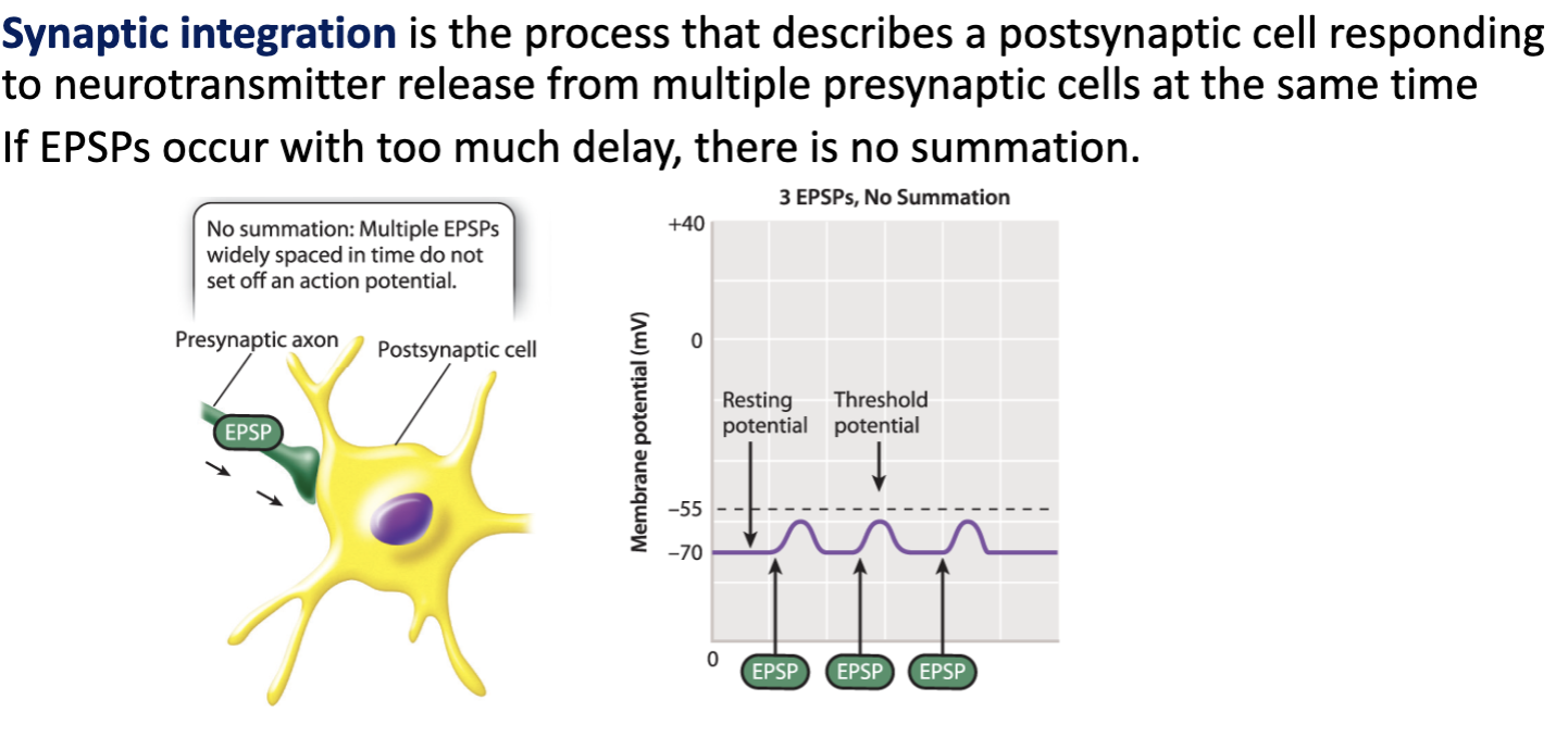
- No summation= multiple EPSPs widely space din time do not set off an action potential
- Temporal summation= multiple EPSPs arrive quickie at a synapse to set off an action potential
- Spatial summation= single EPSPs at two or more different synapses set off an action potential
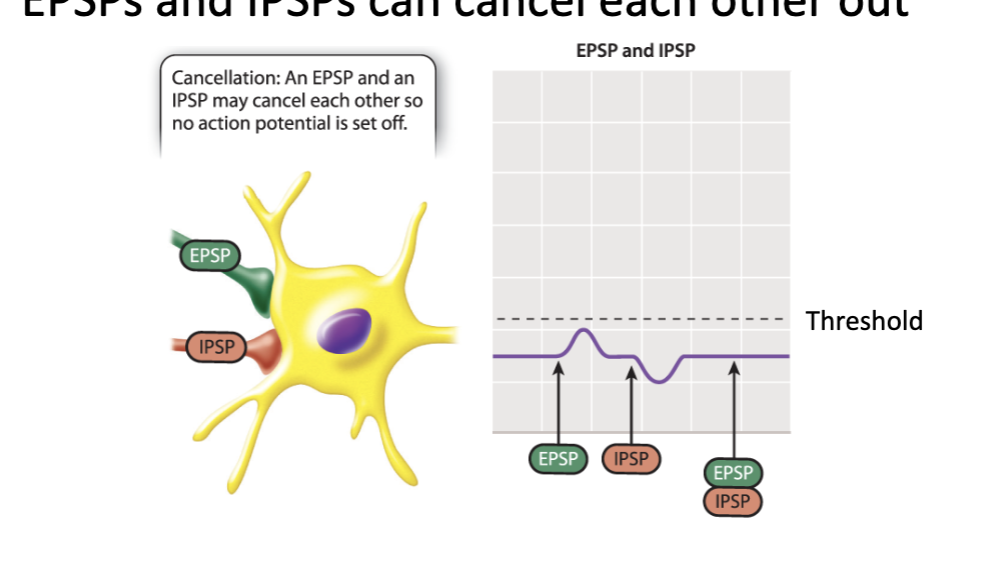
- Cancellation= an EPSP and an IPSP may cancel each other so no action potential is set off
- Refractory period= while the inactivation gate Na+ channel is closed the membrane is in its absolute refractory period, during which time the membrane is unresponsive to another stimulus. A change in voltage will not open these channels
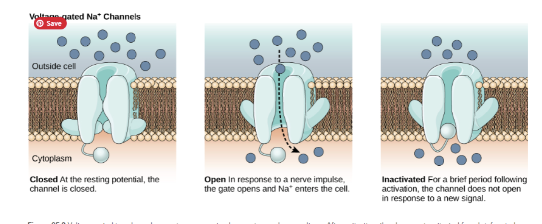
- Once te voltage gated Na channels change to the closed state, but while the voltage gated K+ channels are stiled channels are still open the membrane enters a brief relative refractory period
- A new action potential may be generated but only in response to a really large stimulus
Sensory Systems Eye and Ear
- All sensory information arrives in the central nervous system (CNS) and brain and spinal cord as action potentials
- Sensory reception
- Arrival of stimulus energy ay specialized sensory receptor cells
- Sensory transduction/ amplification
- Conversion of stimulus energy into a change in receptor cell membrane potential: receptor potential
- Integration of stimulus information received by cells of the sensory organ and associated neurons
- Amplification of stimulus into a sufficiently strong signal to be transmitted to CNS
- Interpretation
- Action potentials sent to the CNS are recognized as sensory information of a specific type of circuits in the CNS
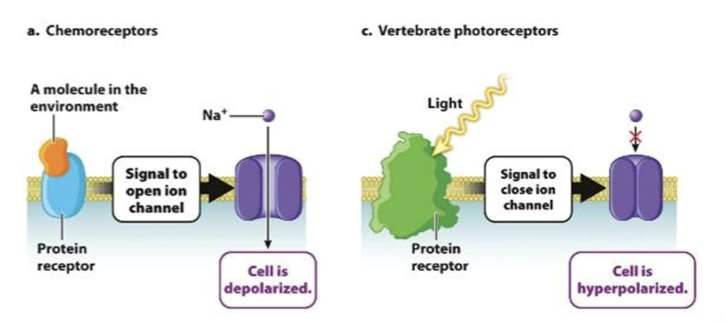
- Sensory receptor- proteins are found in the membranes of sensory receptor cells and respond to stimuli by opening or closing ion channels
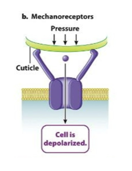
- Ionotropic receptors= open and close due to direct response to a stimulus
- Mechanoreceptor
- Metabotropic receptors= open or close ion channels indirectly via a signaling cascade
- Chemoreceptor
- Photoreceptor
- Sound waves are the stimuli for hearing
- Amplitude= sound wave corresponds to its volume
- Frequency= refers to how frequently a sound wave oscillates per second
- A sound wave that oscillates very quickly will produce a higher pitch while a sound wave that oscillates very slowly will produce a lower pitch
- Process of hearing
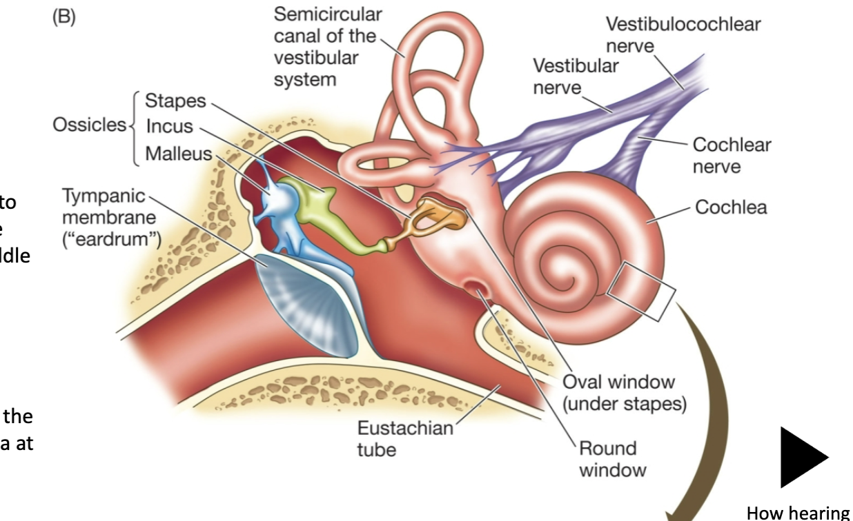
- The tympanic membrane mechanically transmits these pressure waves into movementsof the ossicles in the middle ear
- The ossices transmit their movement into pressure waves in the fluid of the cochlea at the oval window
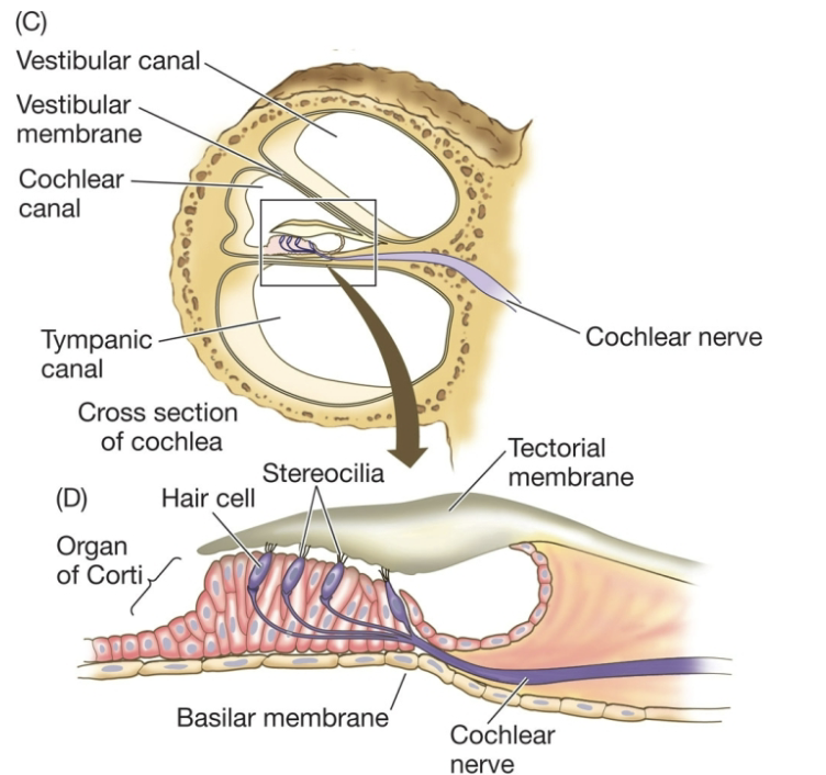
- The cochlea is divided into fluid filled chambers; pressure waves from the ossicles cause the membrane to flex.
- Flexing of the basilar membrane bends stereocilia on hair cells in the organ of Corti
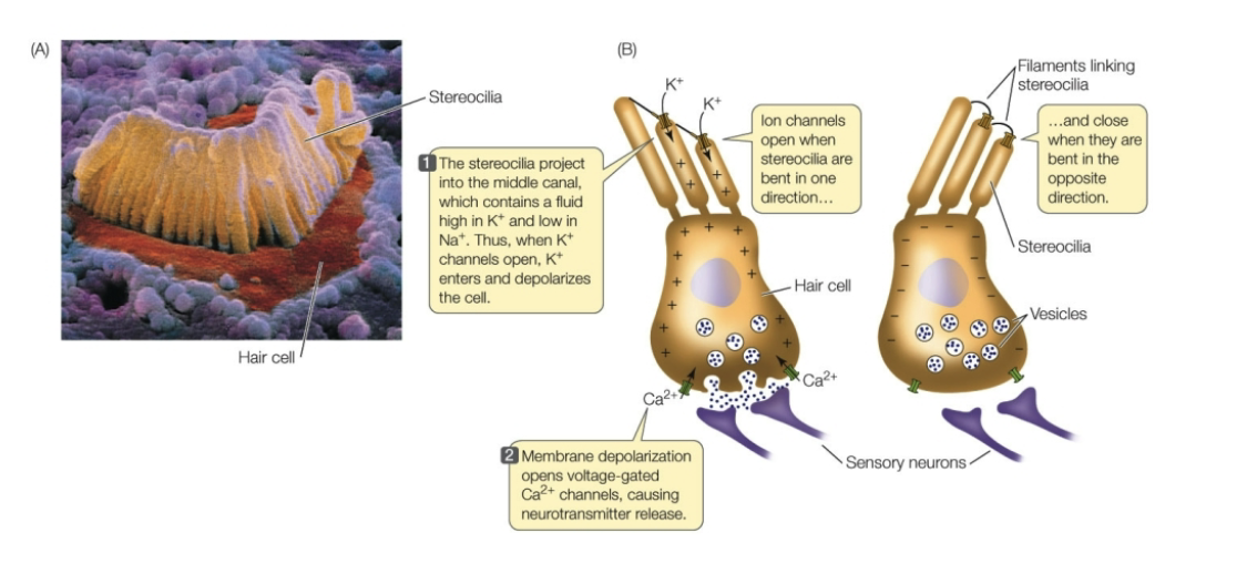
- The hair cells have mechanoreceptors on their stereocilia
- The steocillia project into the middle canal which contains a fluid high in K+ and low in Na+. When K+ opens, the K+ enters and depolarizes the cell
- Membrane depolarization opens voltage gated Ca 2+ channels causing neurotransmitter release
- Low pitch= travel far down the vestibular canal and flex the basilar membrane activating the potentials in low frequency senors
- Medium pitch= pressure waves travel only part way down the upper canal before flexing the basilar membrane and activating mid frequency sensors
- High pitch= pressure waves travel short distance before flexing the basilar membrane and activating high frequency sensors
- Elephants make sounds to be able to travel long distances
- Process of sight
- Sight is based off of opsin which contain photopigments known as retinal
- Wavelengths of light determine color
- Amplitude of light determine brightness
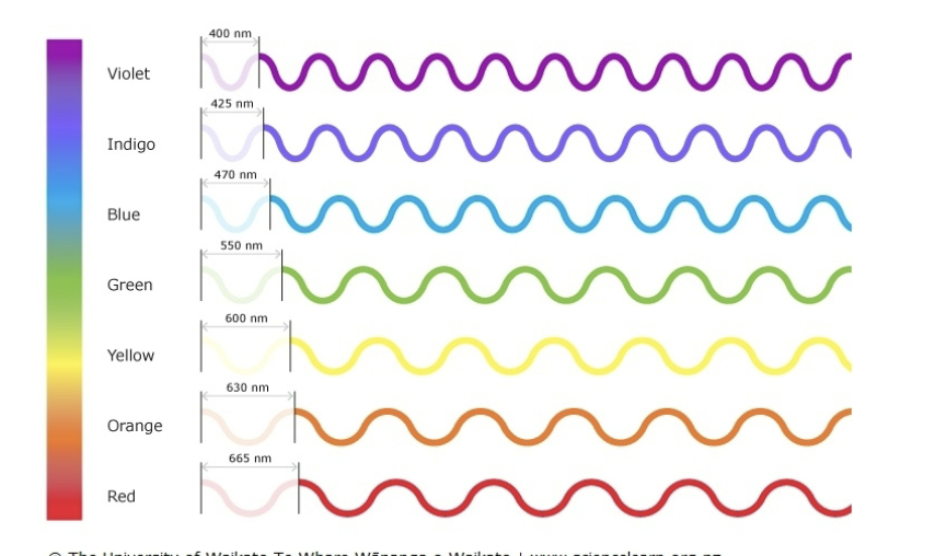
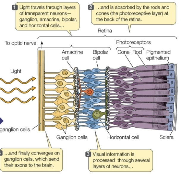
- In the vertebrae retina light is absorbed by photoreceptor cells which have opsin in their membrane.
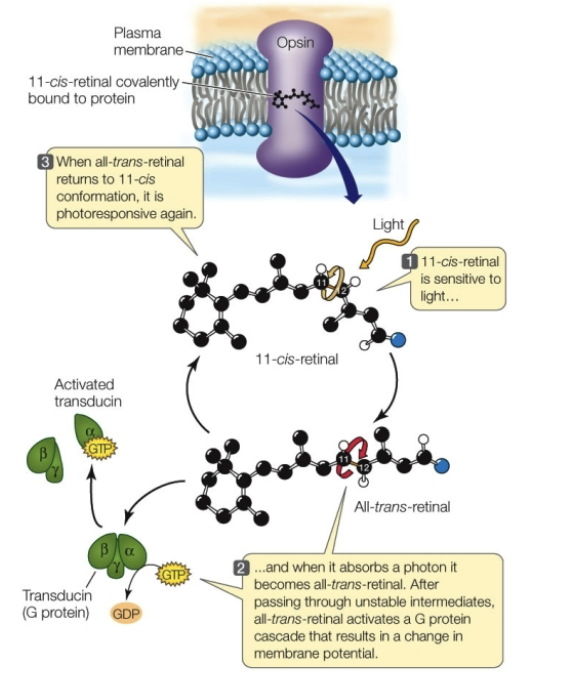
- Rhodopsin= a form of opsin embedded in membranes within photoreceptor cells called rods which sense black and white. Light makes the photopigment in the retina change shape
- Causes a G protein signaling cascade that changes the membrane potential
- Cis retinal is sensitive to light
- And when it absorbs the photon it becomes all trans retinal. Which signals a g protein cascade
- Trans retinal returns to 11 cis conformation, it is photoresponsive again
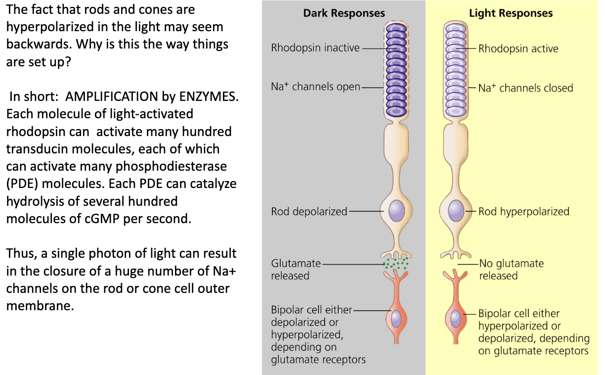
- Light absorption closed sodium channels
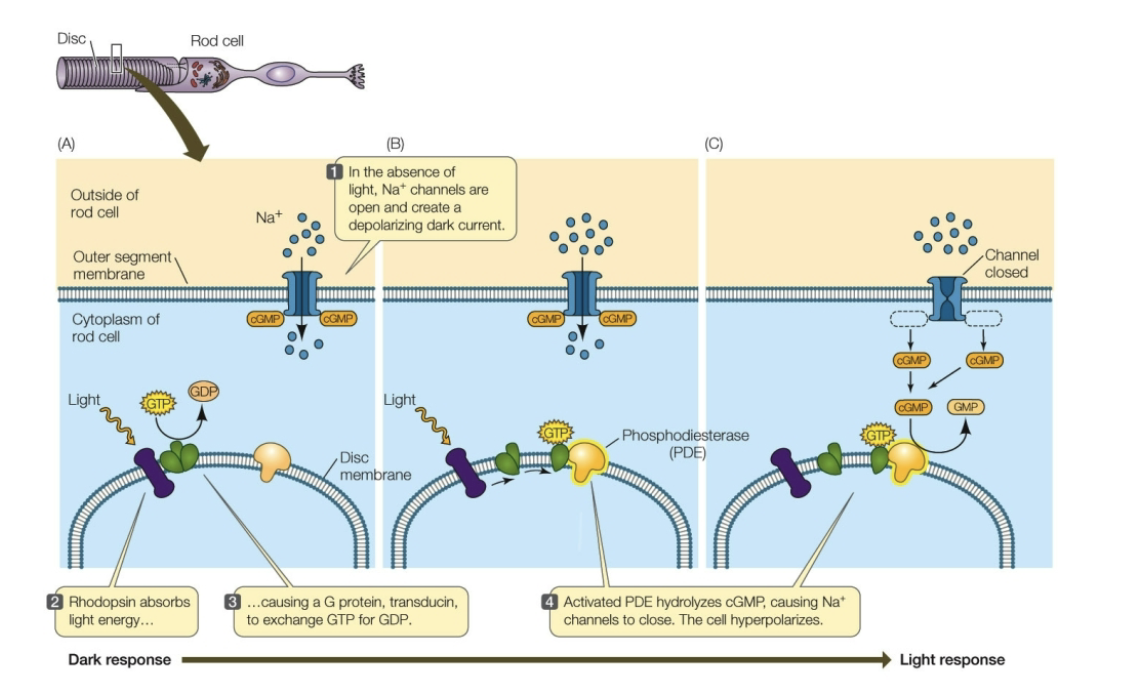
- Tod and cone cells are naturally depolarized and become hyperpolarized with light so with stronger light more hyperpolatrization
- Bipolar cells= synapse with rod or cone cells and relay responses to ganglion cells
- Ganglion cells= send their axons out of the eye in the optic nerve
- Amacrine and horizontal cells= modify electrical signals as they pass fromphotoreceptors to ganglion cells
- Ionotropic receptors= open and close due to direct response to a stimulus
Defies boundaries within images and increase the sensitivity of the eye moving images
Why hyperpolarized?
- Watch ear video
Muscles and movement
- skeleton= a structure or structures that serve functions for support and movement
- Types
- Hydrostatic= consist of fluid filled body compartments
- Soft body invertebrates use this for support and movement
- Combination of muscles anf fluid
- Water is incompressible so pushing against it allows the invertebrate to move
- Exoskeleton= hard substance outside of the body
- External that surrounds and protect most of the body surface
- Provides support and protection from the external environment and predators
- Have to be regrown and shet
- E.g.
- Earwing, beetle, spider, shrimp, etc.
- endoskeleton= hard substance inside the body
- Provide support and protection for some internal organs but do not protect the body surface.
- Found in some species of sponges all echinoderms like starfish and vertebranes
- Hardened by calcium, magnesium phosphate and carbonate
- Tendon and ligaments= made from collagen
- Tendon= muscle to bone, muscle to muscle
- ligaments= bones to bones
- Order of organization
- Myofibril gives rise to muscle fibers which are surrounded by connective tissue. Fibers then give rise to muscles.
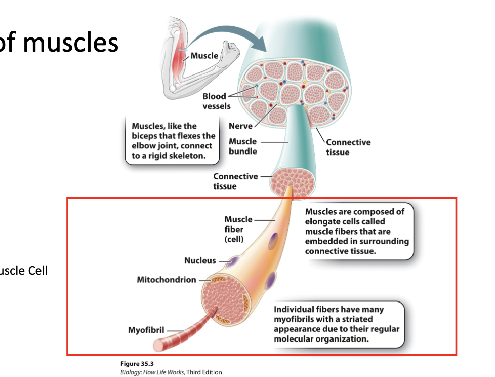
- Z discs= area where to actin filaments connect
- Sarcomere= basic unit of a muscle cell
- Myofibril= long contractile units
- Muscle fiber= muscle cells
- Filaments within muscle
- Thin filament= two actin filaments twisted together
- Tropomyosin runs through the grooved formed by actin helices
- Each myosin molecule consists of two long polypeptide chains coiled together each ending with a globular head
- Myosin molecules are arranged in parallel to form a thick filament, with myosin heads peeking out from the necks along the filament.
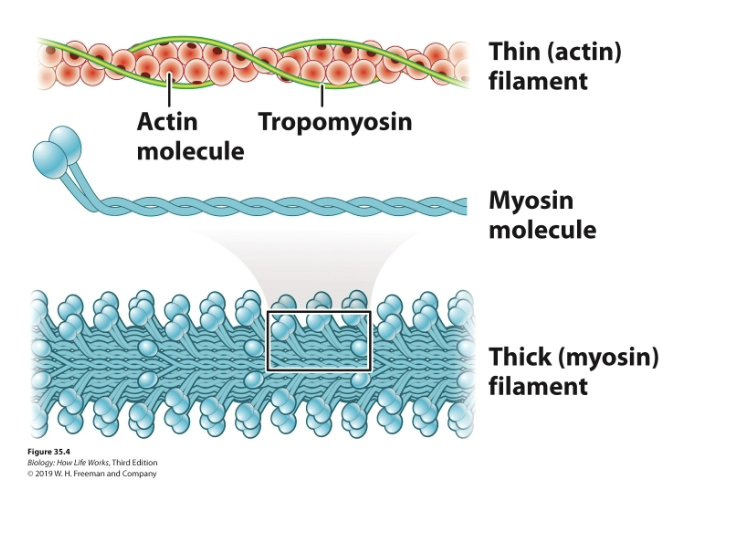
- Actin and Myosin= overlap to form myofibrils
- Tropomyosin and troponin are proteins that block myosin binding sites on monomers of actin filaments
- While Ca2+ binds to troponin, it causes the troposomyosin to move and expose myosin binding sites
- Thin filament= two actin filaments twisted together
Titin acts like a spring and is a giant protein greater than 1 um in length
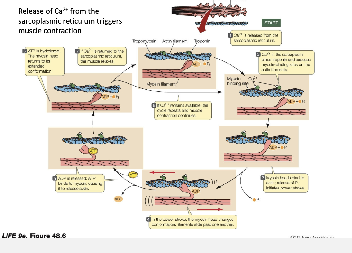
- 1) Calcium is released from the sarcoplasmic reticulum
- 2) Ca2+ in the sarcoplasm binds to troponin and exposes myosin-binding sites on the actin filaments
- 3) Myosin heads bind to actin release Pi initiates power stroke
- 4) In the power stroke, the myosin head changes conformation, filaments slide past one another
- 5) ADP is released; ATP binds to myosin, causing it to release actin
- 6) ATP is hydrolyzed. The myosin head returns to its extend conformation
- 7) if Ca2+ is returned to the sarcoplasmic reticulum, the muscle relaxes
- 8) If Ca2+ remains available, the cycle repeats and muscle contraction continues.
- Watch sarcomere shortening and cross bridge cycle
- Neurons communicate with muscle cells at neuromuscular synapses
- 1) Action potential arrives at axon terminal
- 2) Na+ channels open; depolarization causes voltage gated Ca2+ channels to open
- 3) Ca2+ enters the cells and triggers fusion of acetylcholine (neurotransmitter) vesicles with the presynaptic membrane
- 4) Acetylcholine molecules diffuse across the synaptic cleft and bind to receptors on the postsynaptic membrane
- 5) When receptors bind acetycholine, they open their cation channels and depolarize the postsynaptic membrane
- 6) The spreading depolarization fires an action potential in the postsynaptic membrane
- 7) Acetylcholine is broken down and the components and the components are taken back up by the presynaptic cell. Acetylcholine and vesicles are recycled.
- Actional potential to muscle contraction (look on slide)