Malaria .docx
Human Malaria
- malaria parasite belongs to
- phylum: apicomplexa
- class: sporozoa
- order: hemosporida
- genus: plasmodium
- the genus plasmodium is classified into two subgenera
- Plasmodium
- plasmodium vivax
- plasmodium malariae
- plasmodium ovale
- Laverania
- plasmodium falciparum
- Plasmodium
- plasmodium knowlesi, a parasite of long-tailed Macaque monkeys may also affect man
- Tertian malaria - get fever every other day
- Quartan malaria - get fever every third day
distribution of the malaria
- incidence of malaria is more in
- poor population in rural areas
- bad sanitary condition in urban area
- an epidemic can develop when there are changes in environmental, economic and social conditions such as migrations and heavy rains following draughts
- relative prevalence of the four species of malaria parasites varies in different geographical regions
- Plasmodium vivax
- most widely distributed
- being most common in Asia, North Africa, and Central and South America
- Plasmodium falciparum
- predominant species in Africa, Papua New Guinea and Haiti
- rapidly spreading in Southeast Asia and India
- results in most severe disease
- responsible for almost all malaria related deaths
- Plasmodium malariae
- is present in most places but is rare, except in Africa
- least severe but most persistent
- Plasmodium ovale
- is virtually confined to West Africa
- less prevalent ovale on
- Plasmodium vivax
- In Sri Lanka only Plasmodium vivax and Plasmodium falciparum
vectors
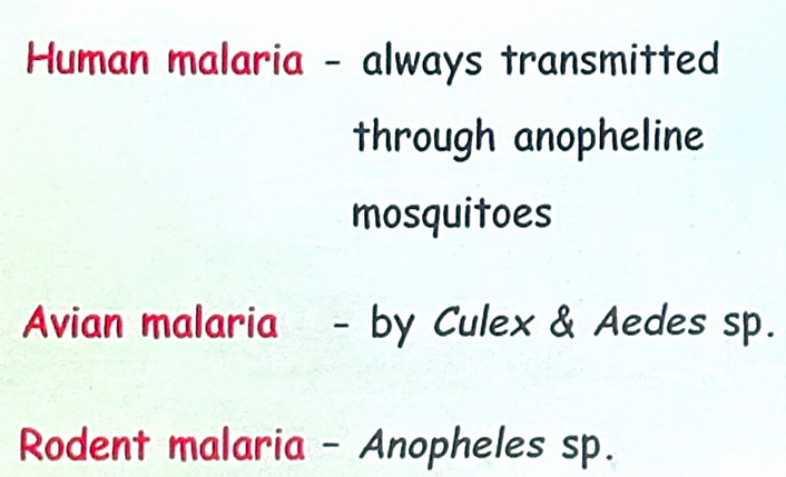
- human malaria is transmitted by over 60 species of female anopheles mosquito
- the male mosquito feeds exclusively on fruits and juices
- the female needs at least two blood meals, before the first batch of eggs can be laid
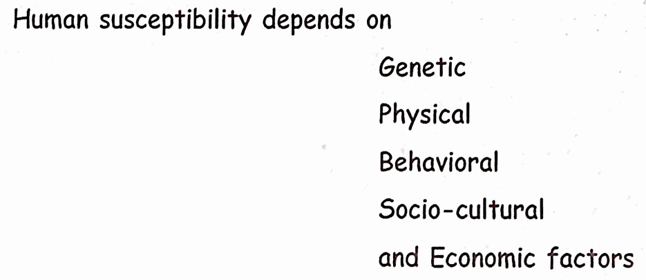
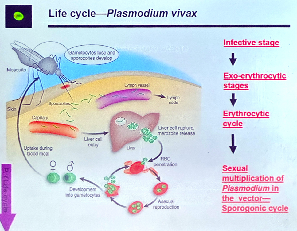
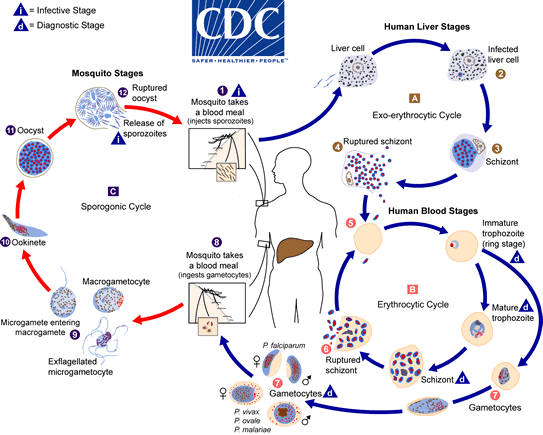
life cycle of Plasmodium (plasmodium vivax)
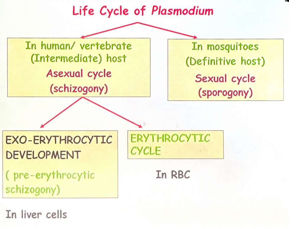
- malaria parasite passes its life cycle in two hosts
- definitive host
- female anopheles mosquito
- intermediate host
- man
- definitive host
- the life cycle of malarial parasite comprises of two stages
- an asexual phase occurring in humans - intermediate host
- a sexual phase occurring in mosquito - definitive host for the parasite
asexual phase
- the malaria parasite multiplies by division or splitting a process - schizogony
- asexual phase occurs in man, it is also called the vertebrate, intrinsic, or endogenous phase
- in humans, schizogony occurs in two locations
- in the red blood cell (erythrocytic schizogony)
- in the liver cells (exoerythrocytic schizogony or the tissue phase)
- because schizogony in the liver is an essential step before the parasites can invade erythrocytes, it is called pre-erythrocytic schizogony
- the products of schizogony, whether erythrocytic or exoerythrocytic, are called merozoites
human transmission
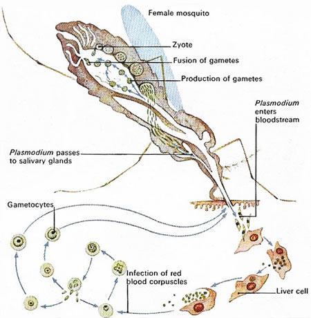
- human infection comes through the bite of the infective female anopheles mosquito
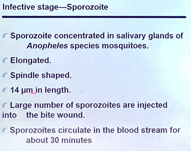
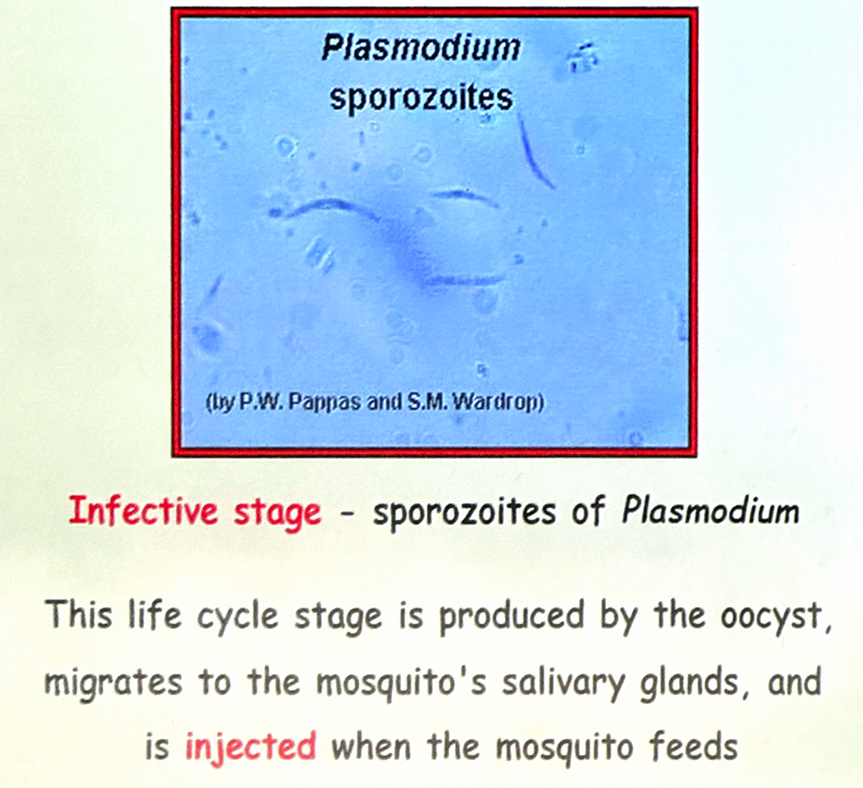
- the sporozoites, which are infective forms of the parasite are present in the salivary gland of the mosquito
- they are injected into blood capillaries when the mosquito feeds on blood after piercing the skin
- usually, 10- 15 sporozoites are injected at a time, but occasionally, many hundreds may be introduced
- the sporozoites pass into the bloodstream
- many are destroyed by the phagocytes, but some reach the liver and enter the parenchymal cells (hepatocytes)
pre-erythrocytic (tissue) stage or exoerythrocytic stage
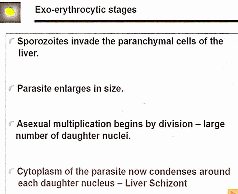
- within an hour of being injected into the body by the mosquito, the sporozoites reach the liver and enter the hepatocytes to initiate the stage of pre-erythrocytic schizogony or merogony
- the sporozoites, which are elongated spindle-shaped bodies 🡪 rounded inside the liver cells
- they enlarge in size and undergo repeated nuclear division to form several daughter nuclei
- each of which is surrounded by cytoplasm
- the hepatocyte is distended by the enlarging schizont and the liver cell nucleus is pushed to the periphery - pre-erythrocytic or exoerythrocylic schizont or meront
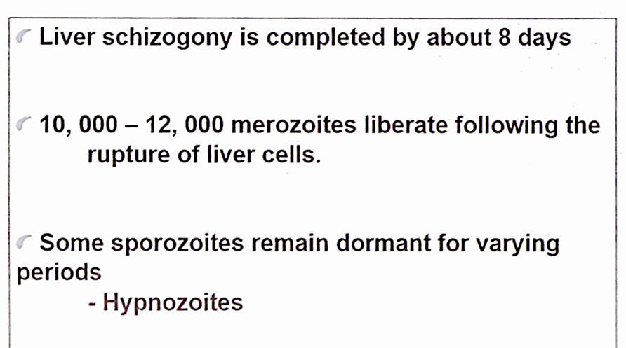
- mature liver stage schizonts are spherical (45-60 µm), multinucleate and contain 2,000-50,000 uninucleate merozoites
- unlike erythrocytic schizogony, there is no pigment in liver schizonts
- these normally rupture in 6-15 days and release thousands of merozoites into the bloodstream
- the merozoites infect the erythrocytes by a process of invagination
prepatent period
- the interval between the entry of the sporozoites into the body and the first appearance of the parasites in blood
latent stage
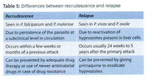
- in plasmodium vivax and plasmodium ovale, two kinds of sporozoites are seen, some of which multiply inside hepatic cells to form schizonts and others persist and remain dormant (resting phase)
relapse
- the resting forms are called hypnozoites
- from time to time, some are activated to become schizonts and release merozoites, which go on infecting RBCs producing clinical relapse
recrudescence
- in plasmodium falciparum and plasmodium malariae, initial tissue phase disappears completely, and no hypnozoites are found
- however, small numbers of erythrocytic parasites persist in the bloodstream
- in due course of time, they multiply to reach significant numbers resulting in clinical disease (short-term relapse or recrudescence)
erythrocytic stage
- the merozoites released by pre-erythrocytic schizonts invade the RBCs
- usually one merozoite invades a red cell
- plasmodium vivax prefers young RBCs
- need 48 hours to complete the cycle
- the receptor for merozoites is glycophorin, which is a major glycoprotein on the red cells
- the differences in the glycophorins of red cells of different species may account for the species specificity of malaria parasites
- merozoites are pear-shaped bodies, about 1.5 µmin length, possessing an apical complex (rhoptery)
- they attach to the erythrocytes by their apex
- the merozoites lie within an intraerythrocytic parasitophorous vacuole formed by red cell membrane by a process of invagination
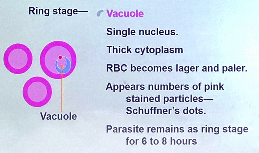
- in the erythrocyte, the merozoite loses its internal organelles and appears as a rounded body having a vacuole in the centre with the cytoplasm pushed to the periphery and the nucleus at one pole
- these young parasites are, therefore called the ringforms or young trophozoites
- the parasite feeds on the haemoglobin of the erythrocyte
- it does not metabolize haemoglobin completely and therefore, leaves behind a hematin-globin pigment called the malaria pigment or hemozoin pigment, as residue
- the malaria pigment released when the parasitized cells rupture is taken up by reticuloendothelial cells
- such pigment-laden cells in the internal organs provide histological evidence of previous malaria infection
- as the ring form develops, it enlarges in size becoming irregular in shape and shows ameboid motility – ameboid form or late trophozoite form
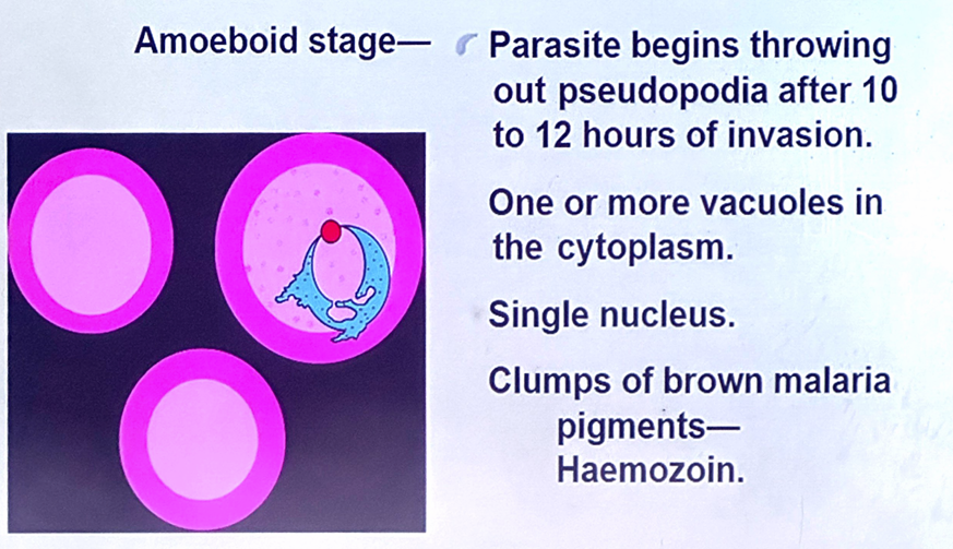
- when the ameboid form reaches a certain stage of development
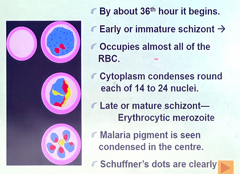
- its nucleus starts dividing by mitosis followed by a division of cytoplasm to become mature schizonts or meronts
- a mature schizont contains 8-32 merozoites and hemozoin
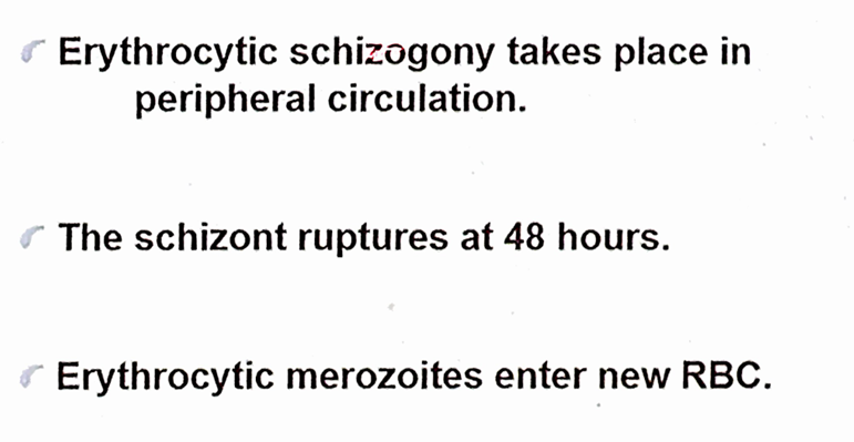
- the mature schizont bursts releasing the merozoites into the circulation
- the merozoites invade fresh erythrocytes within which they go through the same process of development
- this cycle of erythrocytic schizogony or merogony is repeated sequentially, leading to progressive increase in the parasitemia, till it is arrested by the development of host immune response
- the rupture of the mature schizont releases large quantities of pyrogens
- this is responsible for the febrile paroxysms characterizing malaria
gametogony
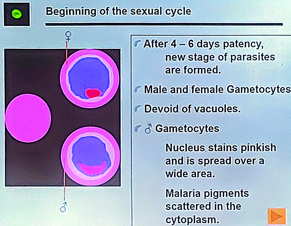
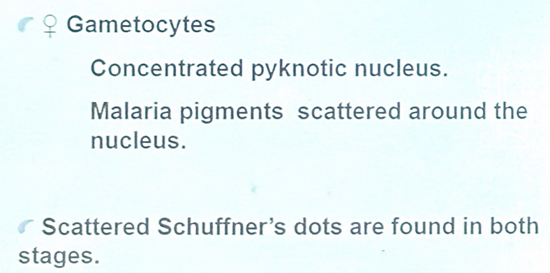
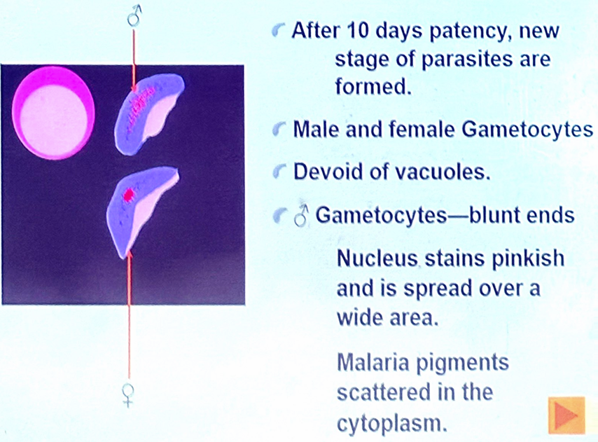
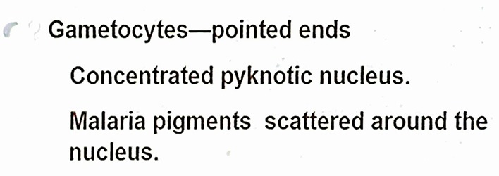
- after a few erythrocytic cycles, some of the merozoites that infect RBCs do not proceed to become trophozoites or schizonts but instead, develop into sexually differentiated forms – gametocytes
- they grow in size till they almost fill the RBC, but the nucleus remains undivided
- development of gametocytes generally takes place within the internal organs
- only the mature forms appear in circulation
- gametocyte appears in circulation after the first appearance of asexual form
- 4-5 days in plasmodium vivax
- 10-12 days in plasmodium falciparum
- a person with gametocytes in blood is a carrier or reservoir
- the gametocytes do not cause any clinical illness in the host, but are essential for transmission of the infection
- a gametocyte concentration of 12 or more per mm3 of blood in the human host is necessary for mosquitoes to become infected
sexual phase
- female anopheles mosquito represents definitive host, in which sexual forms takes place
- sexual forms of the parasite (gametocytes) originate in human RBCs
- maturation and fertilization take place in the mosquito, giving rise to a large number of sporozoites
- phase of sexual multiplication is called sporogony
- called the invertebrate, extrinsic, or exogenous phase
- extrinsic incubation period
- the time taken for completion of sporogony in the mosquito is about 1-4 weeks (extrinsic incubation period), depending on the environmental temperature and the species.
mosquito cycle (sporogony)
- when a female anopheles mosquito ingests parasitized erythrocytes along with its blood meal, the asexual forms of malaria parasite are digested
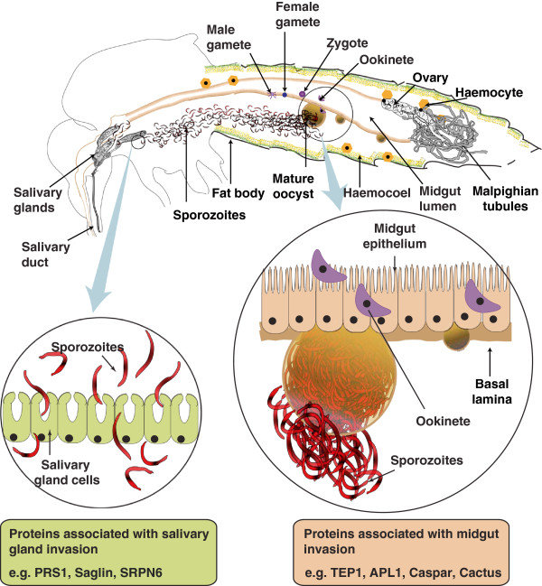
- the gametocytes are set free in the midgut (stomach) of mosquito and undergo further development
- the nuclear material and cytoplasm of the male gametocytes divides to produce eight microgametes
- long, actively motile, whip-like filaments (flagellating male gametocytes)
- the female gametocyte does not divide but undergoes a process of maturation to become the female gamete or macrogamete
- it is fertilized by one of the microgametes to produce the zygote
- fertilization occurs in 0.5-2 hours after the blood meal
- the zygote
- initially a motionless round body
- gradually elongates and within 18-24 hours
- becomes a vermicular motile form with an apical complex anteriorly - ookinete (travelling vermicule).
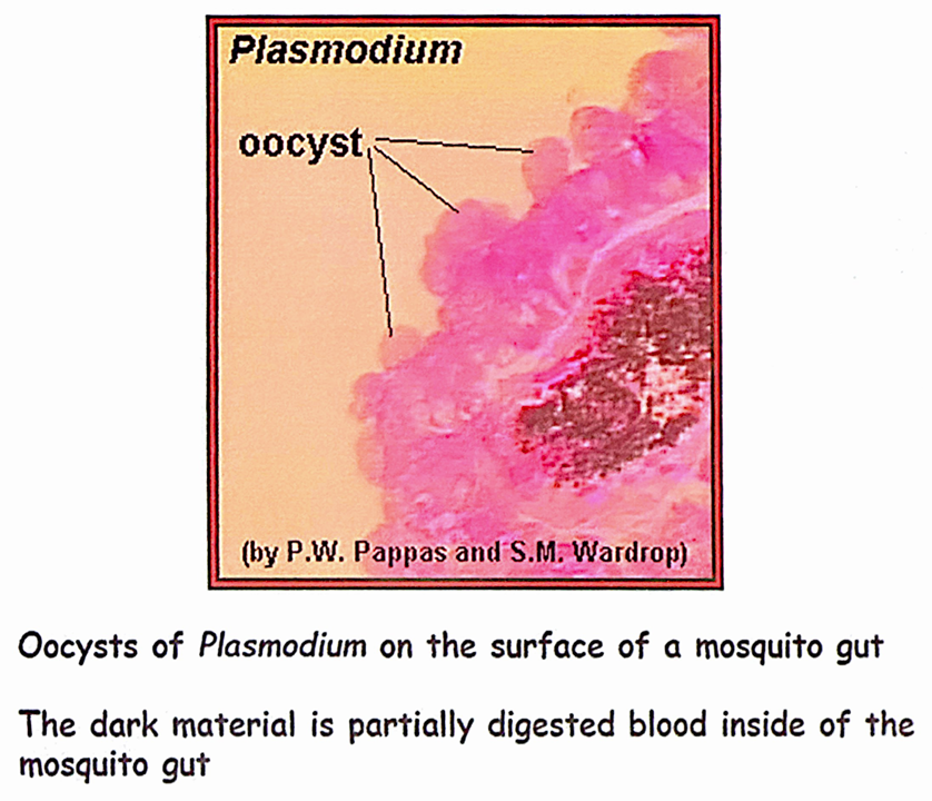
- it penetrates the epithelial lining of the mosquito stomach wall and comes to lie just beneath the basement membrane
- it becomes rounded into a sphere with an elastic membrane – oocyst
- multiplicatory phase, within which numerous sporozoites are formed
- the mature oocyst, which may be about 500 µm in size, bulges into body cavity of mosquito and when it ruptures
- the sporozoites enter into the hemocele or body cavity, from where some sporozoites move to the salivary glands
- the mosquito is now infective and when it feeds on humans, the sporozoites are injected into skin capillaries to initiate human infection
sporogonic cyle
- only female anopheline mosquitoes transmit human malaria
- male and female gametocytes enter the mid gut with the blood meal
- male gametocyte 'exflagellation' → male gametes
- female gamete gets fertilized by male gamete in the mid gut of the mosquito
- maturation of the oocysts depend on
- longevity of the mosquito species
- temperature and humidity of the environment
lifecycle of Plasmodium (plasmodium falciparum)
asexual phase
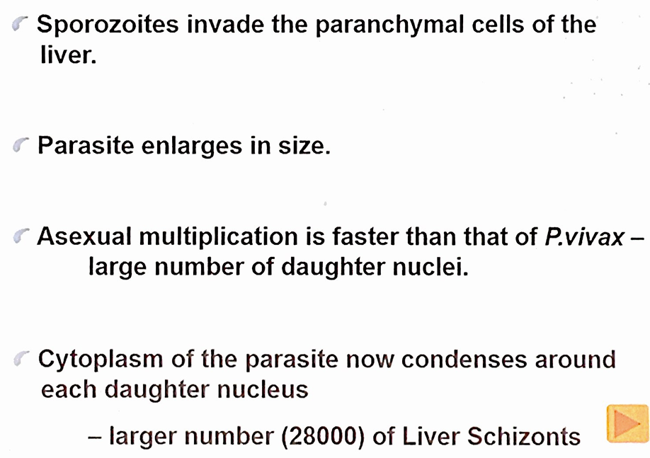
exo-erythrocytic stages
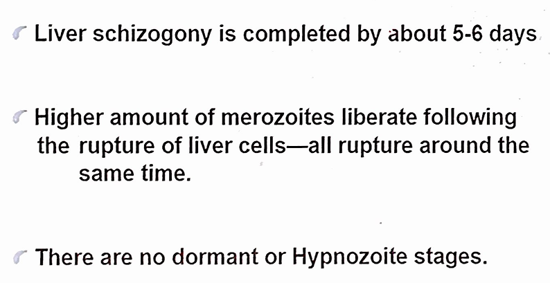
erythrocytic cycle
- in plasmodium falciparum, erythrocytic schizogony always takes place inside the capillaries and vascular beds of internal organs
- therefore, in plasmodium falciparum infections, schizonts and merozoites are usually not seen in the peripheral blood
- usually one or more merozoites invades a red cell
- plasmodium falciparum prefers RBCs of all ages
- need 48 hours to complete the cycle
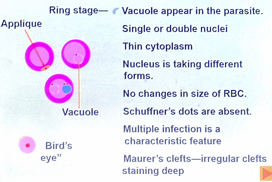
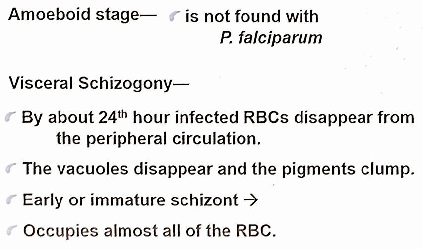
- cytoplasm condenses round each of 8 to 24 nuclei
- late or mature schizont erythrocytic merozoite they are sequestrated in the capillaries and venules of deep organs
- the schizont ruptures at 48 hours
- erythrocytic merozoites enter new RBCs in capillaries of deep organs.
- the schizont ruptures at 48 hours
- erythrocytic merozoites enter new RBC
gametogony
lifecycle of Plasmodium (plasmodium malariae)
asexual phase
- exo-erythrocytic
- development takes a longer time
erythrocytic cycle
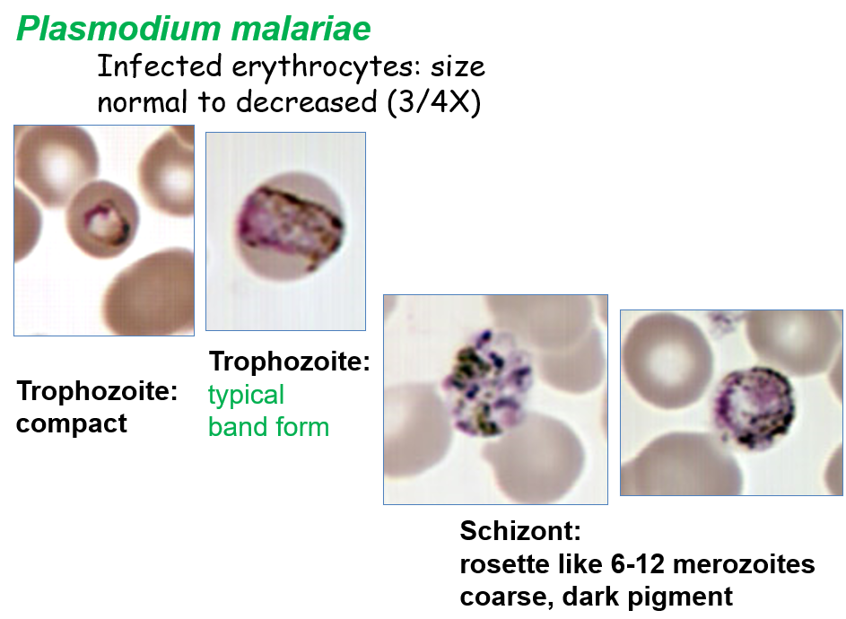
- infected RBCs do not enlarge
- Zeiman's stippling appears only with special staining
- ring stage having thicker cytoplasm than in other species
- trophozoite takes a characteristic 'band' form
- prolonged erythrocytic cycle - 72 hours
- schizonts have characteristic 'daisy head' pattern
- pattern of life cycle is similar to that of plasmodium vivax with some exceptions
lifecycle of Plasmodium (plasmodium ovale)
- relapse could occur following formation of hypnozoites
asexual phase
erythrocytic cycle
- is similar to that of plasmodium vivax
- the infected RBCs are enlarged
- stippling is more intense than schuffner's dots
- infected RBCs wall become weakened and may have irregular shapes
transmission of malaria
- subcutaneous inoculations of sporozoites by anopheline mosquitoes
- blood transfusion
- through the contaminated syringes of drug addicts
- congenital or trans-placental
pathophysiology and pathology of malaria
- almost all severe forms of malaria are caused by infection of plasmodium falciparum
- the following serious complications are rarely seen plasmodium vivax and plasmodium knowlesi
- severe anaemia
- respiratory distress
- splenic complications
- shock
- multiple organ dysfunction
activation of cytokine storm
- clinical features of malaria are developed in the erythrocytic stage
- both parasite and host related factors contribute to the pathogenicity of the severe forms of malaria
- rupture of the parasitized RBCs (pRBCs) in each erythrocytic cycle and destruction of pRBC at high levels in the spleen
- lead to
- activation of innate immunity
- release of inflammatory mediators (TNF-α, IFN-γ, IL-1, IL-6)
- activation of cytokine storm
- symptoms associated with the malaria
- clinical features of malaria are initiated mainly due to the excessive release of various cytokines
- classical malaria fever
- headache
- nausea and vomiting
- diarrhea
- anorexia
- tiredness
- aching joints and muscles
- thrombocytopenia
- immunosuppression
- changes of central nervous system
erythrocytic stage of malaria
- erythrocytic stage of malaria induces changes in the
- surface of the parasitized RBCs (pRBCs) and high levels of inflammatory cytokines induces
- endothelial activation
- mainly on the surface of the microvasculature of vital organs
- expression of the endothelial receptors (act as adhesion molecules)
- endothelial activation
- surface of the parasitized RBCs (pRBCs) and high levels of inflammatory cytokines induces
surface of the pRBCs
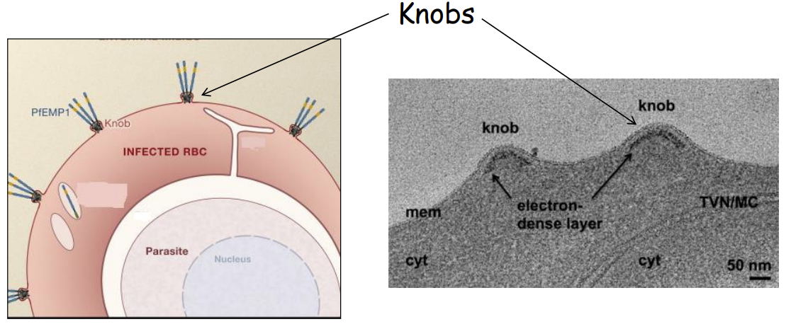
- as plasmodium falciparum matures it release parasite proteins to the surface of pRBC that forms “knobs”
- parasite proteins of the PfEMP-1 (Plasmodium falciparum Erythrocyte Membrane Protein-1) family mainly found on the surface of these knobs
surface of the endothelium microvasculature
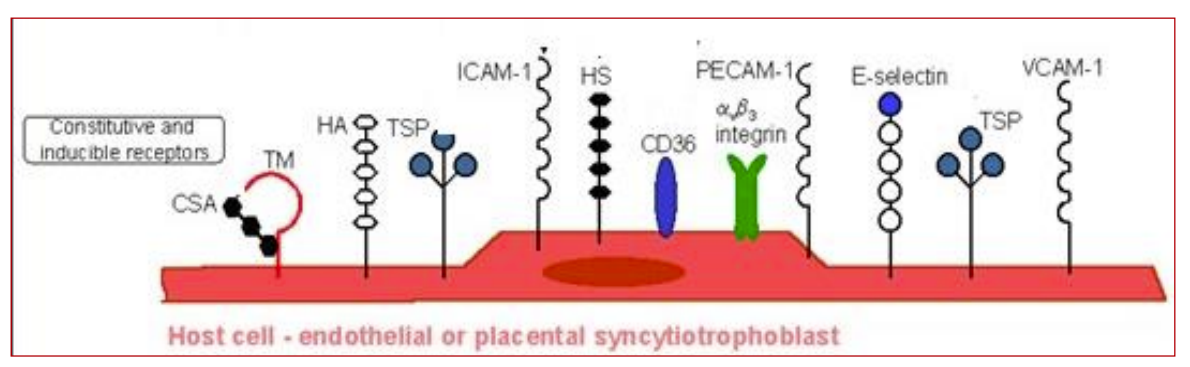
- due to the effects of inflammatory cytokines and other cellular mediators 🡪 increased expression of some of the endothelial cell receptors
- Intra Cellular Adhesion Molecule-1 (ICAM 1)
- Endothelial Protein C Receptor (EPCR)
- CD-36
- E-selectin
- Neural Cell Adhesion Molecule (NCAM)
- CD 56 Chondroitin Sulphate (CSA) – mainly in the placenta
cytoadherence
- severe malaria is mainly caused by plasmodium falciparum because of its ability to induce pRBCs cytoadherence to the vascular endothelium
- pRBCs binds to the endothelial receptors of the microvasculature of the deeper organs
- cytoadherence leads to sequestration of the pRBCs in the microvasculature of vital organs
- allows the maturing pRBCs to get away from the splenic clearance
sequestration
- increased rigidity of both iRBCs and uninfected RBCs (increased deformability of RBCs)
rosettes
- binding of pRBCs with the uninfected RBCs
agglutination
- binding of pRBCs with other pRBCs
clumps
- binding of pRBCs with platelets
microvasculature manifestation consequences
- pathological changes occur due to above processes in the microvasculature leading to
- reduce or obstruction of blood flow
- limits the oxygen supply 🡪 tissue anoxia
- concentration of toxins within the tissue 🡪 release of inflammatory mediators
- endothelial apoptosis
- reduce or obstruction of blood flow
- sequestration and other pathological changes occur in many internal organs including
- brain, lungs, eyes ,kidneys, heart ,GIT (liver), bone marrow , adipose tissue, skin / placenta
pathology cerebral malaria
- is a complex neurological syndrome of severe falciparum malaria
- is often fatal
- presence of plasmodium falciparum in the blood slide examination
- is characterized by
- altered consciousness
- seizures
- neurological defects
brain endothelium changes
- obstruction of blood flow caused by
- cytoadherence
- resetting
- agglutination
- clumping
- decreased RBCs and pRBCs deformability
- breakdown of the blood-brain barrier due to
- reduction of the nutritional supply and hypoxia 🡪 cell death and tissue damage
- excess activation of endothelial cells and excess releases of pro-inflammatory cytokines
- blood-brain barrier breakdown followed by other secondary neuropathologic events in the brain parenchyma
- damage to the blood vessels causes intracranial haemorrhage can lead to cerebral malaria
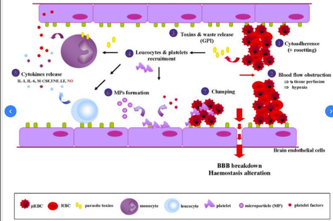
macroscopic and microscopic changes
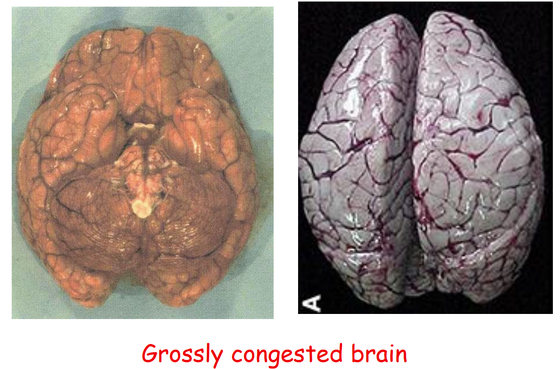
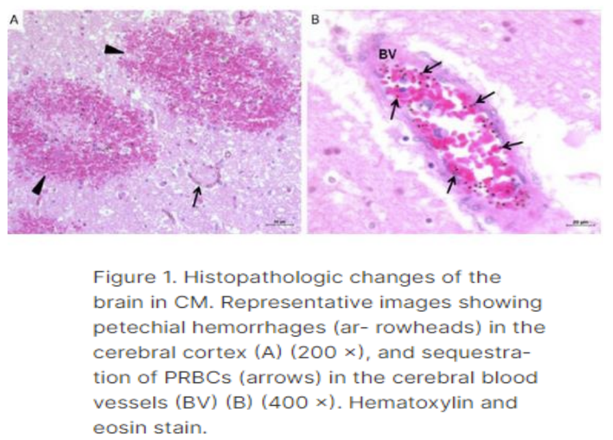
macroscopic changes in central nervous system
- brain swelling with flattened gyri and narrowed sulci
- brain shows gross congestion
- petechial haemorrhages found in white matter of cerebrum, brain stem and cerebellum
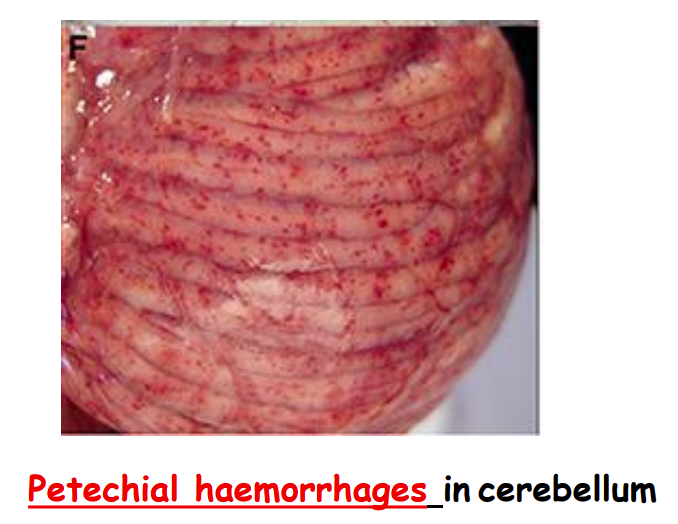
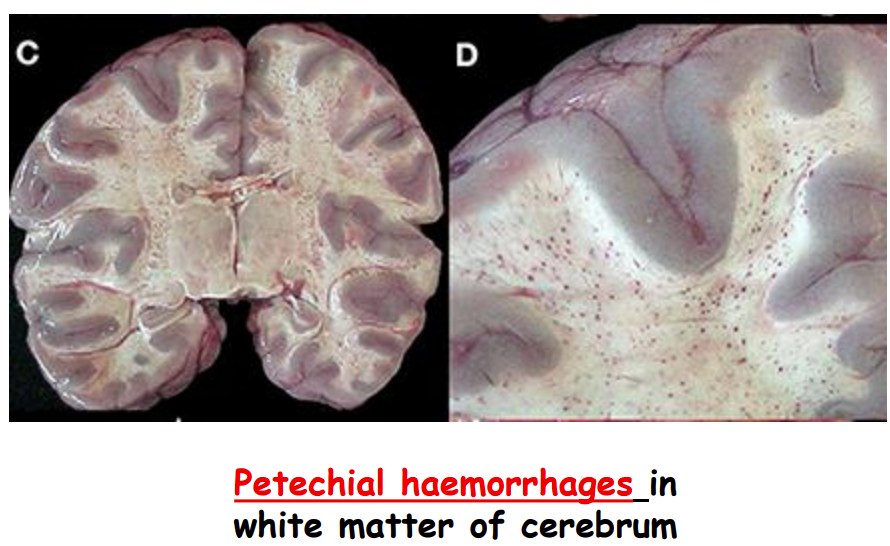
microscopic changes in central nervous system
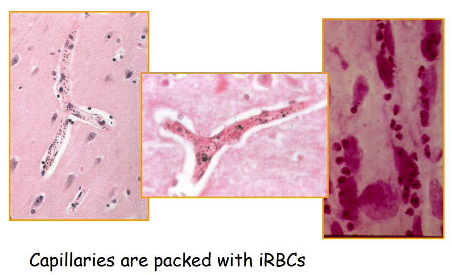
- capillaries are packed with parasitized RBC
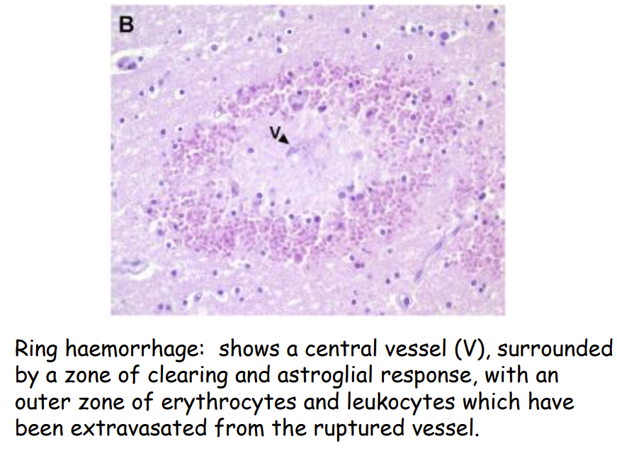
- ring haemorrhages – classical characteristic feature in cerebral malaria
pathology associated with blood
anaemia
- problem in pregnancy & children
- plasmodium vivax predominantly invades young RBC
- parasitaemia rarely exceeds 2%
- plasmodium falciparum affects red cells of all ages
- parasitaemia can be high as 5-10% or more
mechanism of anaemia
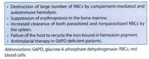
- multifactorial
- haemolysis of pRBCs
- immune and non-immune haemolysis of non-infected RBCs
- increased splenic clearance of pRBC and normal RBCs
- in plasmodium falciparum – per 1 iRBC – 8 non infected RBC
- in plasmodium vivax – per 1 iRBCs - 32 non infected RBCs
- decrease of RBC production in the bone marrow (dyserythropoiesis and bone marrow suppression)
- reduction of red cell survival even after disappearance of parasitaemia
- the red cell becomes more spherical and difficult to circulate
microscopic changes of RBCs
- invasion of the malaria parasites causes extensive changes of the RBCs
- specially in plasmodium falciparum infections
- these includes
- loss of the normal discoid shape
- increased rigidity of the membrane
- increased permeability to a variety of substances
- increased adhesiveness
- most notably to endothelial surfaces
malaria thrombocytopenia
- reduction of platelet count is common in both plasmodium falciparum & plasmodium vivax
- mainly in the early stage of the infection
- due to
- sequestration of platelets in the spleen
- platelet destruction by macrophages
- bone marrow alterations
- antibody-mediated platelet destruction
- oxidative stress
- platelet aggregation
pathology bone marrow
- bone marrow undergoes several changes including
- dyserythropoiesis – production of immature or dysfunctional RBCs
- thrombocytopenia – impaired production of platelets
- leucocytosis (acute condition)
- high white blood cell count
- leukopenia (late stage)
- diminished white blood cell count
- hemophagocytosis
- histiocytes engulf red blood cells, white blood cells, platelets
macroscopic changes in bone marrow
- expansion of the marrow space and reddish appearance in bone marrow
- due to an increase in RBC production / hyperplasia
- in severe cases bone marrow becomes pale in appearance
- due to hypoplasia or aplasia
- fatty infiltration
- in chronic, repeated severe malaria adipose tissue replaces hematopoietic cells within the marrow
microscopic changes in bone marrow
- sequestration of the iRBCs within erythroid precursors and other blood cell lineages
pathology in spleen
- spleen mainly involved in
macroscopic changes
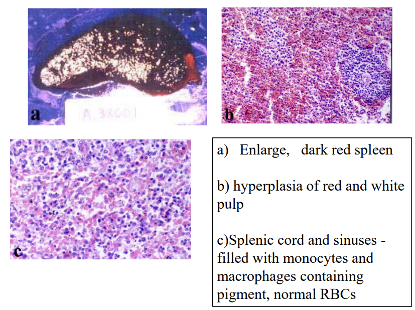
- splenomegaly
- increased immune activity
- sequestration of iRBCs and congestion of spleen blood vessels
- enlarged spleen detected during physical examinations
- dark red or pale appearance of the spleen
- due to the accumulation of iRBCs and other blood cells
- focal necrosis areas
- may develop and appear as pale or discoloured patches on the surface of the spleen
- infarction
- tissue death due to lack of blood supply in certain parts of the spleen
- results in areas with pale of haemorrhagic discolouration
microscopic changes
- sequestration of iRBCs within sinusoides with red pulp
- Hemozoin deposition
- haemozoin accumulates in the macrophages mainly in red pulp
- microscopically visualized as dark brown granules at the cytoplasm of the macrophages
- hyperplasia of red pulp
- accumulation of the inflammatory cells - mainly in the red pulp
- histopathological changes of the splenic architecture
- microscopic examination may reveal the
- loss of lymphoid follicles
- fibrosis of red pulp
- changes of the marginal zone of the spleen
splenic complications
- splenic complications of malaria includes
- hypersplenism (overreactive spleen)
- hyperreactive malarial syndrome
- splenic infarction
- splenic rupture
splenic rupture
- rapid and significant enlargement of spleen
- can rarely result in splenic rupture
- is more common in primary attack and due to fibrosis, less likely happen in case of chronic malaria infections
- is common in infections with plasmodium vivax but can happen in plasmodium falciparum infections
hyperreactive malarial splenomegaly / tropical splenomegaly syndrome
- condition is characterized by abnormal enlargement of the spleen
- response to repeated or persistent malaria infections
- mainly found in malaria endemic countries in Africa, Asia and South America
- due to chronic / repeated stimulation by the anti-malarial immunoglobulins (IgM and other immunoglobulins) for long period
pathology in kidney
- three main clinical forms are associated with malaria
- acute kidney injury
- quartan malaria nephropathy
- black water fever acute
kidney injury / renal failure
- microcirculation disorders due to the
- microvasculature block
- anoxia
- absence of oxygen
- necrosis of the glomeruli and renal tubules
- mainly associated with plasmodium falciparum
quartan malaria nephropathy
- found in plasmodium malariae infection
- deposition of the malaria antigens and immune complex (IgM + compliment + antigens) causes
- inflammation and tissue damage
- haematuria, proteinuria, impaired renal function
black water fever/ malarial hemoglobinuria
- occurs only with infection from plasmodium falciparum
- dark red/black urine
- due to the presence of large amounts of haemoglobin
- rapid severe haemolysis, jaundice, haemoglobinuria and anuria (absence of urine)🡪 lead to death
pathology of the lungs / pulmonary
- found in plasmodium falciparum infection mainly
- lung involvement in malaria is less common compared to other complications
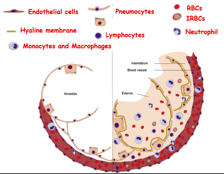
- dysfunction of the endothelial blood vessels (due to obstruction and inflammation) leads to
- vascular permeability 🡪 leakage of fluid and proteins into lung tissue and alveolar space
- severe malaria
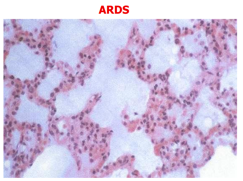
- pulmonary oedema
- acute respiratory distress syndrome
- diffuse alveolar damage
- impaired gas exchange
- respiratory failure 🡪 hypoxemia and respiratory compromise
placental complications in malaria
- malaria during pregnancy is associated to high morbidity and mortality both in mother and the child
- pregnant women more susceptible to malaria
- more frequent in first pregnancy
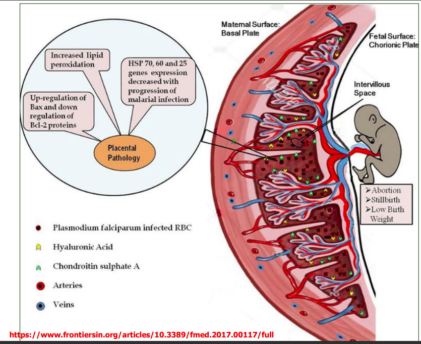
- iRBCs mainly bind with the chondroitin sulphate A (CSA) and also with hyaluronic acid (HA)
- expressed in the placenta
- iRBcs are found in large numbers in the intervillous space
- parasite can avoid maternal immune response when iRBCs located in the placenta
- monocyte and macrophages infiltrates to intervillous space and induces releasing of inflammatory cytokines
- increased inflammatory responses result into oxidative stress induced placental cell death
- severe malaria in pregnant women especially in first pregnancy
- IUGR (Intrauterine growth restriction), LBW (low birth weight) in infants
- miscarriage and still birth
- primigravidae are more susceptible to malaria than multigravidae
- resistance to the malarial infection in multigravidae is due to the development of placental parasite-specific immunity in second and third pregnancies
clinical features of malaria
- malaria has a variety of clinical presentations
- acute
- uncomplicated malaria
- malaria in children
- malaria in pregnancy
- complicated severe malaria
- mixed infection
- relapses
- chronic
- acute
- clinical presentation depends on
- species of the parasite
- immunity of the host
- time period from sporozoites inoculation to appearance of the first symptom
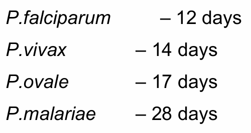
falciparum malaria
- Plasmodium falciparum: Malignant tertian malaria
- Tertian malaria - get fever every other day
- tertian fever pattern is hardly seen
- leads to severe complications
- cerebral malaria
- severe anaemia
- renal failure
- acute respiratory distress syndrome (ARDS)
- acidosis
- hypoglycaemia
vivax malaria
- Plasmodium vivax: Benign tertian malaria
- Tertian malaria - get fever every other day
- is absent in Duffy blood group negative people
- Duffy antigens act as receptors for chemokines and attract immune system cells
- Duffy antigens also act as receptors for the Plasmodium species
- Duffy negative individuals whose erythrocytes do not express the receptor are believed to be resistant to merozoite invasion
- incubation period longer
- relapses present
- tertian fever pattern is frequent
- severe disease is uncommon
ovale malaria
- Plasmodium ovale: Benign tertian malaria
- Quartan malaria - get fever every third day
- infection occurs in Africa
- overlying plasmodium vivax areas
- is absent in duffy blood group negatives
- clinically similar to plasmodium vivax
- incubation period is relatively long
- early phase is absent or unrecognized
- relapses are present
- low parasitaemia - difficult diagnosis
malariae malaria
- Plasmodium malariae: Benign quartan malaria
- Quartan malaria - get fever every third day
- least aggressive of all forms of malaria
- early phase very mild renal involvement—mostly in children
uncomplicated malaria
malaria fever presentation
- comes on with the rupture of erythrocytic schizonts
- malaria fever stages
- cold stage
- hot stage
- sweating stage
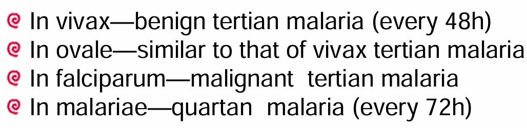
cold stage
- febrile episode
- starts with shaking chills (chills & rigor)
- usually at mid-day (between 11 a.m. to 12 noon)
- lasts for 15 minutes - 1 hour
hot stage
- cold stage symptoms followed by high grade fever and rigors
- temperature exceeds 39 degrees
- lasts 2 - 6 hours
sweating stage
- hot stage symptoms followed by profuse sweating fever gradually subsides over 2-6 hours
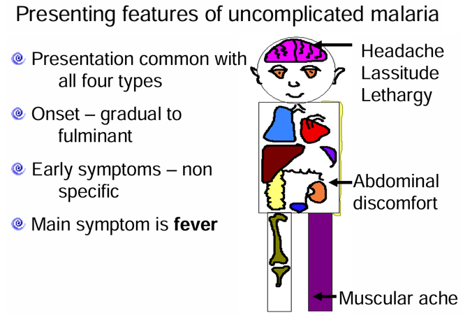
atypical malaria fever presentation
- atypical presentation is common among
- people of endemic zone
- immuno-compromised people
- extremes of age
- pregnancy
- early infection
- plasmodium falciparum infection
malaria in children
infant
- severe falciparum malaria is rare
- if it occurs with infancy the mortality is very high
1-3 years
- severe malaria is common among this group
older children
- severe disease is rarely seen
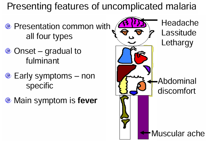
malaria in pregnancy
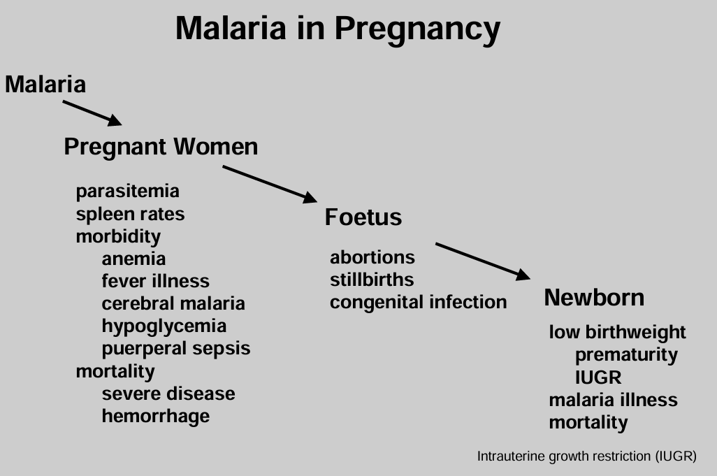
- primigravidae (a woman who conceives for the first time)
- develops severe falciparum malaria
- multigravidae (woman that is or has been pregnant for at least a second time)
- develops severe vivax malaria
- in a low endemic area
- malaria affects birth weight of first three pregnancies
- can develop symptoms in 2nd and 3rd trimesters
- in a high endemic area
- usually asymptomatic
complicated severe malaria
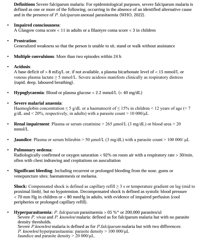
- always caused by plasmodium falciparum
- usually due to delayed and ineffective treatment
- commoner among the non-immune 1-3 year old children
- plasmodium vivax can cause splenic rupture rarely
- plasmodium malariae can cause fatal nephrotic syndrome
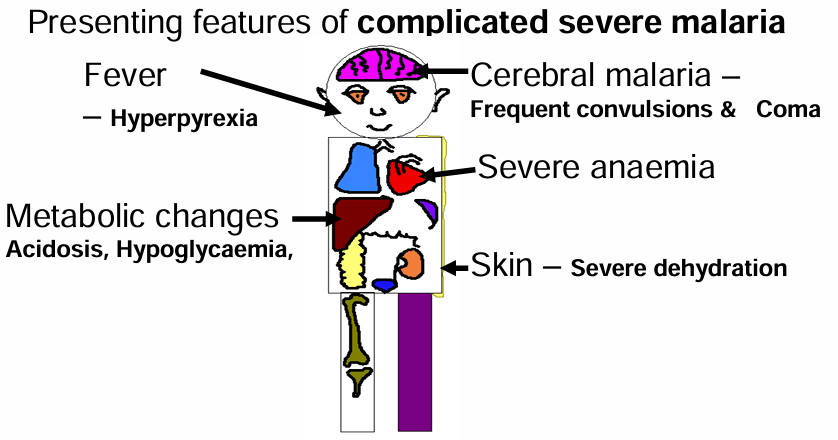
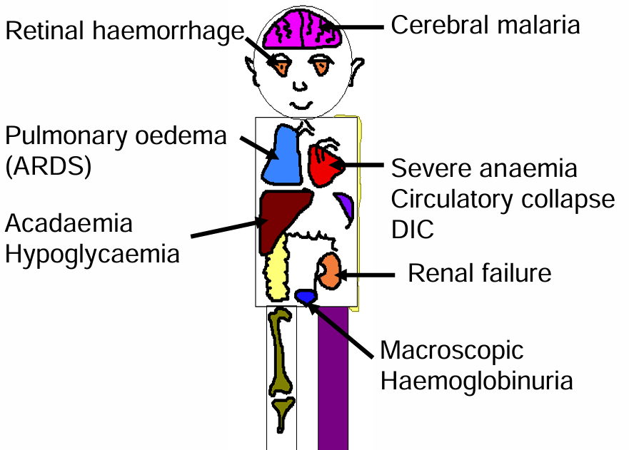
features of complicated severe malaria
- cerebral malaria (coma for > 30 minutes)
- severe anaemia (Hb< 40mg / 2.2mmol /dl)
- circulatory collapse (SBP < 50 - 1 to 5 yrs & SBP < 70 - >5 yrs)
- spontaneous bleeding – disseminated intravascular coagulation
- acidosis (rapid breathing with clear lungs)
- macroscopic haemoglobinuria – blackwater fever
complicated severe malaria clinical symptoms
cerebral malaria clinical symptoms
- is the most prominent feature of falciparum malaria
clinical symptoms
- coma
- altered consciousness or no loss of consciousness (sometimes)
- symmetrical encephalopathy
- five distinct fundoscopic abnormalities
- retinal whitening
- retinal haemorrhages
- focal whitening of vessels
- papilledema
- cotton wool spots
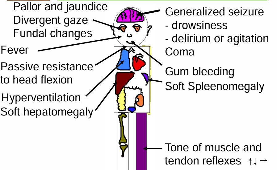
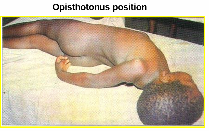
poor prognostic signs of cerebral malaria
- deep coma convulsions
- opisthotonus position
- respiratory distress
- hyper-parasitaemia
- hypoglycaemia
- severe anaemia
anaemia clinical features
- pallor
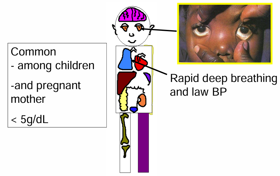
acute respiratory distress clinical features
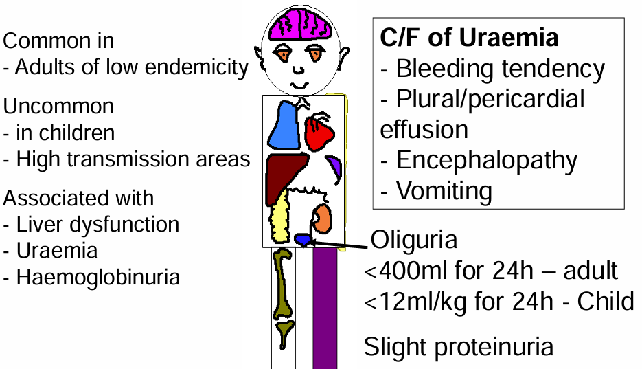
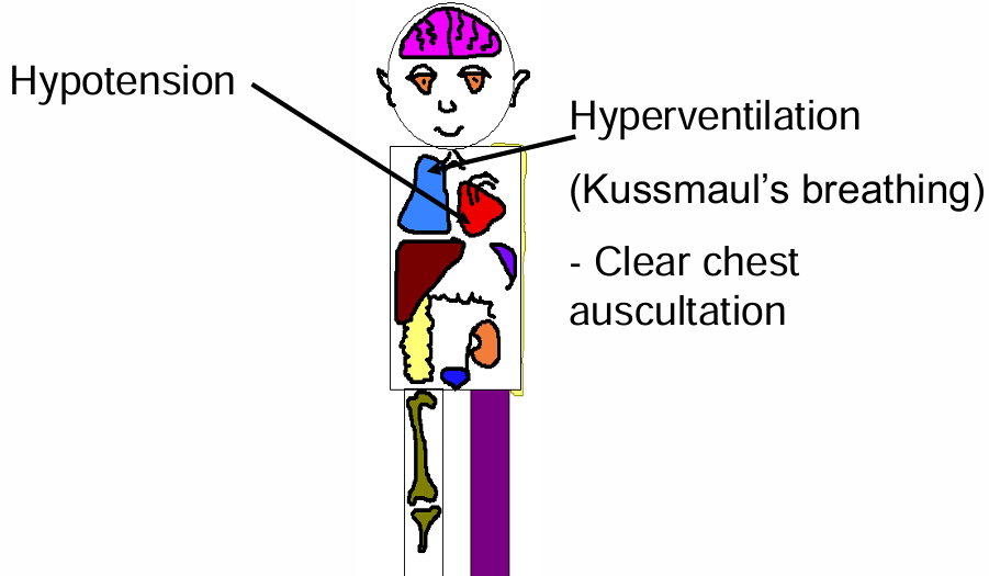
metabolic acidosis clinical features
- hypotension
- hyperventilation
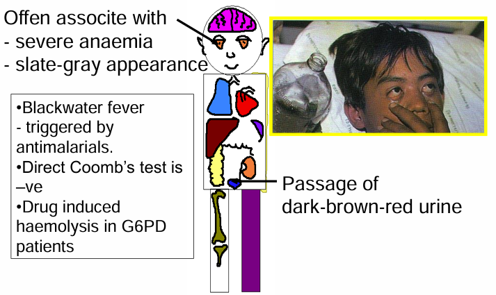
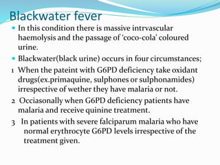
blackwater fever clinical features
- severe anaemia
- slate – grey appearance
- dark – brown – red urine
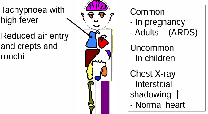
acute pulmonary oedema clinical features
- tachypnoea
- high fever
- reduced air
- crepitations and ronchi lung sounds
- interstitial shadowing - X ray
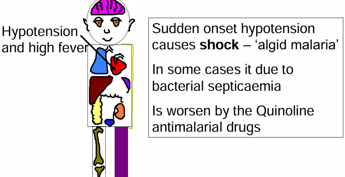
algid malaria clinical features
- algid maria
- prostration, cold and clammy skin, and low blood pressure
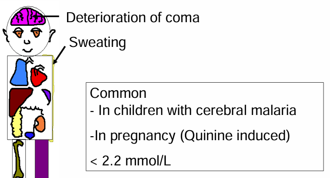
hypoglycaemia clinical features
- deterioration of coma
- sweating
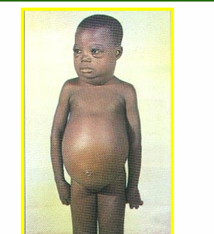
chronic malaria
- hepatosplenomegaly (tropical splenomegaly syndrome (TSS))
- nephrotic syndrome – plasmodium malariae
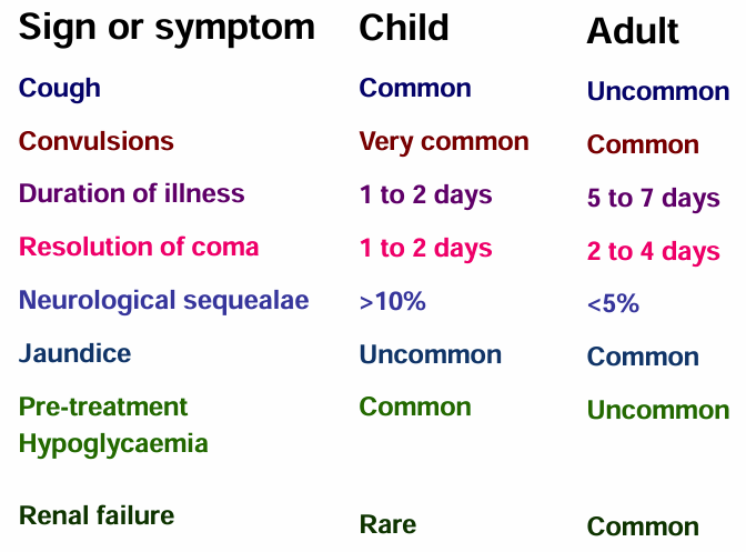
severe malaria in children and adults
management of malaria
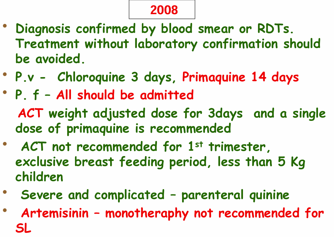
ministry of health 2008 circular
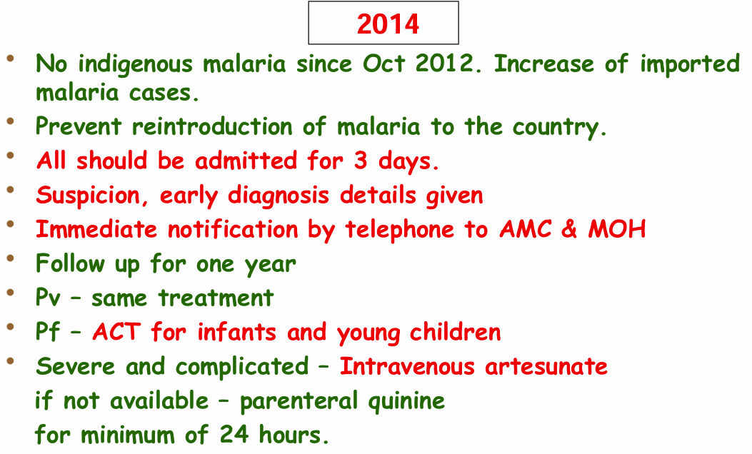
ministry of health 2014 circular
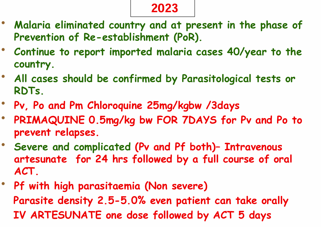
ministry of health 2023 circular
management of malaria
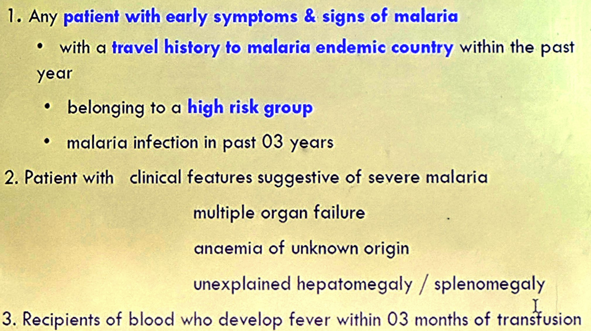
- history taking & clinical examination (signs & symptoms)
- travel history with duration
- past history of malaria
- taken antimalaria before
- any recent blood transfusions
- any close contact members with malaria
- laboratory diagnosis
- parasitological
- immunological
- rapid diagnostic tests
- ELISA based tests
- empirical chemotherapy
- indications
- severe morbidity with high suspicion
- absence of laboratory facilities with high suspicion
- procedure
- collect a blood smear
- full course of treatment for vivax infection
- indications
- after confirmation of malaria
- plasmodium vivax infection
- clinical cure – getting rid of symptoms (fever)
- radical cure ((due to hypnocytes) clinical relapse)
- plasmodium falciparum infection
- clinical cure
- transmission blocking - getting rid of gametocytes
- plasmodium vivax infection
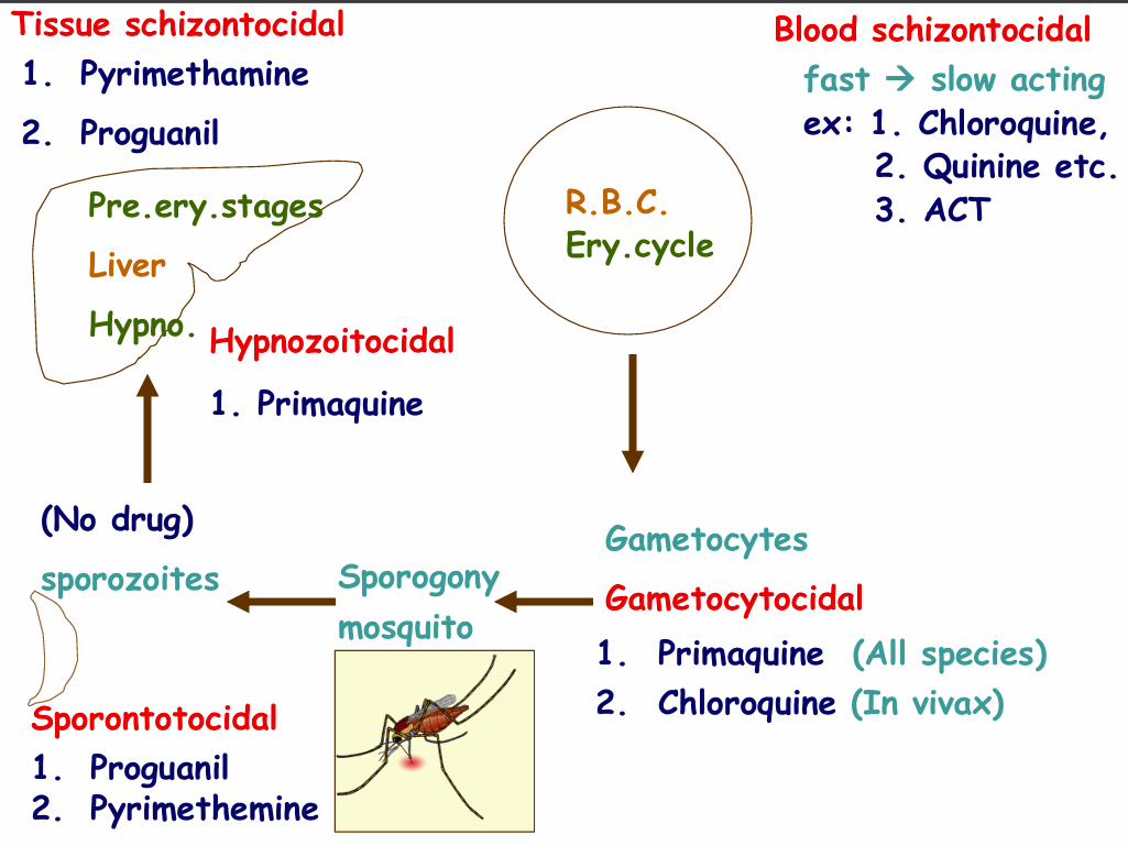
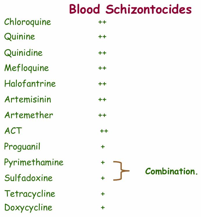
anti-malarial drugs
- clinical cure in patients
- prevention of relapses in plasmodium vivax & plasmodium ovale
- prevent transmission
- prophylaxis (prevention)
anti-malarial drugs history
- assess previous treatment with anti-malarial
- previous history of adverse reactions to anti-malarial
- identify early and late treatment failures
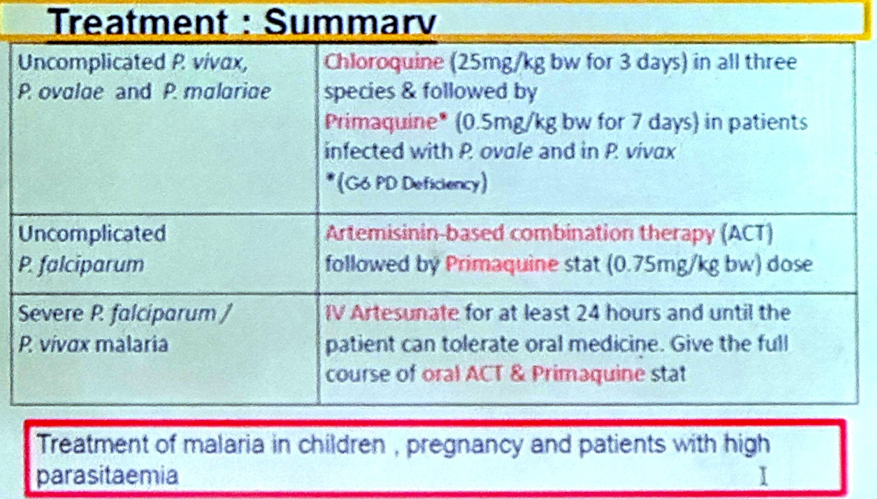
CURRENT REGULATIONS / REMOVED REGULATIONS
ACT (Coartem®) tropic
indications
- plasmodium falciparum infection
contradictions
- pregnant mothers - first trimester
- children weight <5kg (2014 circular)
- lactating mothers (2014 circular)
plasmodium vivax infection
uncomplicated children weight <5kg
- chemotherapy starts after 1 year of age
clinical cure
- oral 25 mg/kg chloroquine base over three days
radical cure
- oral 0.75 mg/kg primaquine base daily over a period of 7 days (2023 circular)
- oral 0.25 mg/kg primaquine base daily over a period of 14 days (2014 circular)
uncomplicated pregnant mother (T1/T2/T3)
- chemotherapy starts 6 weeks after delivery
clinical cure
- oral 25 mg/kg chloroquine base over three days
radical cure
- oral 0.75 mg/kg primaquine base daily over a period of 7 days (2023 circular)
- oral 0.25 mg/kg primaquine base daily over a period of 14 days (2014 circular)
uncomplicated lactating mother
- chemotherapy starts after exclusive breast feeding (4-6 months)
clinical cure
- oral 25 mg/kg chloroquine base over three days
radical cure
- oral 0.75 mg/kg primaquine base daily over a period of 7 days (2023 circular)
- oral 0.25 mg/kg primaquine base daily over a period of 14 days (2014 circular)
uncomplicated others
- chemotherapy starts after completion of chloroquine therapy
clinical cure
- oral 25 mg/kg chloroquine base over three days
radical cure
- oral 0.75 mg/kg primaquine base daily over a period of 7 days (2023 circular)
- oral 0.25 mg/kg primaquine base daily over a period of 14 days (2014 circular)
uncomplicated G6PD deficiency
- chemotherapy starts after completion of chloroquine therapy
- administration is done under special supervision
clinical cure
- oral 25 mg/kg chloroquine base over three days
radical cure
- oral 0.75 mg/kg primaquine base weekly over a period of 8 weeks
plasmodium falciparum infection
uncomplicated pregnant mother (T1)
clinical cure
- oral 10 mg/kg quinine dihydrochloride 8 hourly for 7 days + clindamycin for 7 days (2014 circular)
transmission blocking
- no primaquine therapy
- achieved via obstructing patient-vector contact
uncomplicated pregnant mother (T2/T3)
clinical cure
- oral weight adjusted dose of artemisinin-based combination therapy (ACT) over 3 days
transmission blocking
- no primaquine therapy
- achieved via obstructing patient-vector contact
uncomplicated lactating mother – exclusive breast feeding
clinical cure
- artemisinin-based combination therapy (ACT)
transmission blocking
- no primaquine therapy
- achieved via obstructing patient-vector contact
uncomplicated others
clinical cure
- oral weight adjusted dose of artemisinin-based combination therapy (ACT) over 3 days
transmission blocking
- 0.75 mg/kg primaquine single dose after ACT
- achieved via obstruct patient-vector contact
severe infection
clinical cure
- intravenous artesunate 2.4mg/kg at 12 and 24 hours
- if not available parenteral quinine
- parenteral treatment for a minimum of 24 hours, then a full course of ACT
transmission blocking
- 0.75 mg/kg primaquine single dose after ACT
- achieved via obstruct patient-vector contact
severe infection in pregnancy
clinical cure
- 1st trimester - parenteral quinine and then oral quinine for a total of 7 days
- 2nd and 3rd trimester – parenteral artesunate/quinine and later ACT
transmission blocking
- no primaquine therapy
- achieved via obstruct patient-vector contact
plasmodium vivax infection & plasmodium falciparum infection
uncomplicated mixed infection
clinical cure
- oral weight adjusted dose of artemisinin-based combination therapy (ACT) over 3 days
transmission blocking
- oral 0.75 mg/kg primaquine base daily over a period of 7 days (2023 circular)
- oral 0.25 mg/kg primaquine base daily over a period of 14 days (2014 circular)
follow-up - warded patients
- daily blood smears for 3 days
- if parasitaemia persists more daily films
- Anti-Malarial Campaign and their field staff will follow up for one year period
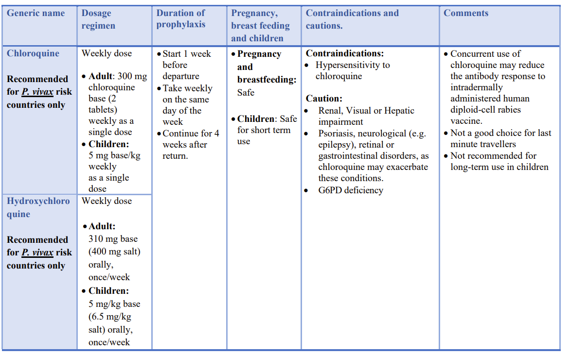
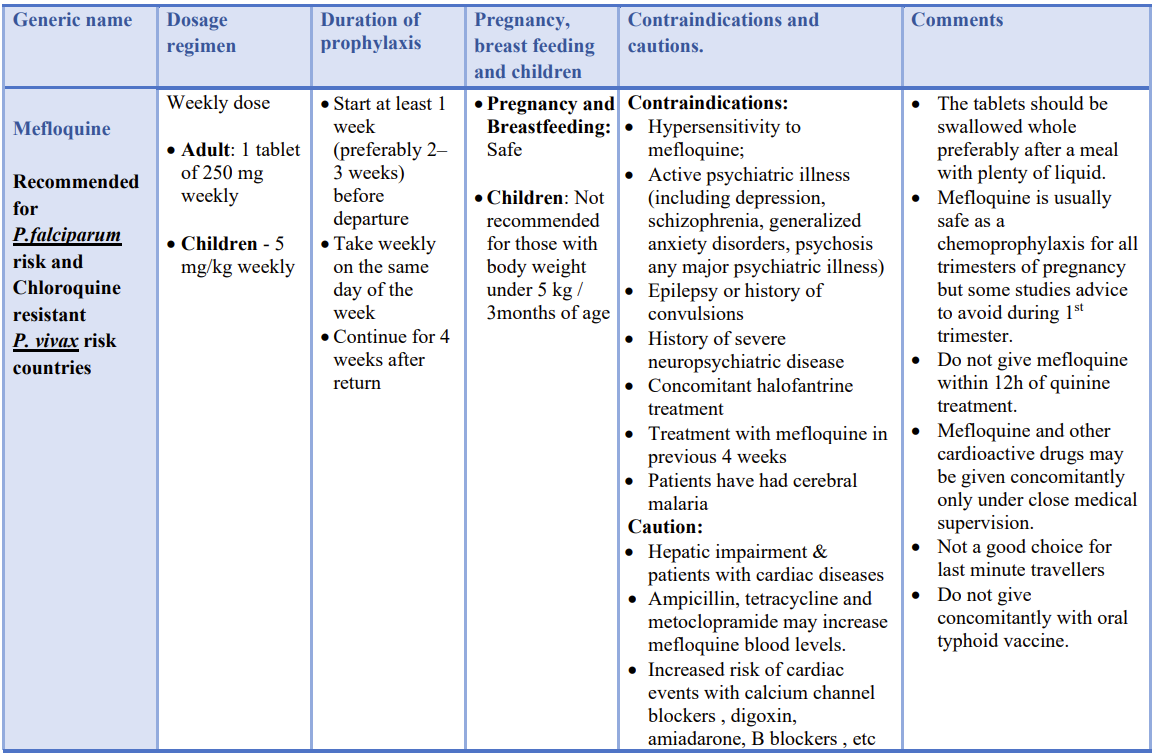
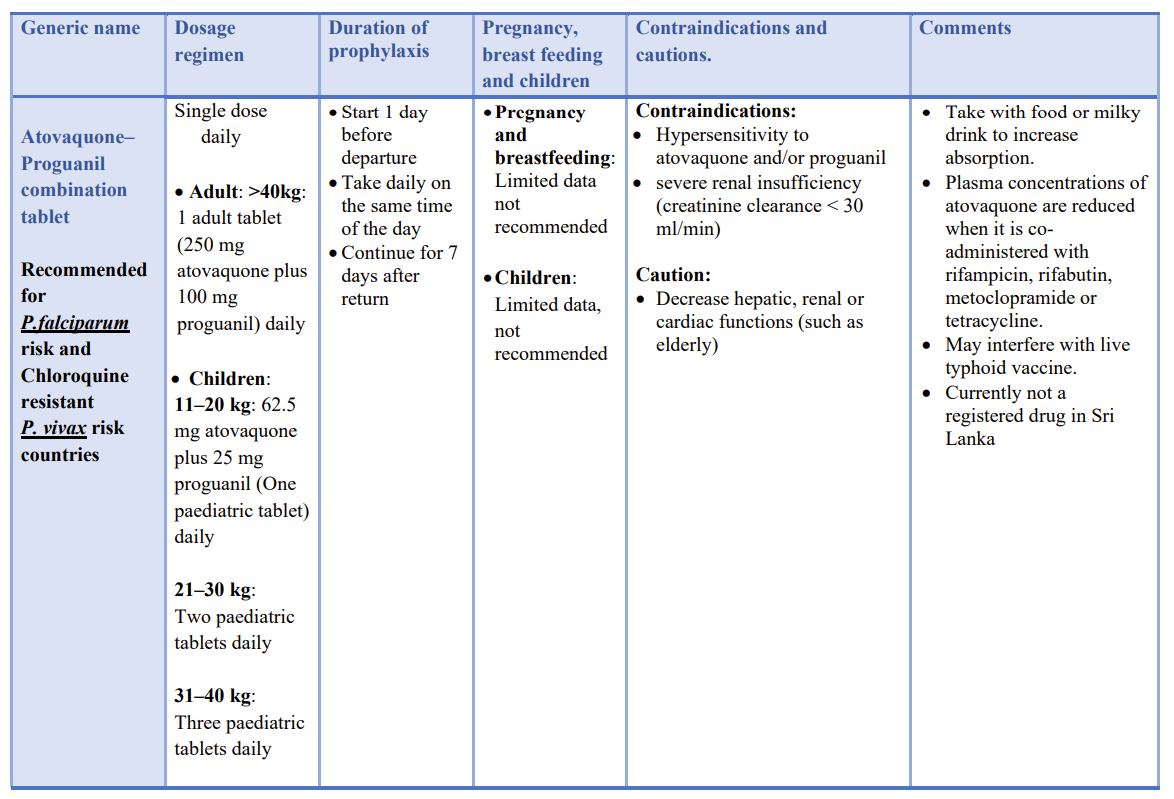
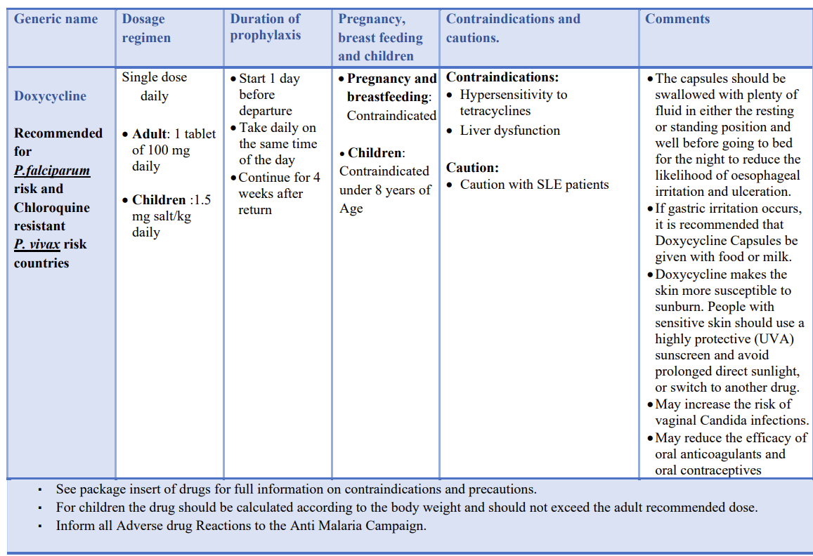
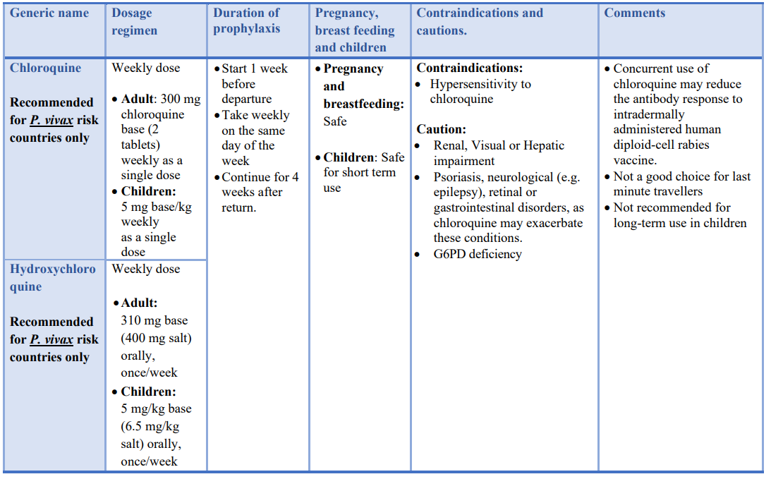
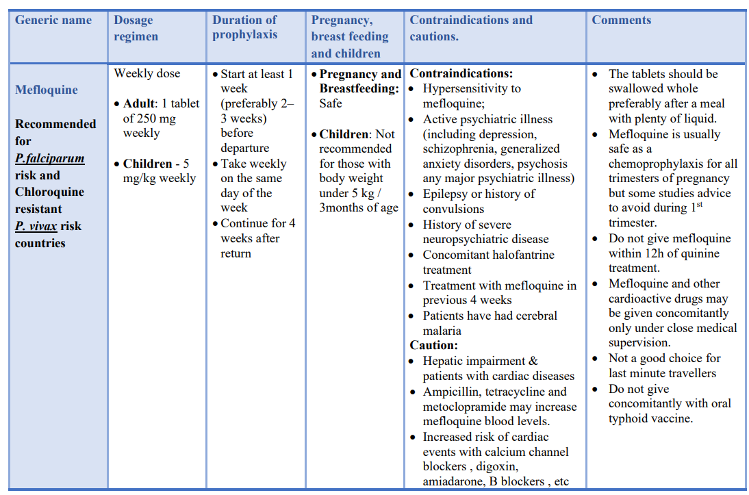
chemoprophylaxis for malaria
- chemoprophylaxis is not needed for
- visitors to Sri Lanka
- anyone living within the country including pregnant women
- chemoprophylaxis is recommended for
- travellers to malaria endemic countries
- contact Anti-Malarial Campaign if travelling to such countries
- Chloroquine
- recommended for Plasmodium vivax risk countries only
- Hydroxychloroquine
- recommended for Plasmodium vivax risk countries only
- Mefloquine
- recommended for Plasmodium falciparum risk and Chloroquine resistant Plasmodium vivax risk countries
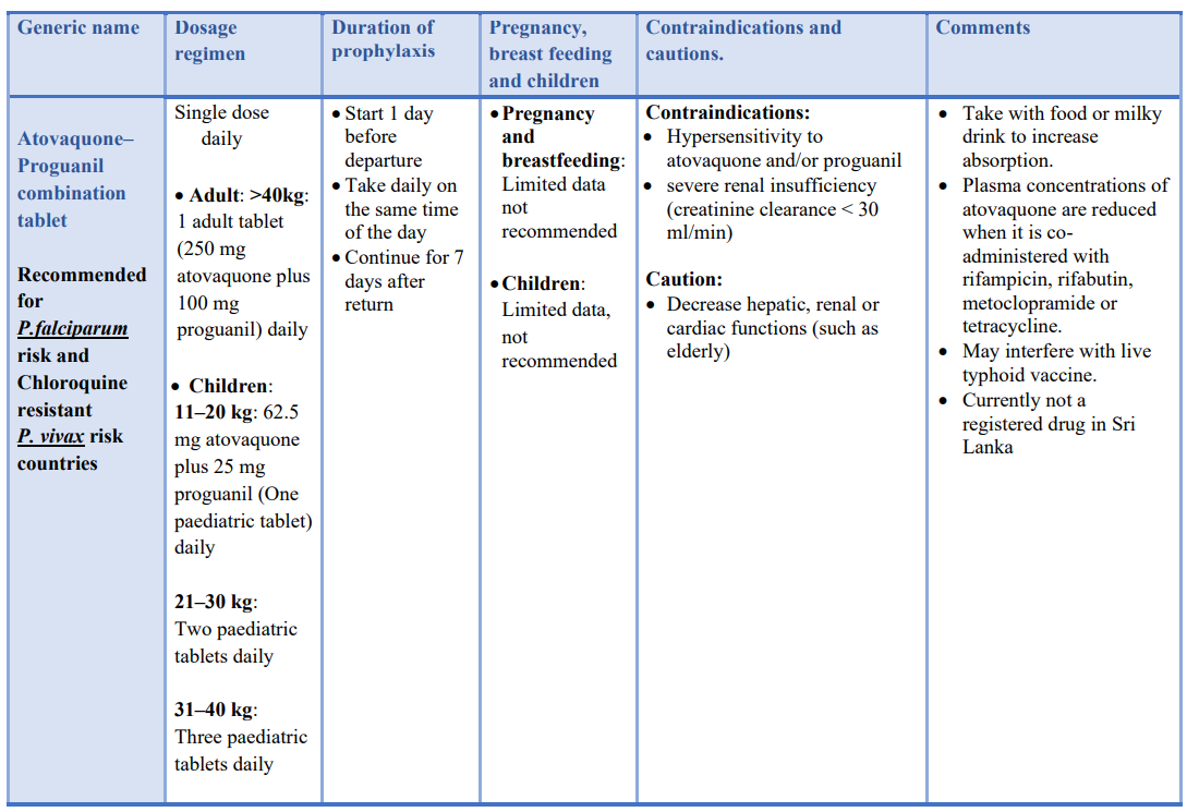
- Atovaquone– Proguanil combination tablet
- recommended for Plasmodium falciparum risk and Chloroquine resistant Plasmodium vivax risk countries
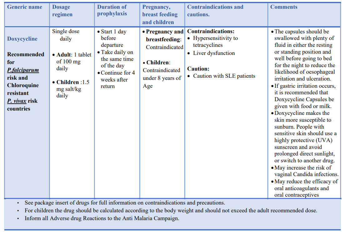
- Doxycycline
- recommended for Plasmodium falciparum risk and Chloroquine resistant Plasmodium vivax risk countries
Laboratory Diagnosis of Malaria
clinic-epidemiological diagnosis
- element in the clinical diagnosis of malaria, in both endemic and non-endemic areas
- have a high index of suspicion
- visit to a malaria endemic area?
- history of malaria
- blood transfusions
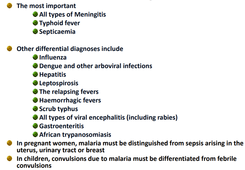
malaria differential diagnosis
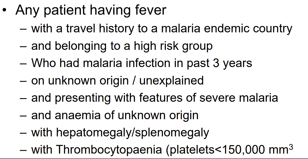
malaria suspicion
laboratory investigation – parasitological
- the two most widely used methods in diagnosis of malaria are
- demonstration of malaria parasite in suspected patients’ peripheral blood
- Thin or Thick blood film screening
- demonstration of malaria antigens in patients’ blood
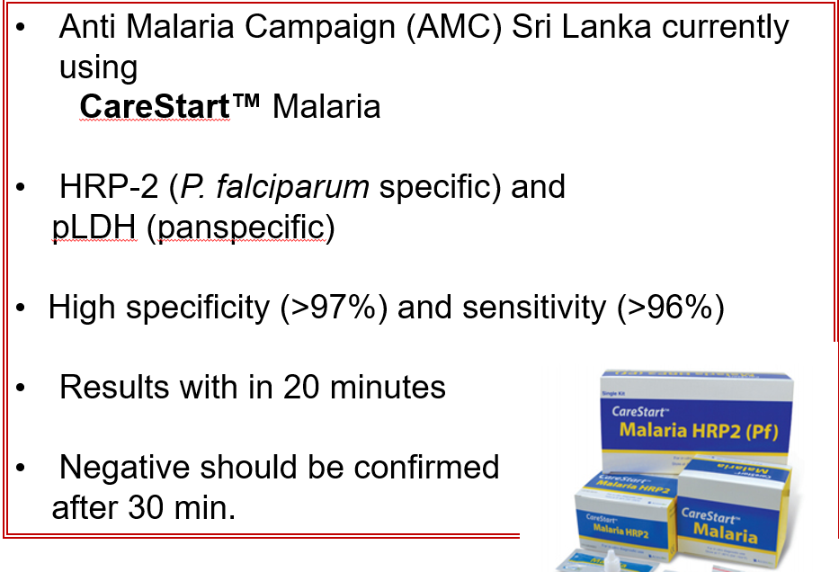
- Rapid Diagnostic Test (RDT)
- if there is a strong clinical suspicion of malaria, and the blood smears / RDT are negative at the time of initial testing, a minimum of three consecutive blood smears / RDTs should be done before concluding that the patient is negative for malaria
- if there is a strong clinical suspicion of malaria, and the blood smears / RDT are positive at the time of initial testing the patient is positive for malaria
- demonstration of malaria parasite in suspected patients’ peripheral blood
- mandatory prior to start anti-malarial treatment
DO NOT TREAT MALARIA BASED ON CLINICAL SUSPICION. LABORATORY CONFIRMATION IS A MUST
demonstration of malaria parasite in blood films
- blood obtained by pricking a finger or earlobe is the ideal sample
- density of developed trophozoite or schizonts are greater in blood from these capillary-rich area
- microscopy remains the gold standard method for
- diagnosis
- identification of parasite species
- estimation of parasite density
- ideal time to collect blood sample
- venous blood
- cord blood
- blood from donor pack
- after peak of fever
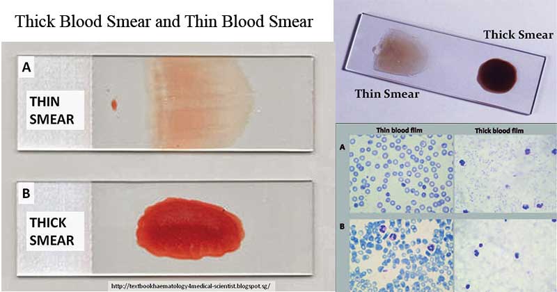
thick blood film
- mainly use to find the parasite present or not
- provides enhanced sensitivity of the blood film
- used in detection of low levels of parasitaemia
- high RBC , high sensitivity
- RBC destroyed in thick blood
thin blood smear
- RBC not destroyed in thin blood
- mainly use to confirm the different plasmodium species present
- used for morphological / stage identification of the parasite species
- provides higher specificity than thick film
- low RBC, low sensitivity
- can use to monitor the treatment response and detect drug resistance
- estimation of parasitaemia is possible
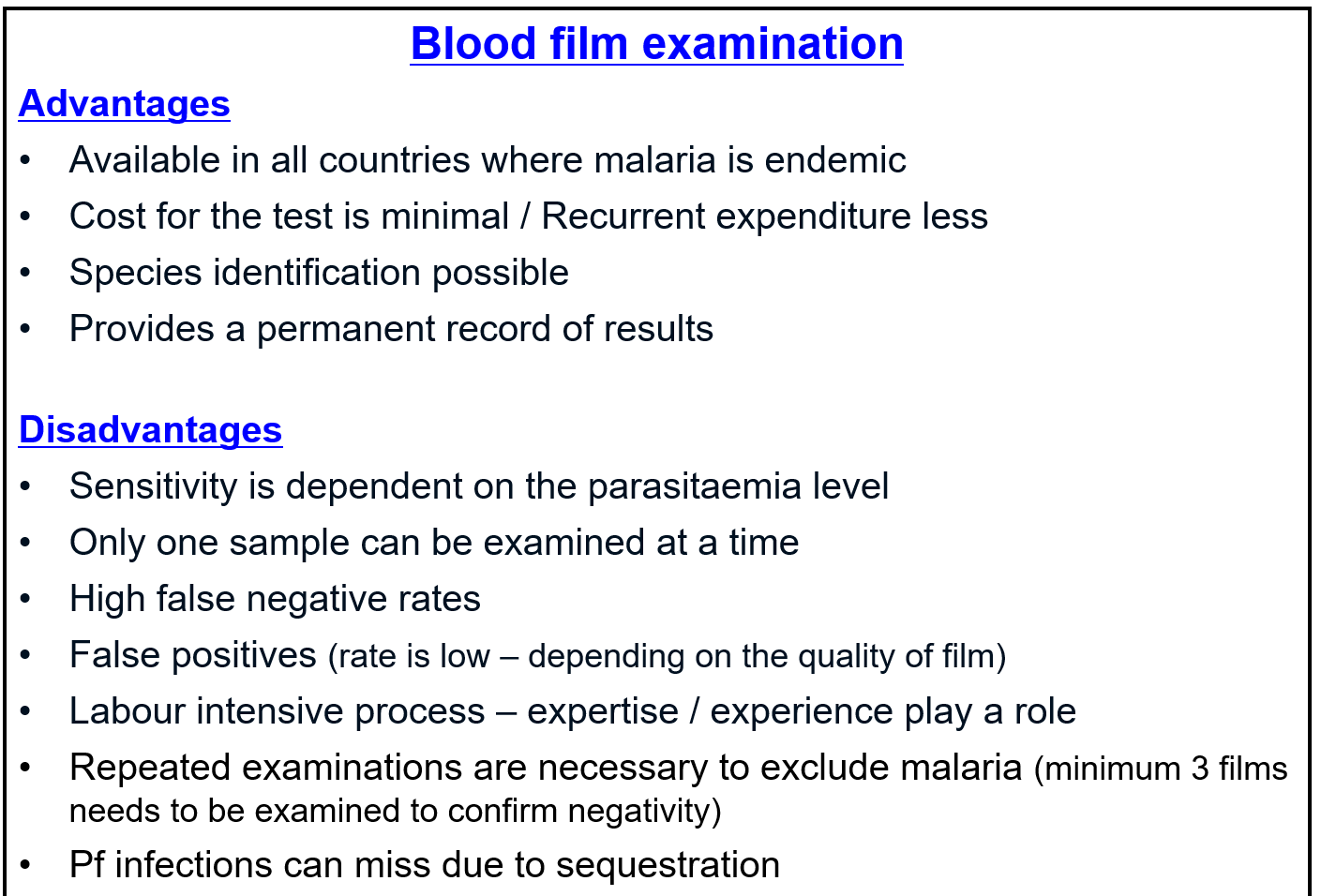
blood film examination advantages and disadvantages
- false negatives due to low parasitemia
- false negatives due to low skill in lab researcher (not identifying the parasite)
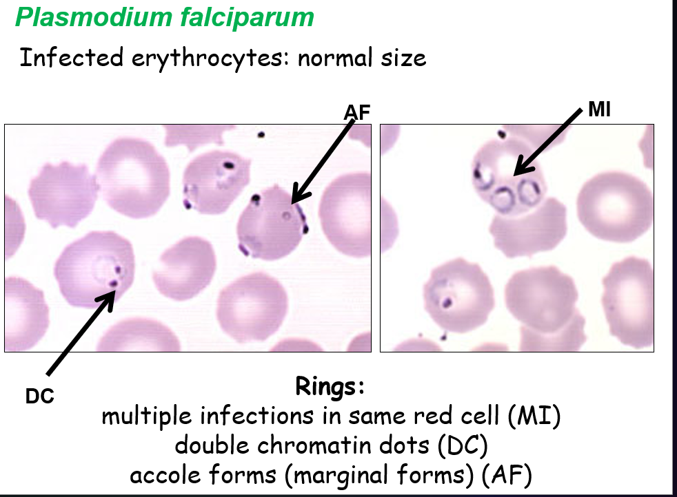
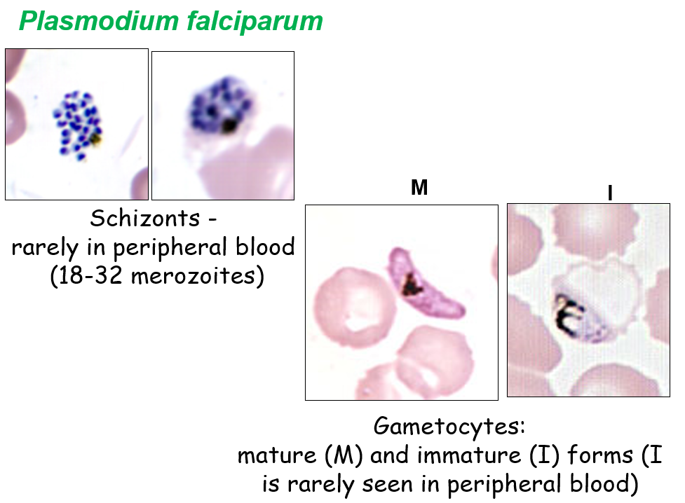
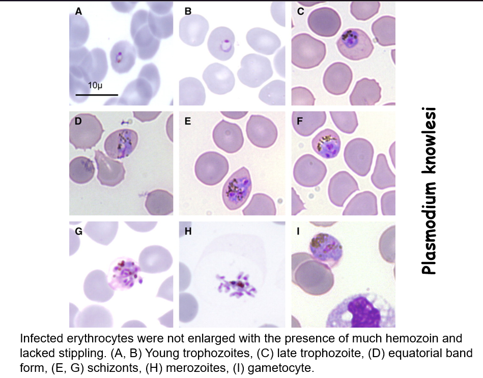
plasmodium parasite under microscope
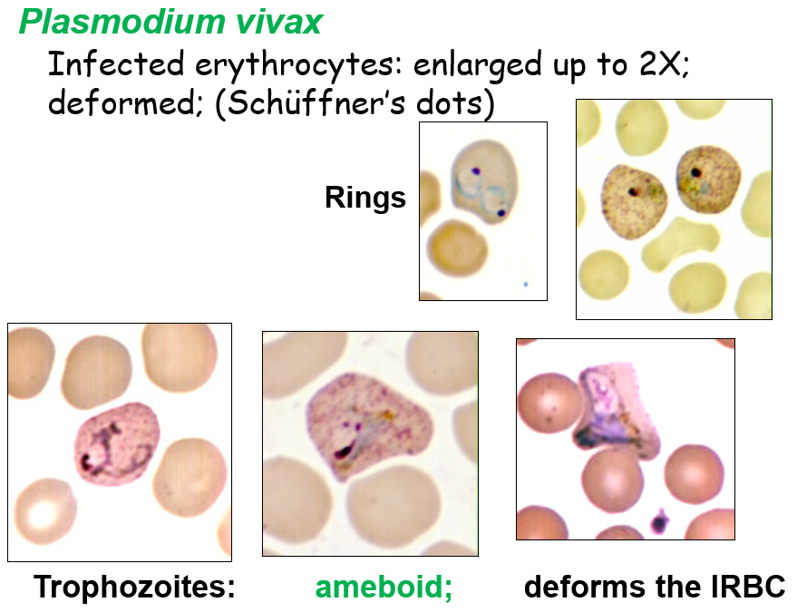
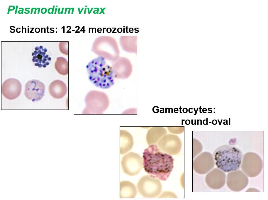
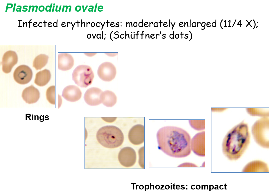
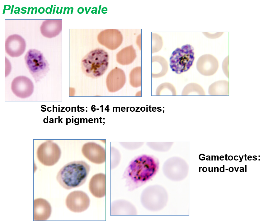
rapid diagnostics tests
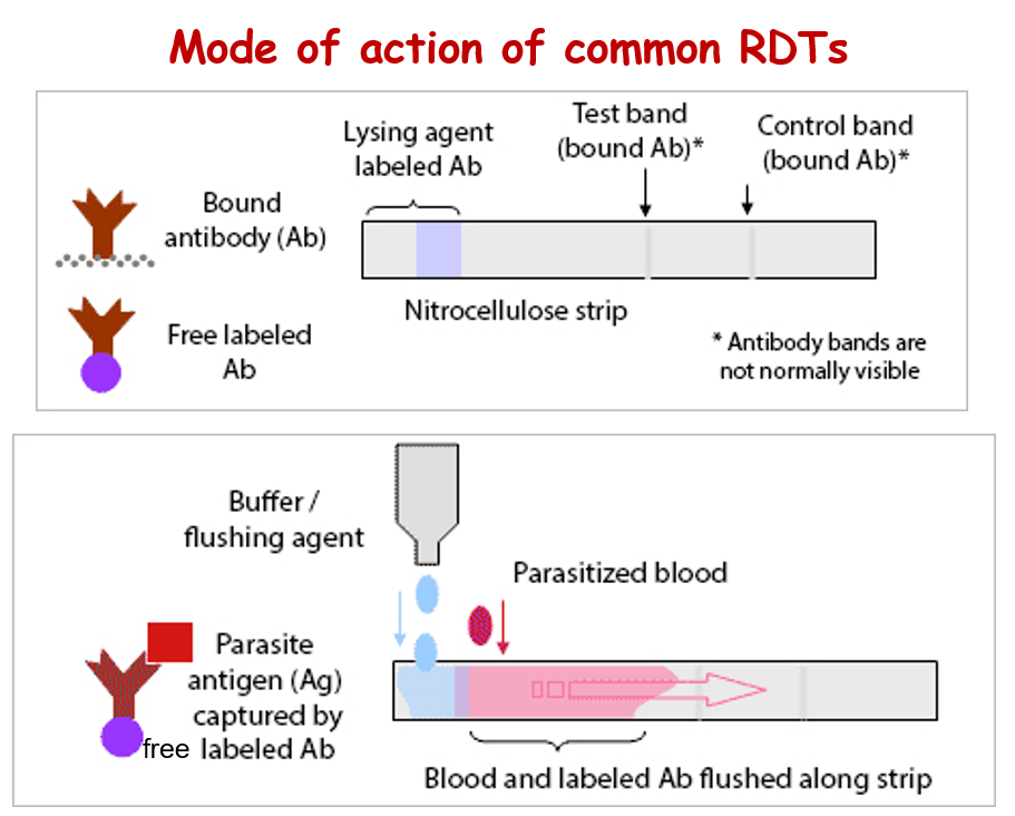
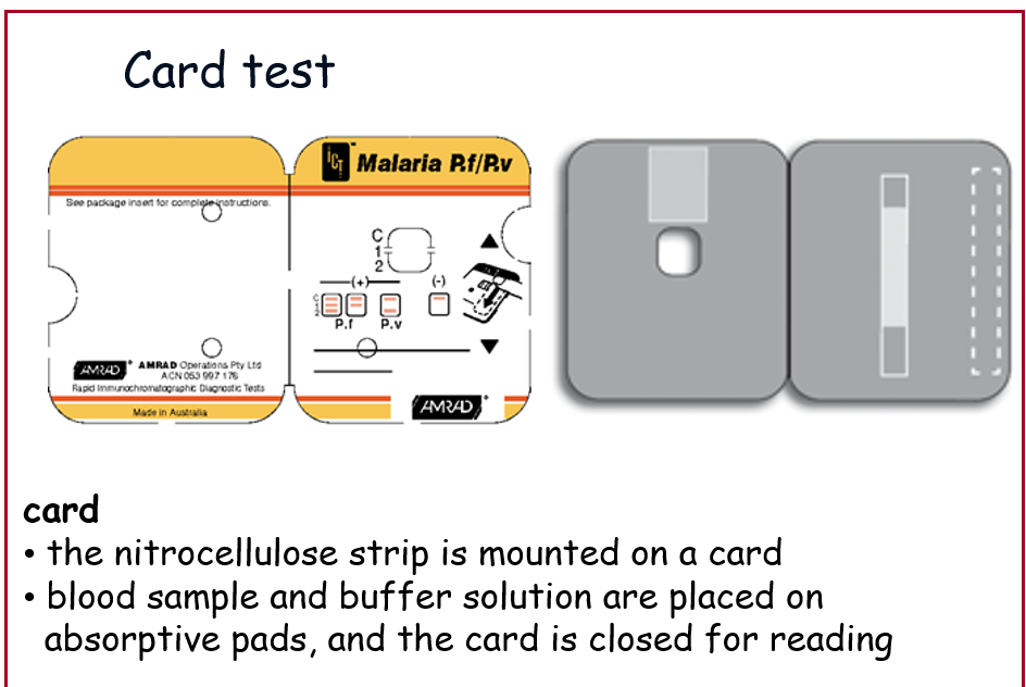
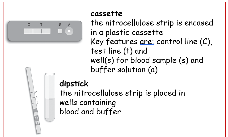
- RDTs detect the parasite specific antigens circulating in blood stream
- RDTs are based on the immunochromatographic lateral flow-strip technology
- several commercial test kits are currently available developed in different test formats
- dipstick, card, cassette
- RDTs
- highly specific
- no false positives - minimal cross reaction
parasite antigens mainly used in commercial RDTs
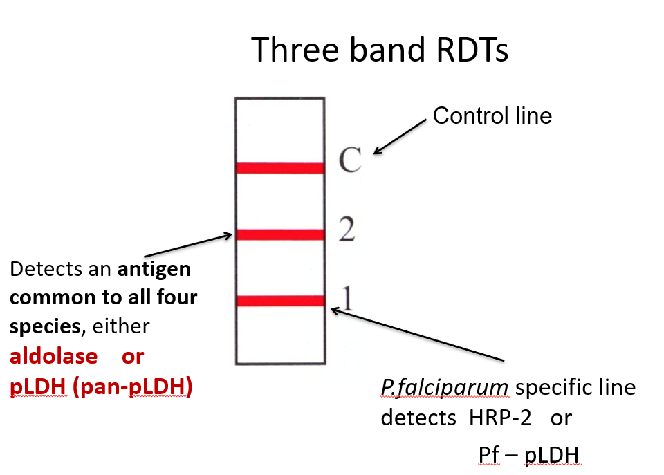
- Histidine-rich protein 2 (HRP-2)
- Parasite lactase dehydrogenase (pLDH)
- found in all 4 human malaria parasites (PAN malaria antigen)
- Aldolase
- all 4 human malaria parasites (PAN malaria antigen)
Histidine-rich protein 2 (HRP-2)
- unique to plasmodium falciparum
- produce by trophozoites and young gametocytes of plasmodium falciparum
- is a water-soluble Ag and found in circulating peripheral blood
- results will be given even the iRBCs are sequestrated
- may remain in the blood at least 28 days after the initiation of antimalarial therapy
- can give false positive even after parasite cleared from the blood
- certain plasmodium falciparum strains may not have the HRP2 antigens
Parasite lactase dehydrogenase (pLDH)
- produced by asexual and sexual stages of all 4 human malaria parasites
- different isomers of pLDH for each of the 4 species exist
- pLDH is produced only by live plasmodium parasites
- test can differentiate untreated from treated malaria
- commercial RDTs use
- pLDH specific for plasmodium falciparum
- pLDH specific for plasmodium vivax
- pLDH common for all human malaria parasites - pan LDH
plasmodium aldolase
- aldolase is an enzyme of the parasite glycolytic pathway
- it is expressed by the blood stages of all four human malaria parasites (PAN malaria antigen)
- commercial RDTs use Plasmodium aldolase (PAN malaria antigen) with PfHRP2
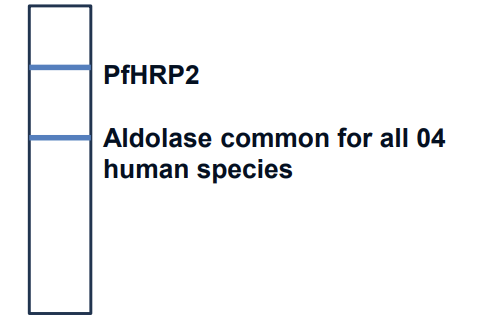
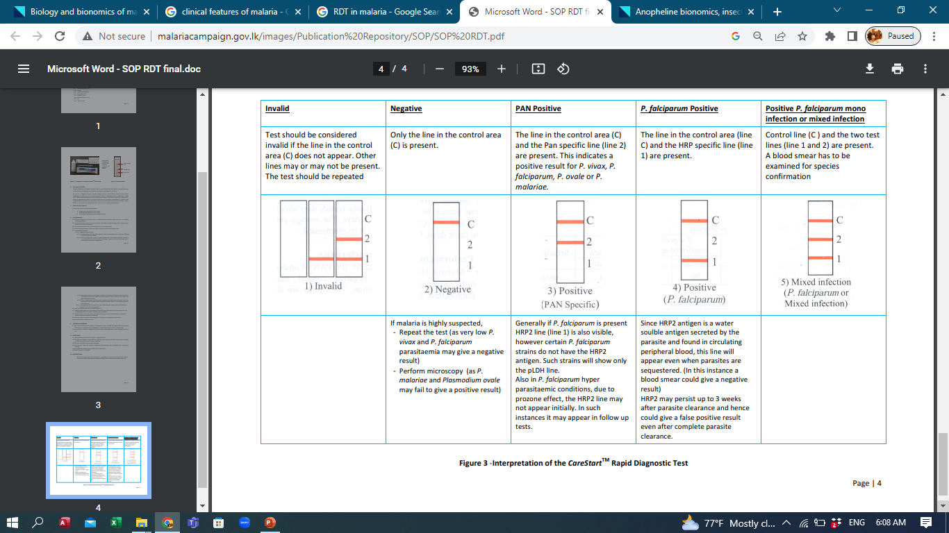
Three band RDTs
interpretation of care stat malaria RDT results
mode of action of common RDTs
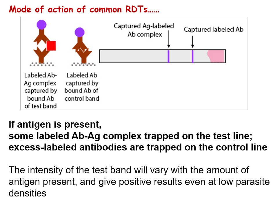
card & cassette / dipstick
molecular diagnostic methods
- use of molecular test for malaria is use primarily for research purposes of epidemiological studies
- it is limited for certain reference laboratories
- based on the detection of the parasite nucleic acid - PCR
- highly sensitive
- very effective in diagnosis of mix infections at low parasite densities that are not detectable by conventional microscopy or even RDT
Malaria Epidemiology
plasmodium species
- plasmodium falciparum
- responsible for almost all severe and complicated malaria cases
- > 98% mortality due to malaria
- most prevalent in Africa
- plasmodium vivax
- most widespread human malaria parasite outside Africa
- plasmodium ovale
- morphologically much similar to plasmodium vivax
- mainly confined to Africa
- causes a relatively mild, low-grade, infection
- it is commonly present in mixed species infections
- plasmodium malariae
- least severe infection with longer asymptomatic period
- plasmodium knowiesi
- infections should be treated aggressively as those infected with falciparum malaria
- plasmodium knowlesi may cause fatal disease
transmission of malaria
- primarily transmitted by the bite of an infected female anopheles mosquito
- by subcutaneous inoculation of sporozoites
- congenital
- blood transfusion
- through contaminated syringes of drug addicts
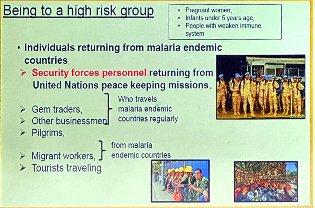
populations at risk
- infants,childrenunder5years of age
- pregnant women - specially primigravids
- patients with HIV/AIDS
- non-immune migrants to malaria endemic areas
- travellers to malaria endemic areas
when to suspect malaria
malaria in Sri Lanka
- malaria was moderately endemic in dry and intermediate zones (2/3 land areas) of the country
- plasmodium vivax and plasmodium falciparum was the most predominant species recorded in Sri Lanka
- vectors in Sri Lanka
- anopheles culicifacies - principal vector
- anopheles subpictus
- anopheles annularis
- anopheles varuna
- anti malari campaign was established in 1911
- malaria was declared as a notifiable disease in 1961
- Dichlorodiphenyltrichloroethane (DDT) insecticide was introduced in Sri Lanka in 1945 as an indoor residual insecticidal spray
- Sri Lanka started malaria eradication program in 1958
- Since October 2012 no indigenous / Iocally transmitted malaria cases were reported from Sri Lanka
- Sri Lanaka eliminated malaria in 2016
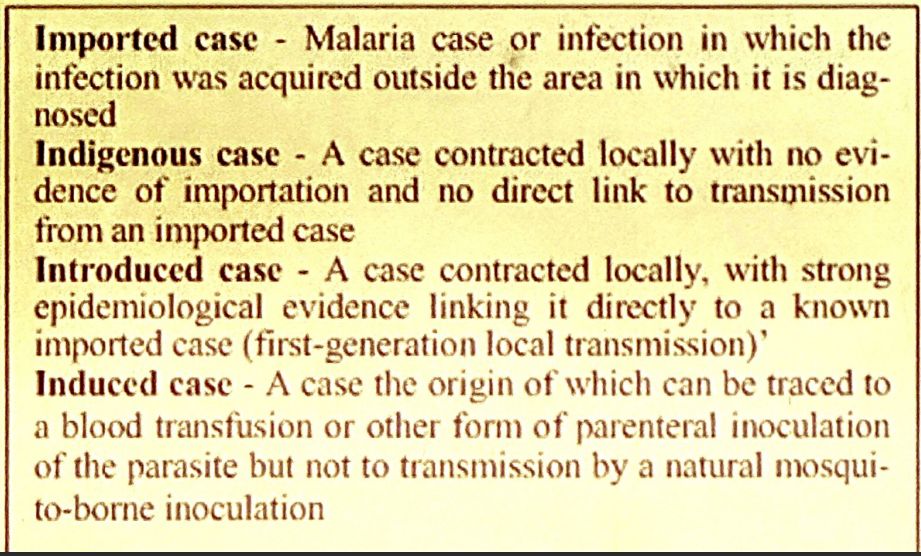
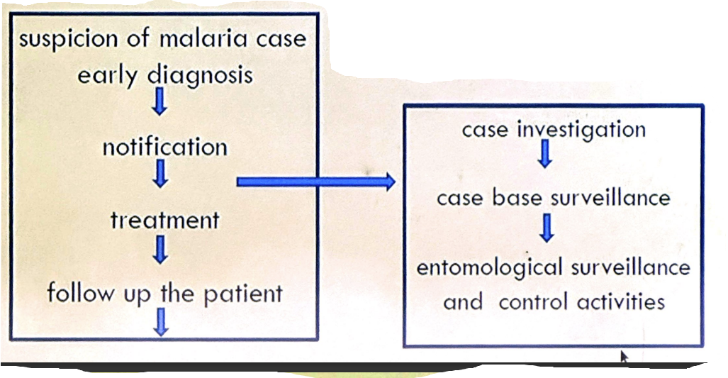
management of imported malaria cases
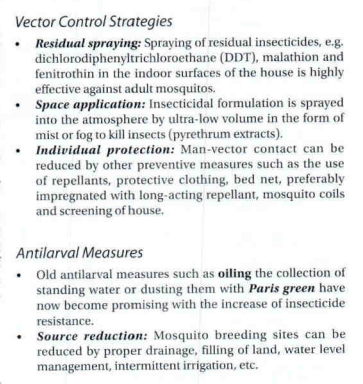
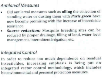
controlling malaria
Chemoproylaxis