subject guide notes
B2.1.1—Lipid bilayers as the basis of cell membranes
membranes are composed of:
lipids (phospholipids, glycolipids & sterols)
proteins
small amounts of carbs in the form of glycoproteins & glycolipids
*remember phospholipids are amphipathic
this forms a lipid bilayer
B2.1.2—Lipid bilayers as barriers
the hydrocarbon tails that extend inwards, are important for determining the permeability of the membrane
since the interior is non-polar & hydrophobic, then non-polar, hydrophobic & lipid-soluble substances (such as steroids) can pass through the bilayer
but ions cannot pass through
uncharged polar molecules (like glucose) are typically hydrophilic
the membrane is impermeable to them
small, uncharged molecules can pass through
so, polar & non-polar molecules can enter & exit cell
so basically, permeability depends on the molecule’s size & if its hydrophilic or hydrophobic
B2.1.3—Simple diffusion across membranes
example of diffusion is movement of non-polar substances (such as O2 & CO2) which are important in gas exchange
oxygen diffuses down its concentration gradient
will diffuse from the alveoli (high concentration) into the erythrocytes/RBCs in the capillaries (low concentration)
oxygen then carried to tissues
in tissues, O2 diffuses from erythrocytes (high concentration) to metabolically active cells (low concentration)
carbon dioxide also diffuses down its concentration gradient
diffuses from tissues (high concentration) into bloodstream (low concentration)
in lungs, diffuses out of blood (high concentration) into alveoli (low concentration)
*remember, due to the hydrophobic nature of the lipid bilayer, only non-polar substances (like O2 or CO2) or very small polar substances (like H2O & alcohol) can diffuse across membranes
B2.1.4—Integral and peripheral proteins in membranes
2 types - integral proteins & peripheral proteins
integral proteins - located in (embedded) into the lipid bilayer
are amphipathic
hydrophobic regions interact with hydrophobic interior of lipid bilayer
hydrophilic regions interact with the hydrophilic heads
most are transmembrane - extend across the membrane
others are only found on side of bilayer
peripheral proteins - typically found on surface of membrane
are hydrophilic (dont hv any hydrophobic regions)
function of membrane proteins
transport proteins - facilitate movement of molecules in & out of cell
includes channel proteins & carrier proteins
recognition - aid in cell-to-cell recognition
important, as it helps the immune system identify pathogens
receptors - functions as receptors for chemical signals & are binding sites for molecules like hormones
enzymes - show enzymatic activity & catalyse reactions
can also aid in cell adhesion to other cells or environment
B2.1.5—Movement of water molecules across membranes by osmosis and the role of aquaporins
water moves by osmosis from an area of low solute concentration (high water concentration) to an area of high solute concentration (but low water concentration)
osmosis continues until equilibrium is achieved (the solute concentration is same on both sides of membrane)
remember - membrane is impermeable to polar solutes
so water can pass through the membrane, just not that easily
aquaporins (type of channel protein) helps water move through the membrane
composed of 4 monomeric subunits & each subunit contains a water channel
they’re lined with specific hydrophilic side chains, allowing water to flow through but not ions
are bidirectional (meaning water can enter & exit the cell)
volume of water needed to be transported determines number of aquaporins
kidney cells hv more amounts of aquaporins as they need to reabsorb higher amounts of water
B2.1.6—Channel proteins for facilitated diffusion
transport proteins (channel proteins & carrier proteins) are important for facilitate diffusion, as they allow molecules (that are blocked by the cell membrane) to move down their concentration gradient
one example of channel proteins are ion channels
allow ions, like sodium & potassium, to move
are highly selective
different channels are needed for different ions (so, not just one channel allows the movement of all ions)
they’re also gated
so when they’re open, the ions can enter the pore & move down their concentration gradient
when they’re closed, there’s no movement of ions
selectivity of ion channels is due to:
the binding sites of the hydrophilic amino side chains that line the channels, are very ion-specific
the size of the pore acts as a filter
most open & close in response to certain stimuli, such as:
changes in voltage across the membrane (voltage-gated channels?)
binding of small molecules to channel proteins (ligand-gated channels?)
mechanical forces, such as pressure
B2.1.7—Pump proteins for active transport
active transport is involved when molecules are transported against their concentration gradient
requires energy, so it’s coupled with energy-releasing/exergonic reaction (like breakdown of ATP)
pump proteins use energy from ATP to transport these molecules against their concentration gradient
B2.1.8—Selectivity in membrane permeability
permeability by simple diffusion isn’t selective
meaning the permeability of the membrane depends on the size & hydrophilic or hydrophobic properties of the molecules
as long as a molecule fits the criteria, it can pass through membrane, regardless of its toxicity
but facilitated diffusion & active transport allow selectivity permeability of membrane
B.2.1.9—Structure and function of glycoproteins and glycolipids
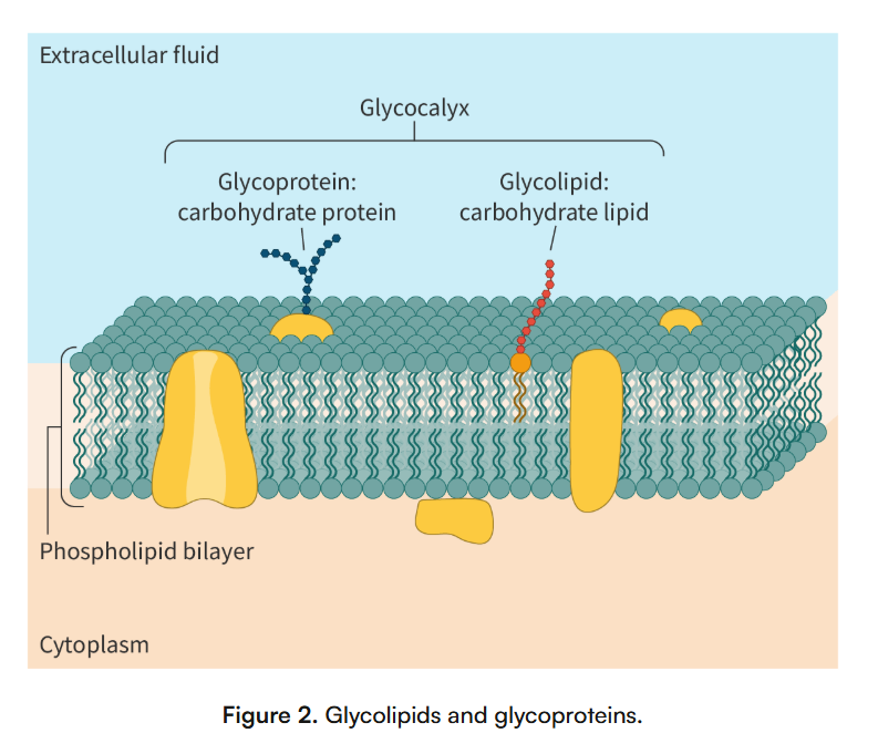
glycolipids - the product of covalent bonding between carbs & lipds
carbohydrate part is polar = extends into extracellular environment
lipid part is non-polar = embedded in membrane
based on their structure, can be classified into:
glycoglycerolipids or glycerol-based lipids
glycosphingolipids or derivatives of sphingosine
form hydrogen bonds with H2O molecules around cell = contribute to membrane stability
glycoproteins - is formed when there is covalent bonding between the oligosaccharides (short carbohydrate chains) & protein molecules
the carb molecules of glycoproteins often stick out into extracellular environment
function of glycolipids & glycoproteins
cell recognition - glycolipids & glycoproteins function as markers on cells surface, helping cells recognize each other
also help immune system cells to identify pathogens/foreign bodies
cell adhesion - both help cells attach to other cells & form tissues
CAMS (cell-adhesion molecules) are cell-surface glycoproteins that play an important role in cell adhesion
cell signalling - function as receptors for enzymes & other molecules, aiding in cell signalling (receiving & transmitting chemical signals)
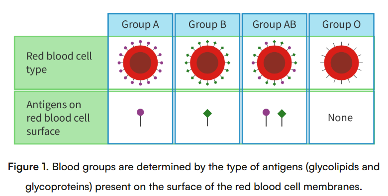
glycocalyx - is formed by the carb groups of glycolipids & glycoproteins
helps in cell signalling, cell adhesion & cell-to-cell recognition
B2.1.10—Fluid mosaic model of membrane structure
hv to be able to draw a a two-dimensional representation of the model and include peripheral and integral proteins, glycoproteins, phospholipids and cholesterol. They should also be able to indicate hydrophobic and hydrophilic regions.
B2.1.11—Relationships between fatty acid composition of lipid bilayers and their fluidity
the hydrocarbon tails of the phospholipid can be composed of saturated or unsaturated fatty acids
saturated fatty acids = higher melting point = provide stability to membrane (especially at high temps)
this is cuz they can fit together more tightly, making membrane more rigid
unsaturated fatty acids = low melting point = ensures fluidity of membrane
this is cuz of the kinks, which prevent the fatty acids from tightly packing together
saturated fatty acids also freeze more easily than unsaturated fatty acids
at low temps, phospholipid molecules come closer together
this decreases the fluidity of membrane
but the kinks prevent the phospholipids from coming too close together (they maintain spaces between the phospholipids)
this maintains the fluidity
cold-blooded organisms (such as frogs) adapt to low temps by increasing the proportion of unsaturated fatty acids in their phospholipid molecules
this maintains fluidity
also, when an animal hibernates, their body temp decreases
so the proportion of unsaturated fatty acids increases
B2.1.12—Cholesterol and membrane fluidity in animal cells
cholesterol is located in both parts of the phospholipid bilayer for the animal cell
at low temps, the cholesterol prevents the fatty acid chains of the phospholipids, from coming too closely together
this helps maintain membrane fluiditiy
at high temps, the cholesterol stabilizes the membrane, reducing fluidity
decreases permeability of membrane to ions & molecules
B2.1.13—Membrane fluidity and the fusion and formation of vesicles
Endocytosis is a process where particles are moved into cell
the cell membrane slowly invaginates the particles, then pinches to form a vesicle containing these particles
can be phagocytosis (ingesting large, solid molecules)
ex: seen in WBC’s
pseudopodia slowly surround the particles
pseudopodia eventually meets & engulfs the particle, forming a vesicle called a phagosome
the phagosome now fuses with a lysosome
digestive enzymes of lysosome digests particle, releasing nutrients
the particle is now digested within the phagosome
or pinocytosis (ingesting liquid)
forms smaller vesicles
Exocytosis - a process where particles are secreted out of cell
material to be removed in enclosed in vesicles
the vesicles then fuse with plasma membrane, releasing the particles
one example: glycolipids produced by endoplasmic reticulum & modified in Golgi apparatus
vesicles created by Golgi apparatus fuse with plasma membrane, releasing the contents to the outside
in exocytosis, as vesicles fuse with plasma membrane, lipids & proteins are added
in endocytosis, the reverse happens during invagination - idk wat this means
B2.1.14—Gated ion channels in neurons
examples of voltage-gated channels: voltage-gated sodium channels & voltage-gated potassium channels
a stimulus causes the voltage-gated sodium channels to open first
sodium ions enter the neuron
this causes interior of neuron to become more positively charged than exterior (depolarisation)
this creates an action potential, which travels down the neuron
voltage-gated sodium channels close & voltage-gated potassium channels open
potassium ions diffuse out of neuron, making interior of neuron less positive (repolarisation)
eventually these channels close
resting membrane potential now established
*note the voltage needs to reach a threshold value in order for the gated channels to open or close
also, voltage-gated channels experience channel inactivation - inactivation particle blocks channel pore
example of neurotransmitter-gated ion channel: nicotinic acetylcholine receptors (nAchR)
binding of acetylcholine molecules = conformational change in channel
allows sodium ions to enter cell
results in depolarisation (interior of cell becomes more positive)
enzyme cholinesterase then breaks down acetylcholine, leading to ion channels to close
resting membrane potential restored when voltage-gated potassium channels open, allowing potassium ions to leave cell
B2.1.15—Sodium–potassium pumps as an example of exchange transporters
energy released from the hydrolysis of ATP is used to drive the movement of sodium & potassium ions against their concentration gradient
how it works
initially, the pump is open to interior of cell
allows 3 NA+ to bind to its 3 binding sites
binding of sodium triggers hydrolysis of ATP
ATP is hydrolysed into ADP & a phosphate group
the phosphate group attaches to the pump, causing it to undergo a conformational change
pump opens to exterior, allowing NA+ to flow out
at same time, 2 K+ attach to their binding sites
causes phosphate group to detach from pump
pump undergoes conformational change again to regain its original form
opens to interior of cell
this pump then helps establish & maintain voltage across membrane
is important for re-establishing membrane potential
B2.1.16—Sodium-dependent glucose cotransporters as an example of indirect active transport
sodium ions bind to binding sites on outer surface of cotransporter
simultaneously, a molecule of glucose also binds to its binding site on cotransporter
results in conformational change that transports sodium ions & glucose molecule to interior of cell
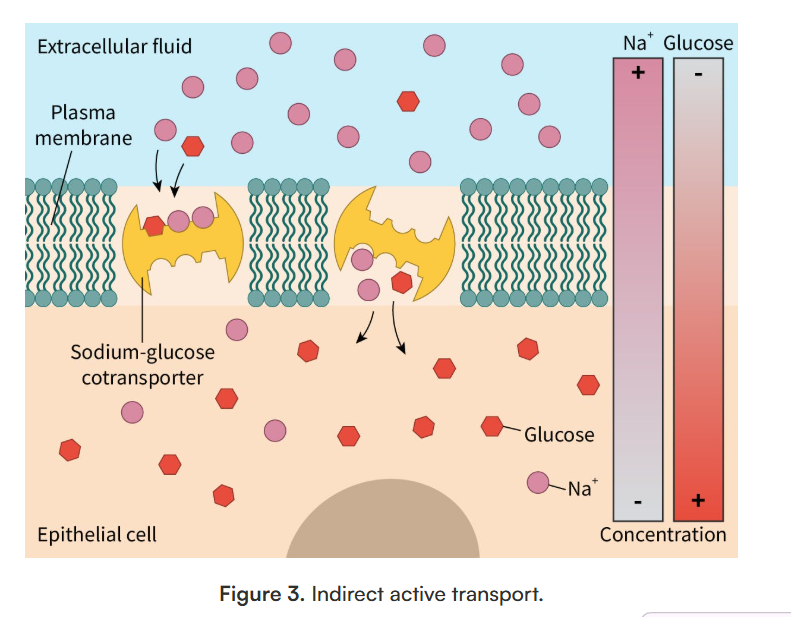
so sodium moves down its concentration gradient (area of high concentration (exterior) to an area of low concentration (interior))
but this movement allows the glucose to move against its concentration gradient (area of low concentration (exterior) to area of high concentration (interior))
B2.1.17—Adhesion of cells to form tissues
cell-adhesion molecules (CAMs) are glycoproteins that’re important for organizing binding of cells to other cells, or to extracellular matrix
different forms of CAMs are utilized for different types of cell-cell junction
main types of cell junctions:
adhesive junctions - present in epithelial cells & cardiac cells
often called anchoring junctions cuz they organize cell-cell adhesion in tissues to ensure structural stability & allow cells to withstand mechanical stress
tight junctions - are in epithelial cells
form tight seal between 2 neighboring cells
this limits unregulated movement of molecules across barrier
gap junctions - present in multiple types of cells
are intracellular channels that physically connect neighboring cells for movement of molecules
aid in cell-cell transfer of small molecules