Lymphatic System
Innate Immunity
Vocabulary
Immune system- The complex collection of cells and organs that destroys or neutralizes pathogens that would otherwise cause disease or death
Natural killer cell (NK)- is a circulating blood cell that contains cytotoxic (cell-killing) granules in its extensive cytoplasm.
Inflammatory response-The inflammatory response (inflammation) occurs when tissues are injured by bacteria, trauma, toxins, heat, or any other cause. The damaged cells release chemicals including histamine, bradykinin, and prostaglandins. These chemicals cause blood vessels to leak fluid into the tissues, causing swelling.
Histamine-a compound which is released by cells in response to injury and in allergic and inflammatory reactions, causing contraction of smooth muscle and dilation of capillaries.
Phagocytes- The phagocytes are the body’s fast acting, first line of immunological defense against organisms that have breached barrier defenses and have entered the vulnerable tissues of the body.
Lymphocytes- coordinate the activities of adaptive immunity
Macrophage- irregularly shaped phagocyte and is the most versatile of the phagocytes in the body.
Neutrophils- Neutrophils are a type of white blood cell that help heals damaged tissues and resolves infections.
Self Recognition- ability to distinguish between self-antigens, those that are normally present in the body, and foreign antigens, those that might be on a potential pathogen
Apoptosis- the death of cells which occurs as a normal and controlled part of an organism’s growth or development.
Introduction
Our immune systems offer us protection against a world full of pathogens. Our immune systems work by providing two types of immunity. In innate immunity, our bodies present the same kinds of defense systems for all types of pathogens. Innate immunity works much like a fence around your property. The fence does not differentiate between friend or foe. It keeps everyone out. The other type of immunity is known as adaptive immunity. Adaptive immunity produces an attack against a specific pathogen. This is much like having an attendant at the gate of the fence around your house. The attendant can identify potential foes and keep them out.
Before birth, the body inventories all of the cells and tissues of the body and classifies them as “self” cells. The presentation of non-self cells can then trigger the immune system. This is known as self-recognition.
The immune system is important in nursing since nurses care for patients with diseases that affect the immune system.
Innate Immunity
Pathogens are bacteria or viruses that enter the body and trigger an immune response. Some of the first lines of defense are the natural barriers of the body. These include the skin and mucous membranes. Skin acts as a physical barrier and mucous contains lysozymes that help to destroy pathogens. Salt and a lower pH also help to combat pathogens.
If pathogens enter the body a number of cells are activated to attack pathogens.
These are white blood cells that ingest pathogens called phagocytes. The process of ingesting pathogens is called phagocytosis. These cells include:
Neutrophils
Macrophages
Dendritic cells
Monocytes
Mast cells
Eosinophil
The process of ingesting pathogens is called phagocytosis.
Macrophages exist in many tissues of the body, including lymph nodes
They have different names, depending on the tissue: Kupffer cells in the liver, histiocytes in connective tissue, and alveolar macrophages in the lungs.
Neutrophils are the most common type of white blood cell in the body, making them a first line to fight infections.
The number of neutrophils in the blood will increase when a person is ill.
Neutrophil levels may decrease if a person has a long-term infection, cancer, an autoimmune condition, or is taking certain medications.
Phagocytic cells, and the cytotoxic Natural Killer cells recognize patterns of pathogen-specific molecules using pattern recognition receptors
Pathogens may also trigger the inflammatory response.
Inflammation is part of a very basic form of immune response which brings fluid and cells into the site to destroy pathogens and isolates the site, limiting the spread of the pathogen.
Inflammation is produced by the release of chemicals from white blood cells called mast cells. One of these chemicals is histamine.
Histamine which increases the diameter of local blood vessels (vasodilation), causing an increase in blood flow. Histamine also promotes capillary permeability causing fluids to leak out which produces swelling.
Other chemicals secreted by immune system cells involved in innate immunity include:
Interferon which inhibits viral replication.
Pyrogens which promote fever that helps to destroy pathogens.
The complement system is also involved in innate immunity.
The complement system includes a series of plasma proteins that can be activated by pathogens. The activated proteins can form complexes that attack pathogens.
Overall, the symptoms of an immune response can be:
Fever.
Chills.
Fatigue/loss of energy.
Headaches.
Loss of appetite.
Muscle stiffness
Adaptive Immunity
Vocabulary
B cells- immune cells that function primarily by producing antibodies.
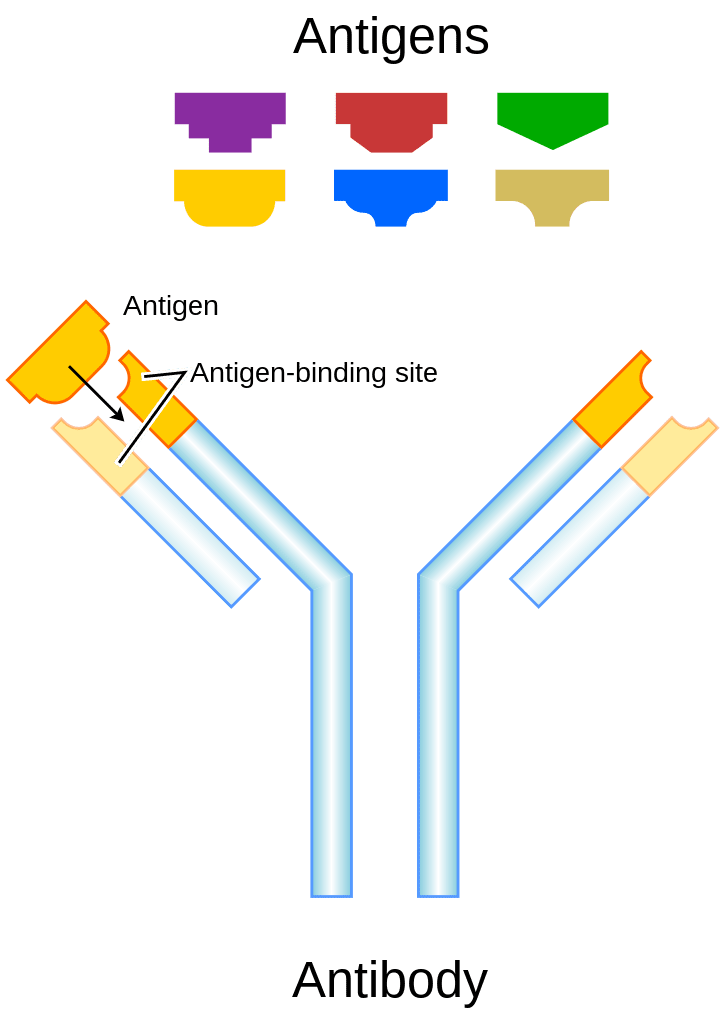
Antibody- is any of the group of proteins that binds specifically to pathogen-associated molecules known as antigens.
Lymphocytes- coordinate the activities of adaptive immunity
Dendritic cells- are antigen-presenting cells that link innate and adaptive immunity and are critical for the induction of protective immune responses against pathogens
Introduction
In addition to innate immunity, the human body has another important system for destroying pathogens called adaptive immunity. Adaptive immunity will produce a specific response to specific antigens on pathogens. Adaptive immunity can also remember the pathogen and exhibit a powerful immune response during subsequent infections of the same pathogens.
Adaptive immunity is a specific immune response to a pathogen which is triggered by the antigen on the surface of pathogens. Adaptive immunity is primarily controlled by white blood cells (leukocytes) known as lymphocytes, which help control immune responses.
Response to these antigens is unlimited and it can respond to nearly any pathogen.
Self Recognition is a feature of the adaptive immune response and is the ability to distinguish between self-antigens and foreign antigens. The immune system catalogues all of the body’s cells and tissues as self so that it only attacks non-self entities.
The adaptive immune response has a unique way to develop trillions of different receptors to recognize pathogens.
The immune system’s first exposure to a pathogen is called a primary response.
Symptoms of a first infection, called primary disease, are always relatively severe because it takes time for an initial adaptive immune response to a pathogen to become effective.
Upon re-exposure to the same pathogen, a secondary adaptive immune response is generated, which is stronger and faster that the primary response.
The secondary adaptive response often eliminates a pathogen before it can cause significant tissue damage or any symptoms.
This secondary response is the basis of immunological memory, which protects us from getting diseases repeatedly from the same pathogen. In other words, memory cells are produced that fully develop upon the next exposure to a pathogen.
The primary cells that control the adaptive immune response are the lymphocytes, the T and B cells. T cells only recognize antigen on the surface of specialized cells called antigen-presenting cells.
For example, a macrophage can ingest a pathogen and break it apart. The macrophage then displays the pathogen’s antigen on MHC or major histocompatibility complex proteins (MHCs).
Other cells such as T cells can then recognize the antigen and trigger the immune response.
The types of T cells that are activated during an immune response include:
Helper T cells (Th), bearing the CD4 molecule, function by secreting cytokines that act to enhance other immune responses.
Cytotoxic T cells (Tc) bear the CD8 molecule and kill target cells by inducing apoptosis using the same mechanism as Natural Killer (NK) cells.
Activated T-cells also produce memory cells that remain inactive until the next time the pathogen appears. They then activate quickly to help destroy the pathogen.
B-cells are also activated via adaptive immunity. B-cells secrete antibodies which disable and destroy pathogens.
Antibodies are also known as Immunoglobulins. There are 5 classes of antibodies which include : IgM, IgD, IgG, IgA, and IgE.
IgG is the largest category and accounts for 80% of all antibodies. IgG antibodies attack viruses and bacteria. IgE antibodies function in allergic reactions. They facilitate the release of histamine and heparin from basophils. IgD antibodies bind to antigens on the surface of B-cells and help in activation of B-cells. IgM antibodies work with IgG antibodies to form immune complexes. IgM antibodies are also resident in plasma as anti-A and anti-B antibodies. IgA is found in secretions such as tears, mucous and saliva and attack pathogens.
Dendritic cells are antigen presenting cells found in mucous membranes, lymphatic organs and the epidermis of the skin. These cells have a branched appearance and can engulf pathogens by way of endocytosis. Dendritic cells contain receptors that recognize non-self antigens that trigger endocytosis when activated.
Reticular cells (sometimes called fibroblastic reticular cells) are antigen presenting cells located in lymphatic organs. These cells are known to help regulate T-cell function.
Immunity can be passive or active and naturally or artificially acquired.
Active immunity is characterized by production of antibodies by the infected person.
Passive immunity is when antibodies come from an outside source.
Naturally acquired immunity occurs from a natural exposure to a pathogen that triggers an immune response. For example, getting a virus and then developing immunity against the virus.
Artificially acquired immunity results from a non-natural exposure to a pathogen. For example, a vaccine triggers an immune response. This would then be artificially acquired active immunity.
Artificially acquired passive immunity would be the administration of antibodies for patients who have damaged immune systems.
Naturally acquired passive immunity would be a fetus or baby getting antibodies from the mom either during fetal development or by way of breast feeding.

Immune System Diseases
Vocabulary
Allergy- an immune response to an inert substance.
HIV- Human Immunodeficiency Virus. A virus that can trigger Acquired Immunodeficiency Syndrome (AIDS).
Introduction
Diseases and disorders of the immune system include deficiencies in the immune response whereby the immune system can exhibit an inadequate response or a response that is over reactive. It is important for nurses to know how to identify some important immune system disease along with general signs of an immune system response.
Allergies
Allergies are characterized in an immune response to an inert substance. The immune system exhibits an overactive response. The substance triggers an immune response that can range from mild to life-threatening.
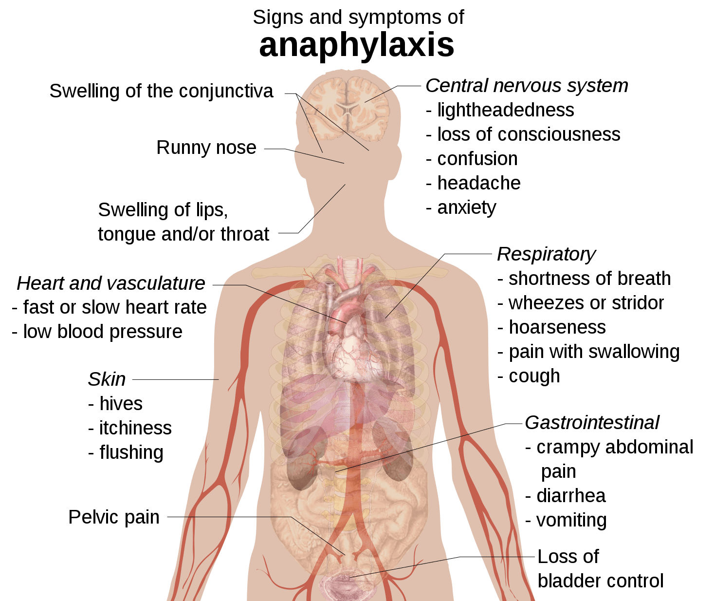
The most severe allergic reaction is the anaphylactic reaction. In this reaction, the B-cells overproduce the IgE antibodies which can trigger mast cells to release substances that cause systemic inflammation. The most common triggers for anaphylactic reactions are penicillin based drugs, food such as peanuts, shellfish and insect stings.
These reactions can be a life threatening medical emergency.
The symptoms include:
Swelling, which may cause swallowing and breathing difficulties.
Shortness of breath.
Hives
Difficulty swallowing
Red rash
Abdominal (belly) pain
Chest tightness
Cramps
Diarrhea
Feeling of doom or dread
Vomiting
Wheezing
HIV
HIV or Human Immunodeficiency Virus is the virus that can cause AIDS or Acquired Immunodeficiency Syndrome. AIDS works to destroy the immune system by wiping out helper T-cells.
Without helper T-cells, other T-cells and B-cells fail to activate.
A person with HIV is considered to have progressed to AIDS when:
The number of CD4 cells falls below 200
Someone with a healthy immune system, CD4 counts are between 500 and 1,600
Without helper T-cells, other T-cells and B-cells fail to activate.
Autoimmunity
In autoimmunity, the person’s own immune system attacks the body.
Examples of autoimmune disorders include:
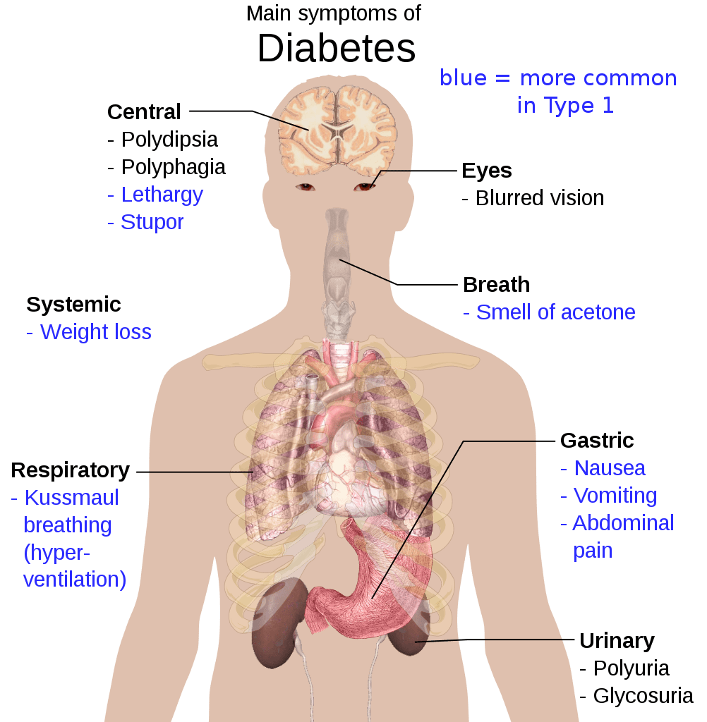
Type 1 Diabetes
Rheumatoid arthritis
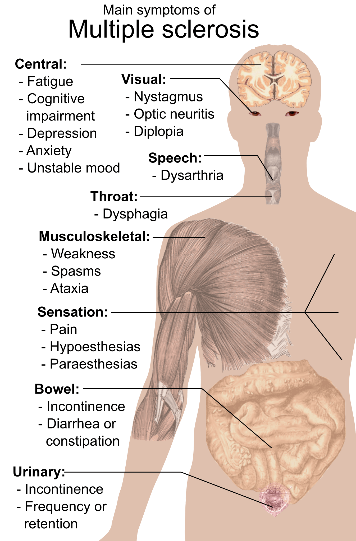
Multiple sclerosis
Lymphatic System
Vocabulary
Lymphatic system- The system of vessels, cells, and organs that carries excess fluids to the bloodstream and filters pathogens from the blood.
Lymph nodes- small, hollow structures that function to remove debris and pathogens from the lymph, and are thus sometimes referred to as the “filters of the lymph.”
Right Lymphatic Duct-one of the major connections between the lymphatic and circulatory systems at the right subclavian vein.
Thoracic Duct- one of the major connections between the lymphatic and circulatory systems at the left subclavian vein.
Introduction
The lymphatic system plays important roles in returning fluid to the circulatory system, transporting dietary fats and in immunity. The lymphatic system works much like a circulatory system that transports its own fluid called lymph through a complex network of vessels and lymph nodes that contain white blood cells to help fight off pathogens.
Lymphatic System
The lymphatic system is a vascular system that contains capillaries, vessels and lymph nodes. The lymph capillaries pick up interstitial fluid lost by the circulatory system. The fluid known as lymph moves through the system and is returned to venous circulation.
The lymphatic system also transports dietary fats from the gastrointestinal system. Small lymphatic structures called lacteals are located in the small intestine in structures called villi. Fats are broken down and packaged as structures known as chylomicrons. The fats then move through the system to the venous circulation.
A good portion of the immune system resides in the lymphatic system as well. Lymph nodes containing white blood cells work to destroy pathogens.
Lymphatic Capillaries
Lymphatic capillaries are distributed throughout the interstitium. Lymphatic capillaries are not found in the central nervous system and bone marrow. They are also not resident in tissues without blood flow such as the epidermis or cartilage. They are designed to allow one way fluid flow into the capillary.
Lymphatic capillaries consist of overlapping simple squamous epithelium. They also form one way valves. This arrangement allows for increased permeability and fluid movement toward the venous circulation.
The lymphatic capillaries form larger structures called lymphatic vessels. The vessels have a similar structure to veins and contain three layers. The three layers consist of an inner endothelium, a middle smooth muscle layer and an outer layer of thin fibrous connective tissue.
Lymphatic Vessels
Lymphatic vessels also contain valves to allow the one way flow of blood. Smooth muscle contraction moves blood from one area separated by a valve to another. Some cells in the lymph vessel walls are capable of generating action potentials that cause the smooth muscle to contract.
Skeletal muscle contraction also moves lymph fluid by means of generating pressure on the outside of the vessels causing them to constrict. The valves only allow one way flow so lymph is moved toward the venous circulation.
Lymph fluid also moves into vessels in the thoracic cavity as a result of dilation of lymph vessels resulting in decreases in thoracic cavity pressure. The thoracic cavity expands during inspiration causing a decrease in thoracic pressure. The vessels react by dilating and creating an area of lower pressure. Lymph fluid then moves toward the area of lower pressure.
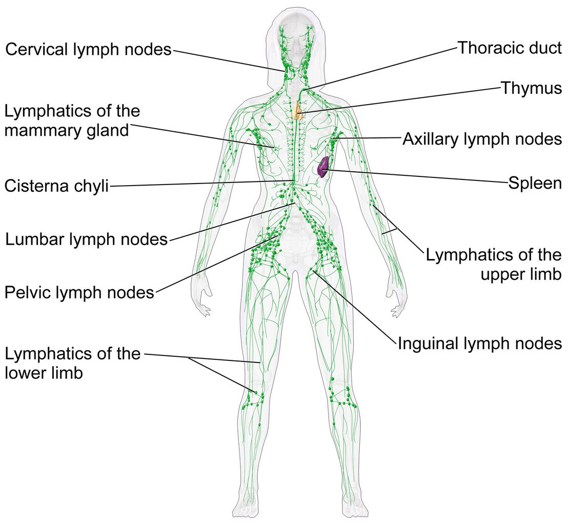
There is an asymmetrical distribution of lymph drainage. The right lymphatic duct drains the right side of the head, neck, and trunk. The thoracic duct drains the remainder of the body.
Lymph Nodes
Lymph nodes are located throughout the lymphatic system. The lymphatic vessels connect with the nodes and fluid moves through them. There are numbers of nodes connected to a vessel so that lymph fluid moves from one node to another. Lymph nodes are small oval structures and are generally not felt during examinations unless enlarged or calcified.
Lymph nodes act as filters and work to remove pathogens such as bacteria and viruses. Although diffusely located throughout the body, lymph nodes tend to conglomerate in certain areas. These include the cervical, axillary, inguinal, popliteal and mammary glands.
Lymph nodes consist of a dense connective tissue covering and a trabeculated internal structure. The nodes contain reticular connective tissue that forms an interconnected web like structure. Vessels entering the nodes are known as afferent vessels. Likewise vessels exiting the nodes are known as efferent vessels.
Lymph nodes consist of an outer cortex and an inner medulla. The cortex contains open areas called sinuses. The medulla contains medullary cords which are branching structures of lymphatic tissue. Open areas called medullary sinuses are also present.
Lymph nodes contain white blood cells called macrophages and lymphocytes. Macrophages are located in the sinuses and phagocytize bacteria and debris. Lymphocytes are located in germinal centers and when activated can move into the bloodstream.
Lymphatic Organs
Two organs are associated with the lymphatic system. These are the spleen and the thymus. The organs contain lymphatic tissue consisting primarily of white blood cells known as macrophages and lymphocytes as well as some other types of cells. There are two general types of lymphocytes. These are the T and B lymphocytes. Both are produced in the bone marrow and carried to the lymphatic system. Activation of the immune system causes these cells to divide and attach pathogens.
Lymphatic tissue also contains reticular cells that produce reticular fibers. White blood cells connect with these fibers so that fluid moving through the tissue is exposed to the cells. The white blood cells can then destroy bacteria and debris.
Lymphatic tissue resides throughout the lymphatic system. When it is not located in a lymph node or organ such as in the mucous membranes of the digestive, urinary, respiratory and reproductive systems it is known as Mucosa associated lymphoid tissue (MALT). The tonsils are another example of MALT.
The spleen is located in the left upper quadrant of the abdominal area generally close to the diaphragm and is about as large as an adult fist. It consists of an outer connective tissue capsule. The inner portion has a trabeculated structure containing areas of red and white pulp. The spleen also contains venous sinuses.
White pulp consists of lymphatic tissue associated with arteries within lymphatic organs. Red pulp contains both white and red blood cells and is associated with veins.
The splenic artery and vein enter and exit the spleen at the hilum. Blood flows into the spleen and through the trabeculated network. The cells in the spleen work to destroy pathogens. Lymphocytes in the spleen can react to pathogens and trigger the immune system. The spleen also acts as a blood reservoir.
Examine the lymphatic system in the diagram below.