Skeletal Muscle Disorders
Key Terms
Atrophy → decrease in muscle fibre size.
Hypertrophy → increase in muscle fibre size.
Sarcopenia → age-related loss of muscle mass.
Cachexia → severe muscle/fat wasting caused by chronic disease.
Muscular dystrophy → inherited disorders causing progressive muscle fibre damage/loss.
Satellite cell → muscle stem cell involved in repair and regeneration.
Eccentric contraction → muscle produces force while lengthening.
Sarcomere → basic contractile unit of muscle.
Dystrophin → protein that helps stabilise the muscle fibre membrane.
CK → enzyme released into blood when muscle is damaged.
Proximal → closer to the body's centre.
Distal → further from the body's centre.
Myotonia → delayed muscle relaxation after contraction.
Fasciculation → involuntary muscle twitch.
Spasticity → abnormal increased muscle stiffness/tone associated with UMN damage.
Cardiomyopathy → disease of the heart muscle.
Cytokine → signalling protein involved in communication/inflammation.
Anabolic → promoting tissue growth/building.
Fibrosis → formation/accumulation of scar tissue.
Orthosis → supportive device used to help control/support a body part.
Gene therapy → treatment that aims to alter or replace genetic material.
Eccentric exercise - the muscle is producing force while it is lengthening
Skeletal Muscle Structure
Micronuclei - On the perimeter of healthy muscle fibres
Structure
Have 2 ends, usually attached to bones via tendons
Epimysium - connective tissue surrounding each muscle
Fascicles - organised by Epimysium forming bundles of muscle cells
Perimysium - connective tissue that separates individual fascicles
Myofiber - Individual muscle cells
Endomysium - connective tissue surrounding connective tissue
3 connective tissues bind muscle cells together and are continuous with tendons
Features of skeletal muscle cells
Sarcolemma - plasma membrane of skeletal muscle
T-Tubule - invagination of sarcolemma, that extents deep into muscle fibre
Sarcoplasmic reticulum - ER of muscles, surrounds each myofibril, ends near T-tubule region called terminal cisternae
Triad - Region where 1 T-Tubule is flanked by 2 terminal cisternae
Myofibril - Bundle of contractile filaments within muscle fibre
Nucleus, mitochondria, cytosol.

Organisation of a Myofibril
Actin - thin filament
Myosin - thick filament
A Band - Quite dark, length of thick filaments
I Band - very light region - with thin but no thick filaments
length decreases during contraction.
Z line - bisects I band. protein disc onto which thin filaments attach
H zone - region where only thick filaments present. length decreases during contraction
M line - Middle of A band. Region where thick filament attach.
Sarcomere - unit of contraction - region between 2 Z lines
Sliding Filament Theory
Components
ATP
Ca2+
Myosin
Actin
Tropomyosin
Troponin
Myosin
thick filaments
Globular head and long tail
Tails or 2 myosin’s wind each other
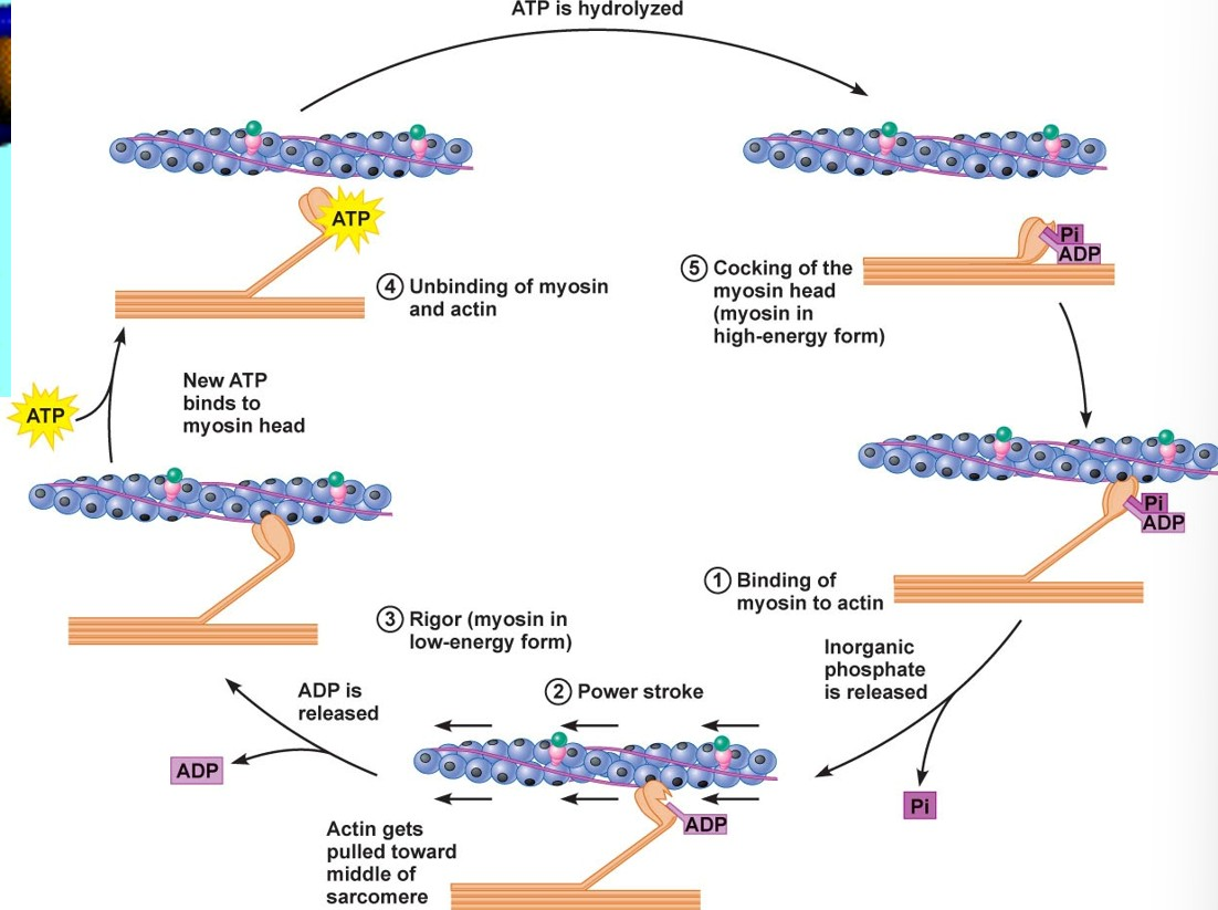
ATP binds at low energy state
ATP hydrolysed to ADP + P1
energy transferred to myosin - high energy state
Process
Thin filaments made of Actin, Tropomyosin and Troponin.
Major component is Actin. Filament organisation-two chains composed of actin monomers wound around each other.
Each Actin monomer has single Myosin binding site (MBS) on external surface.
MBS on Actin is normally covered by the protein Tropomyosin.
Troponin found on thin filaments.
Ca2+ binds Troponin which moves Tropomyosin off the MBS on the Actin molecules.

Muscle fibre types
Slow twitch (type I) - red
small motor unit innervation, smaller, easily excitable, aerobic, high capillary density, high myoglobin, many mitochondria, highly fatigue resistant
Fast twitch (type II) - white
Type IIX/IIB
large motor unit innervation, relatively unexcitable, high glycogen, low myoglobin, extensive sarcoplasmic reticulum, highly glycolytic, low fatigue resistance
Type IIA
intermediate properties but aerobic with high myoglobin
Muscle Plasticity
Muscle plasticity - the ability of skeletal muscle to change its structure and function in response to different demands
Cross-innervation
Different types of muscle fibres can change their characteristics depending on the type of motor neuron supplying them.
Slow-twitch (Type I) fibres → suited to prolonged, aerobic activity and are fatigue-resistant.
Fast-twitch (Type II) fibres → suited to producing high force but fatigue more quickly.
The nervous system therefore plays an important role in determining muscle fibre characteristics.
Training
High repetition + low force
Associated with adaptations useful for endurance.
Increases/maintains features such as:
Mitochondria
Capillaries
Fatigue resistance
Low repetition + high force
Produces adaptations associated with strength.
Increases fibre cross-sectional area (CSA) and strength.
CSA = cross-sectional area
→ essentially the size of the muscle fibre when viewed in cross-section.
Muscle Damage and Repair
Eccentric exercise - the muscle is producing force while it is lengthening
Lifting the bag up → your bicep shortens while working = concentric contraction.
Lowering the bag slowly → your bicep is still working, but it gets longer as the bag comes down = eccentric contraction.
Eccentric contractions can create tiny microscopic tears in muscle fibers and the surrounding connective tissue.
Satellite cells - muscle stem cells located around muscle fibres that help with muscle growth and repair.
After muscle damage:
Muscle fibres are damaged.
Immune cells enter the damaged area.
Satellite cells become activated.
They contribute to repair/regeneration of muscle fibres.
Newly regenerated fibres can initially have centrally located nuclei.
Healthy mature skeletal muscle fibres normally have their nuclei towards the periphery, so centrally nucleated fibres can be a sign of regeneration after damage.
Muscle loss
Muscle loss occurs in many conditions, including:
Metabolic diseases
Type II diabetes
Alcoholic liver disease
COSarcopenia = loss of muscle mass during ageing.PD
Ageing
Cancer
Muscular dystrophy
Muscular atrophy
Muscle atrophy
Atrophy - reduction in the size of muscle fibres.
This is different from muscle loss caused by destruction of muscle fibres
Muscular dystrophy
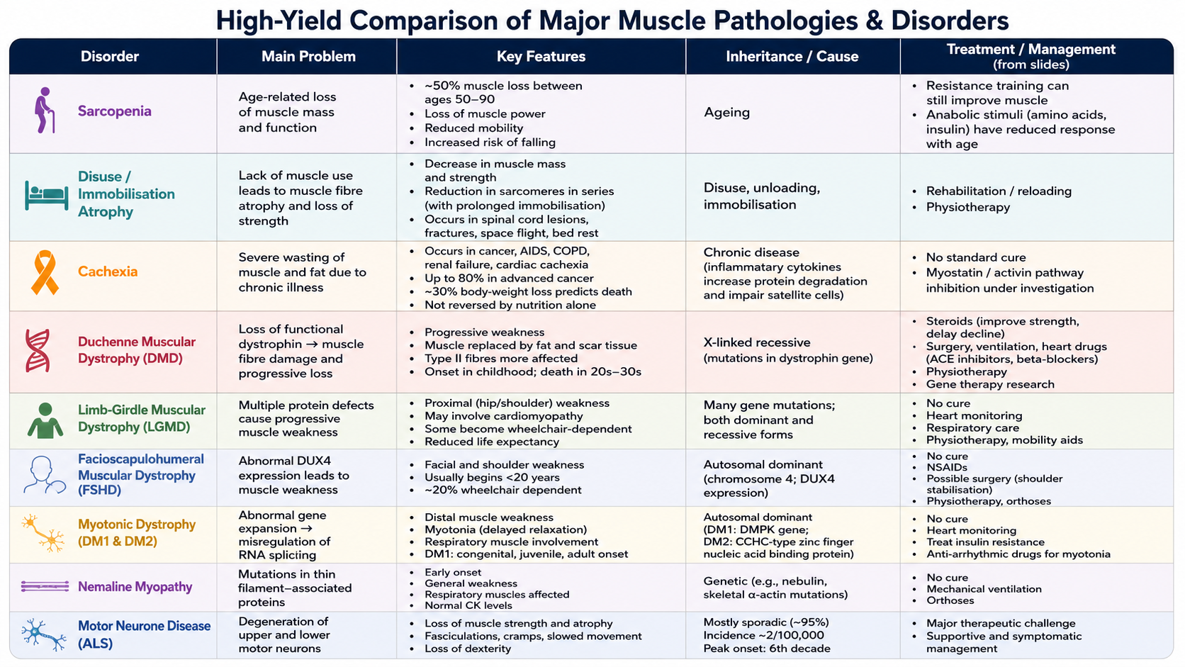
Muscular dystrophy is a group of inherited genetic disorders characterized by progressive muscle weakness and muscle wasting due to degeneration of muscle fibers.
Sarcopenia
Sarcopenia - loss of muscle mass during ageing.
Approximately 50% muscle loss between ages 50–90.
Loss of muscle power.
Reduced mobility.
Increased risk of falling.
Causes
Motor neuron loss
Motor neuron death mainly affects fast-twitch motor units.
This causes fast-twitch fibre atrophy.
Remaining fibres can become re-innervated by slow-twitch motor neurons.
2. Reduced response to anabolic stimuli
Anabolic = promoting growth/building of tissue.
The ageing muscle has a reduced response to:
Amino acids
Insulin
Resistance training
3. Satellite cell changes
Satellite cells become less responsive.
They may have a reduced ability to self-renew.
There may be reduced expression of growth factors.
4. Reduced force production
Even an individual muscle fibre may produce less force.
Possible reasons include:
Fewer cross-bridges
Oxidation of the ryanodine receptor
Ryanodine receptor = a protein involved in releasing calcium from the sarcoplasmic reticulum, which is essential for muscle contraction.
Disuse and immobilisation
Muscle mass and strength can be lost through muscle fibre atrophy during:
Spinal cord lesions
Immobilisation after a bone fracture
Long-term space flight
Long-term bed rest
Sarcomere loss
A reduction in the number of sarcomeres in series can occur with:
Cerebral palsy/spasticity
Walking on high heels
Immobilisation after a bone fracture
Cachexia
Cachexia = severe wasting of the body, particularly muscle and fat, caused by chronic illness
Examples include:
Cancer
AIDS
COPD
Renal failure
Cancer cachexia
It can affect up to 80% of patients with advanced cancer.
Around 30% body-weight loss is a strong predictor of death.
It contributes to around 30% of cancer-related deaths.
Wasting of the respiratory muscles can contribute to death.
It is not reversed by conventional nutritional support.
Possible mechanism
Tumour/inflammatory cells release cytokines.
Cytokines = signalling proteins released by cells that can influence inflammation and other cellular processes.
These cytokines can:
→ increase muscle protein degradation
→ cause muscle wasting
→ impair satellite-cell function
Myostatin
Myostatin (MSTN) - a protein that normally limits muscle growth.
Animals/humans with reduced myostatin activity can have:
Increased muscle mass
Reduced adipogenesis
Adipogenesis = formation of fat cells.
Therefore, inhibiting myostatin was considered as a possible treatment strategy for:
Muscular dystrophy
Muscle atrophy
Metabolic conditions
Cachexia
Sarcopenia
Activin receptor type 2 inhibition
The idea is to interfere with signalling from:
Myostatin
Activin
This could potentially reduce signals that inhibit muscle growth.
Duchenne muscular dystrophy — DMD
Pathology
DMD involves:
Progressive loss of muscle fibres
Replacement of muscle with:
Fat
Scar tissue
Progressive decline in muscle function
DMD is caused by:
Mutations in the dystrophin gene
X-linked recessive inheritance
X-linked recessive - the disease-causing gene is on the X chromosome and typically affects males much more frequently.
Epidemiology
Approximately 1 in 3,500 live male births.
High spontaneous mutation rate.
Death typically occurs in the 20s or early 30s according to the lecture.
Diagnosis
Clinical history
Creatine kinase (CK) levels
Genetic testing
Muscle biopsy
CK = creatine kinase, an enzyme released into the blood when muscle fibres are damaged.
DMD treatment
Current conventional therapies listed in the lecture include:
Steroids
Surgery
Ventilation
Heart drugs:
ACE inhibitors
Beta-blockers
Physiotherapy
Glucocorticoid steroids
These are used chronically in DMD.
The lecture states that they can:
Increase strength
Prolong ambulation
Delay loss of ambulation
Reduce the need for spinal fusion
Treatment is generally initiated around 4–6 years
Side effects can include:
Behavioural changes
Weight gain
Impaired glucose tolerance
Growth suppression
Excessive hair growth
Adrenal suppression
Muscle loss
Dystrophin
Dystrophin - an important protein that helps stabilise the muscle fibre membrane during contraction.
Why is dystrophin important?
Muscle fibres experience mechanical stress every time they contract.
Dystrophin helps the membrane withstand this stress.
Without functional dystrophin:
→ membrane becomes more vulnerable to damage
→ muscle fibres become damaged
→ fibres progressively degenerate
→ muscle is replaced by fat/scar tissue
→ weakness develops.
Limb-girdle muscular dystrophy — LGMD
Limb-girdle muscular dystrophy (LGMD) - a group of muscular dystrophies characterised particularly by weakness around the hips and shoulders.
Many different gene mutations can cause LGMD.
There are both dominant and recessive forms.
Prevalence : approximately 20–40 per million.
Can begin in childhood or adulthood.
Causes proximal muscle weakness.
May involve cardiomyopathy.
Some patients may become wheelchair-dependent.
Life expectancy may be reduced.
Proximal = closer to the centre of the body, e.g. shoulder and hip muscles.
Diagnosis
History
CK levels
Genetic testing
Biopsy
Management includes:
Heart monitoring
Respiratory care
Physiotherapy
Mobility aids
Facioscapulohumeral muscular dystrophy — FSHD
Facioscapulohumeral muscular dystrophy (FSHD) - a muscular dystrophy particularly affecting the face and shoulder muscles.
Key facts
Mutation involving a region of chromosome 4
Associated with abnormal DUX4 expression
Autosomal dominant inheritance
Prevalence approximately 1:15,000
Usually begins before age 20
Facial and shoulder muscle weakness
Around 20% wheelchair dependent according to the lecture
Diagnosis
Clinical history
CK levels
Genetic testing
Sometimes biopsy
Management includes:
NSAIDs
Possible surgery to stabilise shoulder blades
Physiotherapy
Orthoses
Orthosis = a device used to support or control part of the body, e.g. an ankle/foot support.
Myotonic dystrophy
Myotonic dystrophy - an inherited muscle disorder involving muscle weakness and myotonia.
Myotonia - delayed relaxation of a muscle after it contracts.
There are two main forms discussed:
DM1
DM2
Cause
Both involve an abnormal gene expansion.
This causes misregulation of RNA splicing.
Clinical features
Weakness of distal muscles
Respiratory muscle weakness
Myotonia
Nemaline myopathy
Nemaline myopathy = a muscle disorder involving abnormal proteins associated with the muscle's thin filaments.
Cause
Mutations in genes encoding small filament-associated proteins, including:
Nebulin
Skeletal α-actin
Features
Usually early onset
General muscle weakness
Particularly affects respiratory muscles
Normal CK levels
Motor neurone disease — MND
Although MND is primarily a neurodegenerative disease, it causes major muscle pathology because the neurons controlling muscle movement degenerate.
Motor neurons
Upper motor neurons (UMNs)
Originate in the brain/cortex.
Their degeneration is associated with spasticity.
Lower motor neurons (LMNs)
Originate in the spinal cord.
Their degeneration causes:
Muscle wasting
Weakness
Overall idea
Motor neurons act as the communication system between the brain and muscles.
In MND:
Motor neuron degeneration → loss of communication with muscle → weakness + wasting.
Amyotrophic lateral sclerosis — ALS
ALS = amyotrophic lateral sclerosis, a form of motor neurone disease.
Epidemiology
Incidence: approximately 2 per 100,000
Slightly more common in males
Peak age of onset: 6th decade
Age range can be approximately 20–90
Around 95% are sporadic
Sporadic = occurring without an obvious inherited/familial cause.
Symptoms
Loss of muscle strength
Muscle atrophy
Fasciculations — involuntary muscle twitches
Muscle cramps
Slowed movements
Loss of dexterity
Dexterity = ability to perform precise, coordinated movements.
Viral gene delivery
One approach is using adeno-associated virus (AAV).
AAV = adeno-associated virus, a virus used as a vehicle to deliver genetic material.
Advantages:
Non-pathogenic in humans
Very efficient gene transfer
Problem:
AAV has a DNA carrying capacity of about 4,000 base pairs (bp).
Dystrophin is approximately 12,000 bp.
Therefore, the complete dystrophin gene is too large to fit easily into AAV.
Gene delivery can be:
Intramuscular
Intravascular
Summery