Acetylcholine Part 1
Introduction to Cholinergic Transmission
Cholinergic transmission represents a foundational signaling system within the peripheral nervous system (PNS) and central nervous system (CNS), making it a primary target for widespread therapeutic pharmacology.
Key focus areas of cholinergic study include:
The molecular structure, classification, and physiological roles of acetylcholine (ACh) receptors.
Biosynthetic pathways and enzymatic degradation mechanisms governing ACh turnover.
Electrophysiological and systemic physiological actions of ACh.
Specific drug classes designed to modulate cholinergic neurotransmission for clinical intervention.
History and Discovery of Acetylcholine
First Identified Neurotransmitter: Acetylcholine was the first chemical neurotransmitter ever identified, providing definitive confirmation of chemical synaptic transmission as opposed to purely electrical nerve conduction.
Key Historical Milestones:
1914: Sir Henry Hallett Dale first isolated acetylcholine and characterized its biological actions.
1921: Otto Loewi demonstrated chemical transmission by establishing that stimulating the vagus nerve in a isolated frog heart released a diffusible substance (Vagusstoff) that slowed the rate of a second frog heart. This substance was later confirmed to be acetylcholine.
1936: Sir Henry Hallett Dale and Otto Loewi were jointly awarded the Nobel Prize in Physiology or Medicine for their discoveries relating to chemical transmission of nerve impulses.

Muscarinic and Nicotinic Pharmacological Actions
Dale differentiated two distinct pharmacodynamics responses produced by acetylcholine: muscarinic actions and nicotinic actions.
Muscarinic Actions
Muscarinic activity refers to actions that can be reproduced by the administration of muscarine, an alkaloid isolated from the poisonous fly agaric mushroom (Amanita muscaria).

These responses are selectively blocked and abolished by low concentrations of atropine, an alkaloid extracted from Atropa belladonna (deadly nightshade).

Muscarinic activity corresponds to postganglionic parasympathetic nerve stimulation (e.g., slowing of heart rate, increased glandular secretions, smooth muscle contraction).
Nicotinic Actions
Nicotinic activity refers to responses elicited by larger doses of acetylcholine that mimic the physiological effects produced by nicotine.
These effects correspond to activation of autonomic ganglia, the adrenal medulla, and skeletal neuromuscular junctions.
Dale's Classical Experiment

Dale demonstrated the dual actions of ACh by monitoring blood pressure (in ) in experimental animal models across four experimental phases:
Trace A ( ACh): Causes a transient, sharp fall in blood pressure resulting from arteriolar vasodilation and cardiac slowing (bradycardia).
Trace B ( ACh): A larger dose produces a more pronounced bradycardia, driving blood pressure down further.
Trace C ( Atropine followed by ACh): Prior administration of atropine completely blocks the hypotensive and bradycardic muscarinic effects of ACh.
Trace D ( Atropine followed by ACh): In the presence of atropine blocking muscarinic receptors, a massive dose of ACh () unmasks its nicotinic actions. This produces a profound rise in blood pressure and marked tachycardia due to direct stimulation of sympathetic autonomic ganglia (causing widespread arteriolar vasoconstriction) and stimulation of the adrenal medulla to secrete epinephrine/adrenaline.
Anatomical Distribution of Acetylcholine
Acetylcholine serves as the primary neurotransmitter across multiple divisions of the peripheral nervous system (PNS) and plays a key modulatory role in the central nervous system (CNS):
Somatic Nervous System (SNS): Neurotransmitter at all skeletal neuromuscular junctions (NMJ).
Autonomic Nervous System (ANS) Ganglia: Neurotransmitter at all preganglionic synapses in both the sympathetic and parasympathetic divisions.
Parasympathetic Effector Junctions: Neurotransmitter released by postganglionic parasympathetic fibers onto visceral effectors (e.g., cardiac muscle, smooth muscle, exocrine glands).
Central Nervous System (CNS): Functions predominantly as a neuromodulator across subcortical and cortical networks rather than a classical fast point-to-point primary excitatory or inhibitory transmitter.
Synthesis, Storage, and Enzymatic Clearance of Acetylcholine
Biosynthesis and Storage
Substrates:
Choline: Derived primarily from diet via phospholipid head groups (e.g., phosphatidylcholine).
found in animal derived foods, dairy products and more
Acetyl-CoA: Synthesized in neuronal mitochondria from pyruvate produced during glycolysis.
Uptake Mechanism: Choline is transported into the presynaptic terminal via a specialized high-affinity sodium-dependent choline transporter (CHT1). This choline transport step is the rate-limiting step in acetylcholine synthesis.
Enzymatic Assembly: In the presynaptic cytoplasm, choline acetyltransferase (ChAT) transfers the acetyl group from acetyl-CoA to choline, forming acetylcholine.
Vesicular Storage: ACh is packaged into synaptic vesicles by the vesicular acetylcholine transporter (VAChT). A single presynaptic action potential triggers the exocytotic release of a very small fraction of the total vesicular storage pool.
Enzymatic Breakdown and Cholinesterases
Released ACh binds to postsynaptic receptors and is almost instantaneously inactivated by enzymatic hydrolysis into choline and acetate within the synaptic cleft.
Cholinesterases belong to a family of globular serine hydrolases(most functionally diverse enzyme classes in all forms of life), divided into two distinct biological types:
Acetylcholinesterase (AChE): High specificity for acetylcholine. Possesses one of the fastest catalytic rates known in biochemistry, hydrolyzing over molecules of ACh per second per active site. Found bound to the extracellular matrix, cell membranes, postsynaptic basement membranes, or circulating free in cerebrospinal fluid (CSF).
Butyrylcholinesterase (BuChE / Pseudocholinesterase): Possesses broader substrate specificity. Located primarily in blood plasma and liver. While its exact physiological role remains unclear, it plays an essential role in the metabolic clearance of exogenous compounds and therapeutic drugs that mimic or contain choline esters (e.g., succinylcholine).
Hormone vs. Neurotransmitter Status: Plasma levels of acetylcholine are virtually undetectable due to rapid hydrolysis by circulating cholinesterases. Consequently, unlike noradrenaline, acetylcholine operates exclusively as a localized neurotransmitter and never as a circulating hormone.
Pharmacological Modulation of Cholinergic Transmission

Presynaptic Interventions
Choline Uptake Inhibition: Hemicholinium blocks the high-affinity choline transporter, depriving the presynaptic terminal of substrate and halting ACh synthesis.
Vesicular Loading Inhibition: Vesamicol selectively blocks the vesicular acetylcholine transporter (VAChT), preventing ACh storage inside synaptic vesicles.
Exocytosis Inhibition: Botulinum toxin (BT) cleaves SNARE proteins necessary for synaptic vesicle fusion, blocking exocytotic ACh release into the cleft.
Postsynaptic and Cleft Interventions
Receptor Antagonism: Tubocurarine competitively blocks postsynaptic nicotinic ACh receptors, stopping activation by endogenous ACh.
Depolarizing Receptor Agonism: Suxamethonium (succinylcholine) binds postsynaptic receptors as an agonist, causing persistent depolarization and desensitization blockade.
Acetylcholinesterase Inhibition: Neostigmine inhibits AChE, slowing ACh degradation and raising transmitter concentration within the synaptic cleft.
Physiological and Environmental Factors Interfere with Transmission
Elevated Extracellular Magnesium (): In pathological renal conditions associated with hypermagnesemia, excess extracellular competes with for entry through presynaptic voltage-gated calcium channels during action potentials. Blocked calcium entry prevents excitation-secretion coupling and prevents ACh release.
Aminoglycoside Antibiotics: High concentrations of streptomycin bind directly to voltage-gated calcium channels on motor nerve terminals, inhibiting calcium influx and causing neuromuscular paralysis.
Nicotinic Acetylcholine Receptors (nAChR)
Discovery and Biophysical Characterization
Isolation: First isolated in 1970 by Jean-Pierre Changeux using the electric organ (electroplax) of the electric ray (Torpedo marmorata).

Electroplax Physiology: The electric organ contains extremely dense arrays of nAChRs. Synchronous depolarization of the organ produces electric discharges reaching , comparable to falling active electrical appliances into water.
General Pharmacology: Activated by nicotine and competitively inhibited by curare.
Molecular Structure and Subunit Diversity
Pentameric Channel Complex: nAChRs are ligand-gated ion channels constructed from five homologous protein subunits arranged radially around a central transmembrane ion pore.

Human Subunit Genes: Humans possess 16 distinct nAChR subunit genes:
9 subunits:
4 subunits:
1 subunit
1 subunit
1 subunit
Transmembrane Domain Architecture: Each individual subunit contains 4 hydrophobic membrane-spanning helical domains (). The hydrophobic segment of each of the five subunits lines the interior walls of the ion-conducting pore.
Channel Function and Kinetics:
Almost all functional nAChRs contain combinations of and subunits, with the notable exception of the homomeric receptor in the brain.
Adult human muscle subtype structure: .
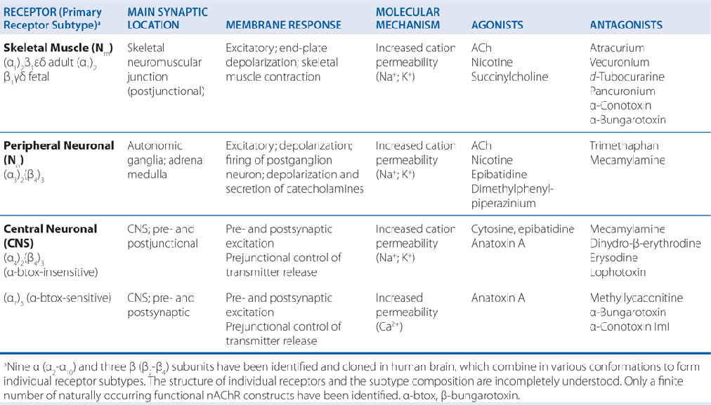
Autonomic Ganglia Pharmacology and Clinical Therapeutics
Therapeutic Range and Toxicity
Because nAChRs are expressed pervasively throughout somatic motor, autonomic, and central neural tissues, most non-selective nAChR agonists and antagonists possess limited medical utility.
nAChR-targeting drugs generally exhibit a narrow therapeutic window where the dose range yielding acceptable efficacy overlaps closely with doses causing unacceptable systemic toxicity.

Ganglionic Stimulants
Nicotine and Lobeline: Tertiary amine alkaloids extracted from tobacco (Nicotiana tabacum) and Indian tobacco (Lobelia inflata), respectively.


Both agents induce widespread generalized stimulation of autonomic ganglia. Nicotine alone is used clinically as replacement therapy in smoking cessation.

Nicotine Mechanism and Systemic Actions
Desensitization & Receptor Up-regulation: Sustained exposure to nicotine results in rapid receptor desensitization. Paradoxically, chronic nicotine exposure induces an up-regulation (increase in total number) of cell-surface nAChRs, representing an adaptive cellular compensatory response.
Biphasic Action: The physiological response to nicotine reflects a complex balance between transient neuronal excitation and persistent desensitization block. Small doses stimulate the central nervous system, whereas high doses depress CNS activity.
Systemic PNS Effects: Induces tachycardia, increased cardiac output, elevated arterial blood pressure, reduced gastrointestinal motility, and increased diaphoresis. Many systemic cardiovascular actions stem indirectly from stimulated catecholamine (epinephrine and norepinephrine) secretion from the adrenal medulla.
Ganglionic Blockers and Antagonists
Mecamylamine: A secondary amine competitive antagonist (counteract) of nicotinic receptors, originally introduced in the 1950s for hypertension and nicotine dependence.
Low doses paradoxically increase self-administered tobacco smoking, as individuals smoke more to overcome competitive receptor blockade.
High doses produce widespread autonomic shutdown, eliminating the reinforcing reward of smoking without reducing underlying psychological craving.
Hexamethonium: Structurally notable as the original ganglionic blocking drug introduced into surgical practice to induce controlled hypotension and reduce blood loss.
Experimental Importance: Ganglion-blocking drugs remain key reference tools in experimental neuropharmacology research, despite having limited modern clinical utility due to widespread side effects.
Neuromuscular Blocking Agents
Neuromuscular blockers disrupt signal transmission at the motor endplate. They act presynaptically by impairing ACh synthesis or exocytosis, or postsynaptically by blocking endplate nAChRs.
Exclude botulinum toxin, all clinically utilized neuromuscular blockers act on postsynaptic targets. They are categorized into non-depolarizing and depolarizing agents.
Non-Depolarizing Neuromuscular Blockers
Tubocurarine: The primary active alkaloid purified from curare.

Tubocurarine has been largely replaced in modern medicine by synthetic congeners with improved safety profiles, including pancuronium, vecuronium, and atracurium.
Mechanism: Competitive antagonist at motor endplate nAChRs, preventing endogenous ACh binding and producing flaccid muscular paralysis.
Critical Clinical Distinction: Non-depolarizing muscular blockers cause complete paralysis without altering sensory perception or brain consciousness. They possess no intrinsic analgesic or anesthetic properties. Paralysed patients remain conscious and fully capable of sensing pain unless co-administered general anesthesia and analgesia.
Depolarizing Neuromuscular Blockers
Succinylcholine (Suxamethonium): The only depolarizing nicotinic agonist in active clinical use.
Structure: Composed of two acetylcholine molecules joined covalently head-to-head by their acetyl groups.
Mechanism: Acts as a persistent agonist at the motor endplate nAChR, keeping the membrane continuously depolarized. Prolonged endplate depolarization keeps voltage-gated sodium channels inactivated, preventing action potential propagation and producing muscle relaxation.
Clinical Kinetics: Characterized by rapid onset of muscle relaxation (under 2 minutes) and brief duration of action (approximately 10 minutes), as the drug is rapidly hydrolyzed by plasma butyrylcholinesterase.