Neurophysiology: Resting Potential and Action Potential
Key Terms & Definitions
Neurophysiology – study of life processes in neurons (electrical & chemical signaling).
Polarized cell – electrical charge difference inside and outside the cell
Neurons are more negative on inside then outside
• Resting membrane potential (RMP): -50 \text{ to } -80\;\text{mV} , when neuron is inactive, baseline of polarization in cells
Negative sign shows that cell is negative inside
Ions: electrically charged molecules
• Cations (+): positively charged molecules
• Anions (–): , negatively charged ions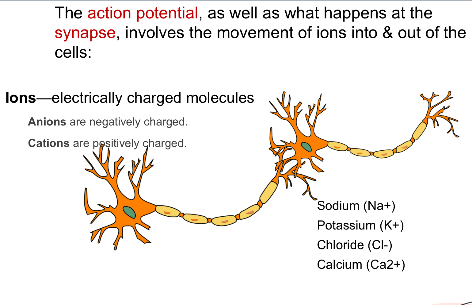
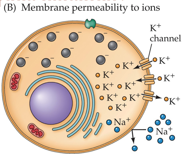
Ion channel – proteins located across the membrane of cells, pore; gated or leak.
More potassium will be inside the cell, more sodium outside
Selective permeability – membrane at rest lets \text{K}^+ (Potassium) move more freely than \text{Na}^+ (Sodium)
Two forces motivate ion movement
Diffusion: Ions spread to a uniform concentration along the gradient
HIGH CONCENTRATION TO LOW
Electrostatic pressure: Ions flow to areas that are oppositely charged sections
Sodium–potassium pump – active transporter moving 3\,\text{Na}^+ out / 2\,\text{K}^+ in (uses ATP). As K+(pottassim) accumulates in the cell, they will move across their concentration gradient and leave the cell
Electrostatic pressure will pull Pottasium back
Sodium accumulates inside the cell as is cannot go back and forth
Equilibrium potential (E_ion) – voltage where diffusion & electrostatic forces balance for a given ion. (Corresponds to resting potential)
Action potential starts in the axon hillock
Depolarization – Decrease in membrane potential
inside of cell becomes less negative (closer to 0)
cause action potential
Hyperpolarization – Increase in membrane potential
inside of cell becomes more negative
makes action potentials more difficult temporarily
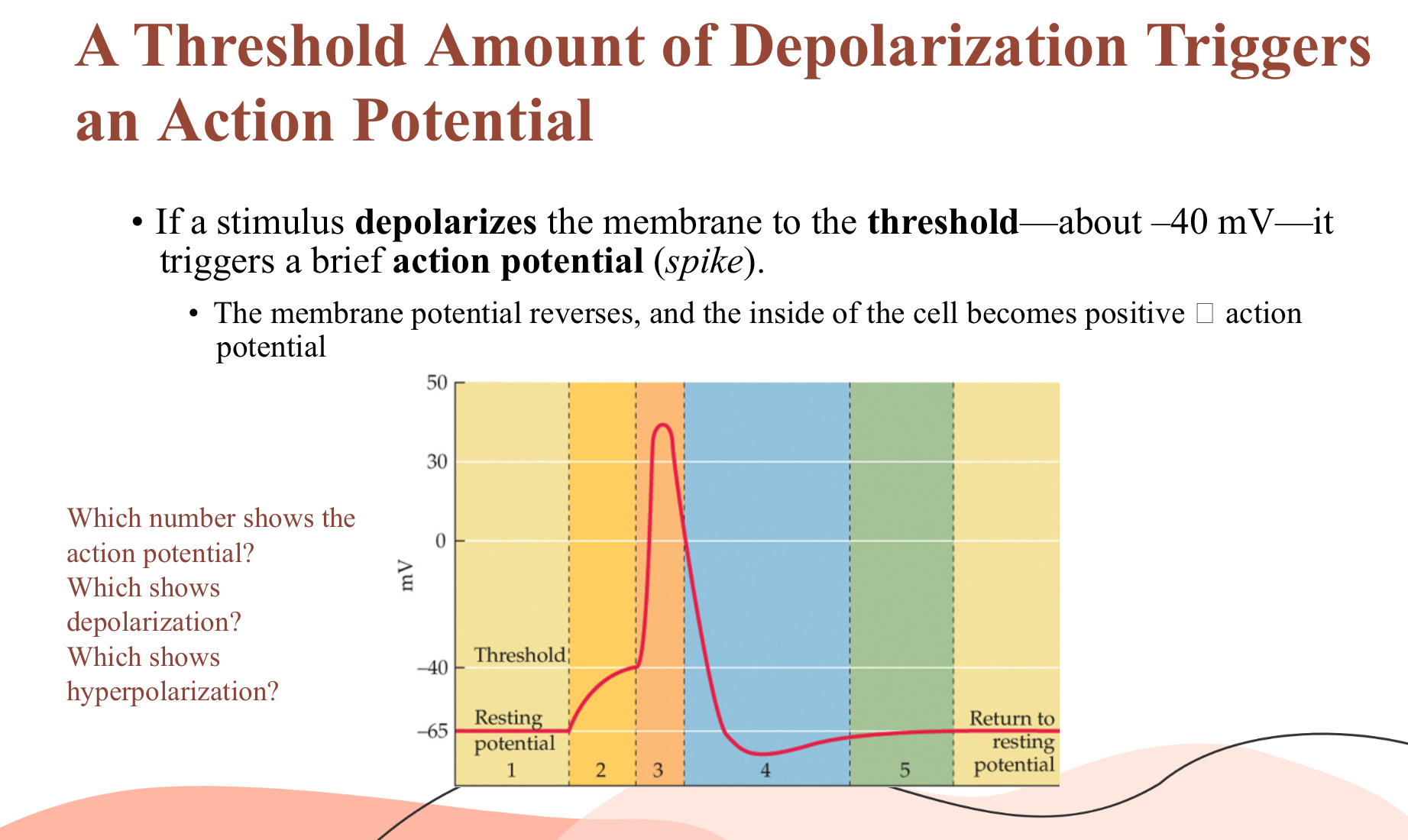
Threshold – critical V_m \approx -40\;\text{mV} that triggers an action potential
Action Potential – brief change in the cells charge; “all-or-none.”
Action potential begins with sodium ions Na+ flowing into neuron
Neurons allows Na+ in and becomes more positive, this helps to reach thereshold causing action potentials
Voltage gates Na+ channels change shape and open in response to positive charge
As they open more Na+ comes in which continues until the membranes potent ion reaches Na+ equilibrium of +40 mV
As more Na+ comes in, cell becomes more positive, Na+ will rush out of the cell across its gradient, and resting potential is restored
Refractory periods
For closely spaced stimuli, only the first one is able to trigger action potential, membrane becomes temporarily unresponsive to stimuli
• Absolute – sodium channels don’t reopen, axon membrane can’t trigger action potential
• Relative – Neuron becomes hyperpolarized (extremely negative), membrane can only respond to stimuli that is stronger than first action potential
Saltatory Conduction
Action potentials are regenerated (propagated) along the axon
Each section next to another is depolarized and a new action potential occurs
Action potentials only travel in one direction due to refraction state
Action potentials have an “all or none” property: neuron either fires and action potential or it does not
amplitude of the action potential does not care (will always be the same) about stimulus size or amount of stimulation
Neurons encode information by changes in number of action potentials ,
stronger stimuli = more action potentials but all the same size
Saltatory conduction – AP “jumps” node-to-node in myelinated axons.
EPSP – excitatory postsynaptic potential (local depolarization, \text{Na}^+ in).
IPSP – inhibitory postsynaptic potential (local hyperpolarization, \text{Cl}^- in or \text{K}^+ out).
Summation
• Spatial – multiple sites simultaneously.
• Temporal – rapid succession at one site.
Synaptic Transmission (Chemical Synapse)
Action potential arrives at axon terminal.
Voltage gated \text{Ca}^{2+} (Calcium) channels open → Ca²⁺ influx.
Ca²⁺ triggers vesicle to fuse into the membrane→ NT release into cleft.
Receptor that has been waiting for NT to bind to postsynaptic membrane, ion channels open causing an epsp or an ipsp
EPSP or IPSP spreads toward postsynaptic axon hillock, summation could lead to postsynaptic neuron firing action
Degradation ornreuptakr stops transmission
Neurotransmitter receptors: Proeteins in the cell membrane, detect and respond to specific NTs
When NT binds to receptor it inhibits or excites neuron
Ligand: molecules that bind to receptors to activate or block reaction
The action of synaptic transmitters is stopped rapidly
Degradation: Breaks down neurotransmitters
Reuptake: Transporter protein located along the postsynaptic terminals absorbs NT’s and recycles them
Postsynaptic Potentials
Neurons receive synapses from many cells, the accumulation of these stimuli determine whether the neurons axon hillock;s membrane potential will reach the threshold to fire and action potential
EPSP (excitatory postsynaptic potential)
Small depolarizing potential in the postsynaptic membrane
• Ligand-gated Na⁺ (sodium) channels open → depolarization.
• Moves cell toward threshold.Makes cell more positive
IPSP (Inhibitory postsynaptic potential)
a hyperpolarizing potential on the postsynaptic membrane
• Ligand-gated Cl⁻ (chloride ) channels open → hyperpolarization.
• Moves cell away from threshold.
Properties: graded, decremental, can summate (no refractory).
Summation Mechanics (two was neurons integrate inputs) review this
Spatial summation: summing of potentials that reach the axon hillock from the many locations across the cell body
Temporal summation: summing of potentials that reach 5he axon hollow based on time of arrival.
Closer the time potentials arrive the more urgency is sensed and greater their impact and likelihood of firing an action potential
Decision point: if combined V_m ≥ threshold → AP generated.
Action Potentials vs Synaptic Potentials
Feature | Action Potential | Synaptic Potential |
|---|---|---|
Location | Axon (hillock → terminals) | At the synapse in postsynaptic cell (Dendrites, soma, spines,) |
Size | Fixed (~100 mV) | Variable (mV–sub-mV) |
Nature | All-or-none | Graded- depends on amount of neurotransmitter released and receptors activated |
Trigger | Threshold depolarization | NT binding |
Channels | VG Na⁺/K⁺ | Ligand-gated (ionotropic/GPCR) |
Propagation | Active, non-decremental | Passive, decremental |
Refractory | Yes | No |
Summation | Not applicable | Spatial & temporal |