chapter 7- Skeletal system
The skeletal system is composed of bones, cartilages, and ligaments joined tightly to form a strong, flexible framework for the body.
•Functions of the skeletal system include:
◦Support, protection, and movement
◦Blood cell formation
-The process of blood cell formation is called hematopoiesis, or hemopoiesis.
→hemo- = blood; -poietic = forming
-Red bone marrow produces red blood cells (erythrocytes), white blood cells (leukocytes), and blood platelets.
◦Inorganic salt storage
-The most abundant salt in bone is a type of calcium phosphate called hydroxyapatite.
-Bone tissue also contains smaller amounts of magnesium, sodium, potassium, and carbonate ions.
◦Energy storage
-Yellow bone marrow stores fat and does not produce blood cells.
Bones can be classified according to their shape.
•Long bones are longer than they are wide, often with an expanded end.
◦Includes bones in the arms and legs.
•Short bones have equal lengths and widths.
◦Includes carpal and tarsal bones.
◦A special type of short bone is a sesamoid bone, or round bone.
-Embedded in a tendon.
-The patella is a sesamoid bone.
•Flat bones are plate-like structures with broad surfaces.
◦Examples include the ribs, sternum, the scapulae, and some cranial bones.
•Irregular bones have a variety of shapes, and most are connected to several other bones.
◦Examples include vertebral bones and many facial bones.
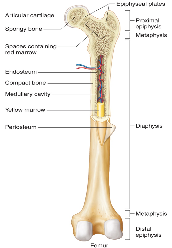
Femur:
Periosteum- outer covering of tissue. on the inside contains bone cells. Where ligaments and tendons insert to. Where blood vessels and nerves that enter and exits the bone. Nutrient foramina.
Diaphysis (shaft)- longest part of the bone. Composed of primarily of compact bone
Proximal epiphysis- at the top. Composed of spongy bone (honeycomb appearance)
Distal epiphysis- at the bottoms. Composed of spongy bone
metaphysis- transition zone from the diaphyssis to the epiphysis.
Red bone marrow- makes blood cells. Composed of spongy bone.
Medullary Cavity- contain blood vessels and yellow bone marrow (fat). Hollow cavity.
Endosteum- lines the medullary cavity on the inside
Epiphyseal plates- Little lines. The growth plates for bone. Where bone grows in length.
Articular cartilage- hyaline cartilage. Serves as a cushion between two bones.
Short, flat, and irregular bones typically consist of a mass of spongy bone either covered by a layer of compact bone or sandwiched between plates of compact bone.
The microscopic structure of bone is composed of three different types of bone cells.
•Osteoblasts are bone forming cells.
◦osteo = bone; blast = form, produce
◦These cells deposit bone matrix around themselves through a process called bone deposition (secret calcified ground substance). Cells completely surround themselves with bone matrix and become entrant and lead to inactive state
•Osteocytes are former osteoblasts that have become trapped in the matrix they deposit. T
◦osteo = bone; cyte = cell
◦They reside in tiny cavities called lacunae, which are interconnected by slender channels called canaliculi (canaliculus is singular).
-lac = lake, hollow; una = little
-canal = canal; icul = little
The microscopic structure of bone is composed of three different types of bone cells.
•Osteoclasts are large, multinucleated cells that break down bone matrix through a process called bone resorption. much larger than osteocytes. Calcium levels need to be maintained.
◦osteo = bone; clast = destroy, breakdown
◦These cells form through the fusion of multiple white blood cells called monocytes.
◦Release an acid that dissolves the inorganic component of the bone matrix.
◦Release an enzyme that breaks down collagen.
The microscopic structure of compact bone consist of a continuous extracellular matrix composed largely of collagen fibers and inorganic salts.
•Collagen gives a bone strength and a degree of flexibility, and inorganic salts give a bone hardness and resistance to being crushed.
— Osteogenesis imperfecta- brittle bone disease.
•The salt is mostly small crystals of a type of calcium phosphate called hydroxyapatite.
◦Salts account for 70% of extracellular matrix by weight.
- Most abundant mineral; calcium phosphate.
- If we do not absorb enough calcium, bones can soften. disorders such as Rickets; Were a child does not get calcium/Vitamin D and bones will soften and can lead to bow legged.
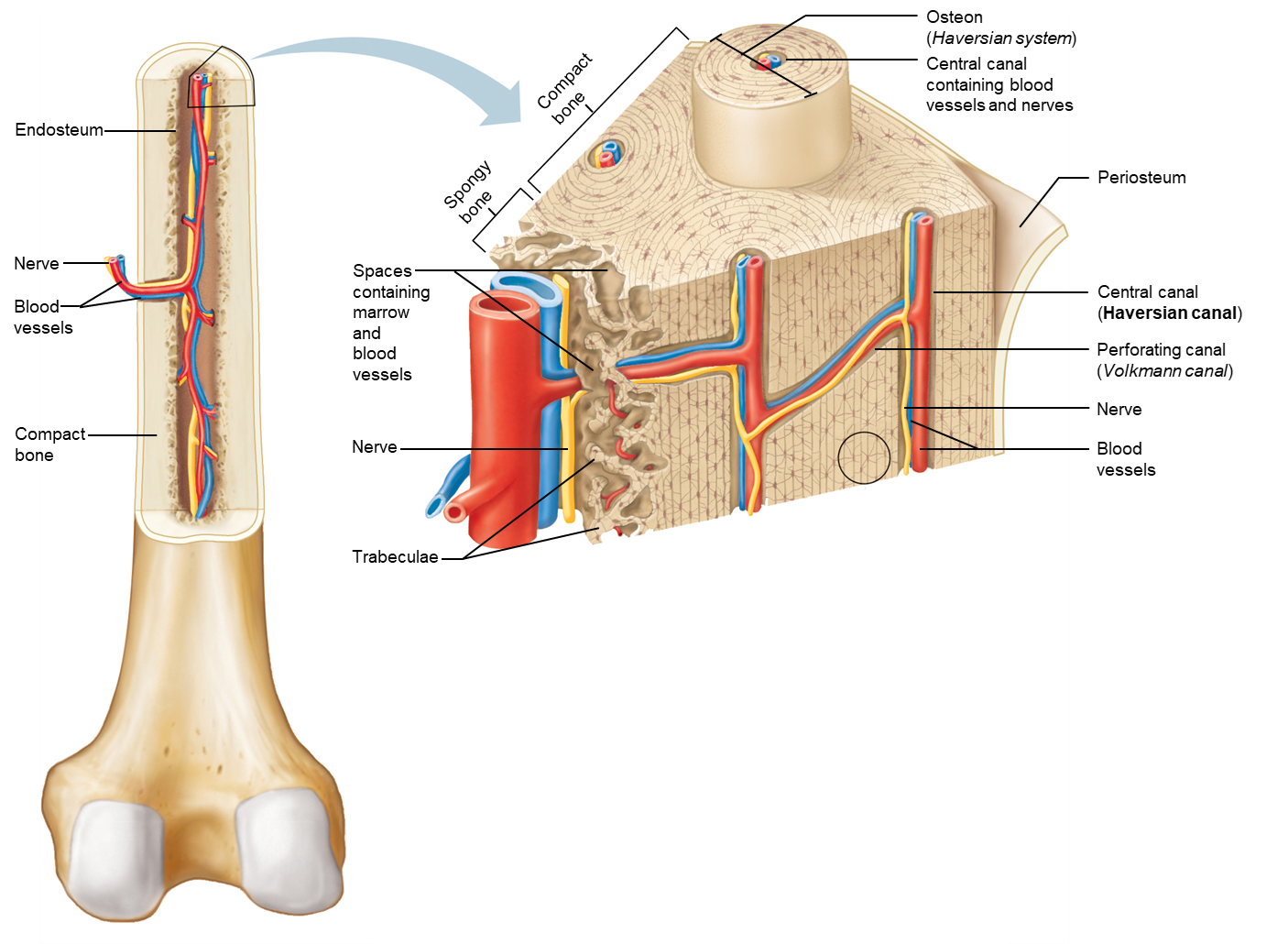
Compact Bone:
Compact bone: More compact, organized, more dense, and stronger. It is organized in elongated cylinders; Osteons. Thy can as canal in middle, called central canals. Central canals have blood vessels and nerves. Adjacent osteons are connected by a second canal; perforated canals.
Spongy bone:
Not as organized and very porous
Has red bone marrow; honeycomb
Osteocytes and Calcified ground substance Organized into thin plates; Trabeculae. The point in various directions and give bones strength and reinforcement in various directions as well.
The formation of bones, osteogenesis or ossification, begins during the first few weeks of prenatal development, and continues to grow and develop into adulthood.
•Bones form in one of two ways:
◦Intramembranous ossification involves bones forming bones from membrane-like layers of embryonic connective tissue called mesenchyme.
-Flat bones of the skull, clavicles, sternum, and some facial bones (mandible, maxillae, and zygomatic bones)
— Original layer of mesenchyme, then differentiate into osteoblasts. Then osteoblasts begin to form calcium matrix, surround the mere with matrix and become osteocytes. First layer that forms is a layer of spongy bone. Then surrounding spongy bone is surrounded by layers of periosteums. The osteoblasts that line the periosteum layers open compact bone. Result in= layer of spongy bone, 2 layers of compact bone, and layers of periosteum that surrounds bone.
◦Endochondral ossification begins with pieces of hyaline cartilage that are later replaced with bone tissue.
-Most bones of the skeleton form through this type of ossification.
— Begins with piece of cartilage that serves as a template that a future bone forms; Hyaline Cartilage. In this cartilage, Chondrocytes will begin to die and then cartilage then begins to break down and be replaced by osteoblasts. Then they will form calcified matrix. The primary ossification center will be ossified first, prior to birth. Fetal Development. The Diaphysis will be the primary ossification center of long bone
— Any other area that begins to ossify after a primary ossification begins, it will be classified as Secondary ossification center. They can begin before or after birth and can have multiple secondary ossification centers. For long bones, the Epiphyseal ends will begin the secondary ossification centers. As a child ages, they narrow down to become Epiphyseal plates.
Bone growth in length ceases once the epiphyseal plates close. Composed of Hyalne cartilage. In order for bones to grow, new cartilage has to form in Epiphyseal plots, the cartilage will break down and become calcified matrix.
•An epiphyseal line is a remnant structure of the growth plate. Where growth plates close.
◦Nearly all growth plates are closed by the age of 23 for females, and by the age of 25 for males.
-
Appositional growth is a type of bone growth where the bone grows in diameter. (Width).
•Osteoblast lining the inside of the periosteum new compact bone.
Several factors influence bone development, growth, and repair.
•Nutritional factors
◦Vitamin D is needed for proper absorption of dietary calcium in the small intestine.
-Vitamin D is scarce in natural foods, except for eggs.
→Milk and other dairy products are fortified with vitamin D.
-The body can also make vitamin D when the skin is exposed to sunlight- absorption of UV.
— vitamin d can strengthen our immune system. More like a hormone.
-A lack of vitamin D in the diet can lead to a softening of bones, and potential deformation.
→A softening in bones is called rickets in children, and osteomalacia in adults.
◦Vitamins A and C are also required for normal bone development and growth.
-Vitamin A is necessary for osteoblast and osteoclast activity during normal development.
-Vitamin C is required for collagen synthesis.
→A deficiency of vitamin C can lead to scurvy.
Several factors influence bone development, growth, and repair.
•Hormonal factors
◦The pituitary gland secretes growth hormone, which stimulates division of cartilage cells in the epiphyseal plates to divide and increase in size.
-Pituitary dwarfism is caused by too little growth hormone during childhood.
→Individuals are very short but have normal body proportions.
-Pituitary gigantism is caused by too much growth hormone during childhood.
→Height may exceed 8 feet!
→In an adult, secretion of excess growth hormone causes acromegaly in which the hands, feet, and jaw thicken.
Several factors influence bone development, growth, and repair.
•Hormonal factors
◦Both male and female hormones (testosterone from the testes and estrogen from the ovaries) promote the formation of bone tissue.
- estrogen is much more potent in stimulating bone growth than testosterone.
-During puberty, both hormones cause long bones to grow considerably.
-Both hormones also eventually stimulate the closure, or ossification of the epiphyseal plates.
Several factors influence bone development, growth, and repair.
•Physical stress also stimulates bone growth.
◦Bones reflect stresses they encounter.
-Bones are stressed when weight bears on them or muscles pull on them.
◦Wolff’s law states that bones grow or remodel in response to demands placed on them.
-Bones strengthen with hypertrophy (growth of tissue) when stress is applied.
-Bones weaken with atrophy (tissue shrinks) when stress is not applied.
In addition to their growth, bones are continually remodeled through life by the absorption of old bone and deposition of new bone.
•These opposing processes are highly regulated so that total mass of bone tissue in adult skeleton normally remains constant, even though around 10% to 20% of bone calcium is exchanged each year.
◦Bone deposition is the buildup of bone tissue by osteoblasts.
◦Bone resorption is the break down of bone tissue by osteoclasts.
•Calcium is required for many metabolic processes, including:
◦Muscle contraction
◦Nerve conduction
◦Blood clotting
Several factors influence bone development, growth, and repair.
•Hormonal factors
◦The thyroid gland releases calcitonin when blood calcium levels are too high.
-Stimulates an increase in osteoblasts activity.
-Stimulates bone deposition.
◦The parathyroid glands releases parathyroid hormone when blood calcium levels are too low. Much more common. Found a lot with lactating women.
-Parathyroid hormone stimulates an increase in the number and activity of osteoclasts.
-Stimulates bone resorption.
Homeostatic mechanism:
Blood calcium level that are too high are detected by thyroid gland
Thyroid gland will respond and release hormone; Calcitonin. Calcitonin can bring blood calcium levels back down by activating osteoblasts. If we activate osteoblasts, it will lead to more bone deposition and bones lay down new bone matrix and take calcium out of blood.
Thyroid gland is the receptor and control center when calcium blood level are too high and response by releasing calcitonin and increase osteoblasts activity.
If blood calcium levels are too low, it will be detected by cells from the Parathyroid glands.
The lands will respond by releasing parathyroid hormones. The hormone will help bring blood calcium levels up by activating osteoclasts.
The osteoclasts will increase the rate at which bone is broken down, it will increase bone resorption and release calcium and calcium is reabsorbed by blood vessel and help bring blood level back up. Help activate osteoclasts and increase bone resorption.
When blood calcium levels are too low, the parathyroid glands are the receptors and the control center. They respond by releasing parathyroid hormone and increase osteoclasts activity which increases bone resorption.
Despite their remarkable strength, bones are susceptible to fractures, or breaks.
•Classification by cause:
◦Traumatic: Fracture caused by injury
◦Spontaneous or pathologic: Fracture caused by disease
•Classification by nature of break:
◦Simple (closed): Fracture protected by uninjured skin
(or mucous membrane)
◦Compound (open): Fracture in which the bone is exposed to the outside through opening in skin (or mucous membrane).
•Can also be described by location of fracture, external appearance, and nature of break
Tranverse fracture: right angle break
Oblique fracture: an angle other than right angle
Spiral fracture: excessive twisting of bone
Comminuted fracture: bone shattered
Tissue fracture: has a crack in the middle
Green stick fracture: bone mends but doesn’t snap in half. More common in children
Takes 6-8 weeks to heal for an average fracture
On average, the human body has 206 bones that are divided between the axial and appendicular skeleton.
The skull is typically composed of 22 bones
•All skull bones are interlocked along sutures, except the lower jaw (mandible)
•The skull = cranium + facial skeleton
◦Cranium contains 8 bones that enclose and protect brain
◦Facial skeleton contains 14 bones that form and shape of face
•Mandible is movable, and held in place next to cranium by ligaments
•Orbit of eye is formed by some facial and cranial bones
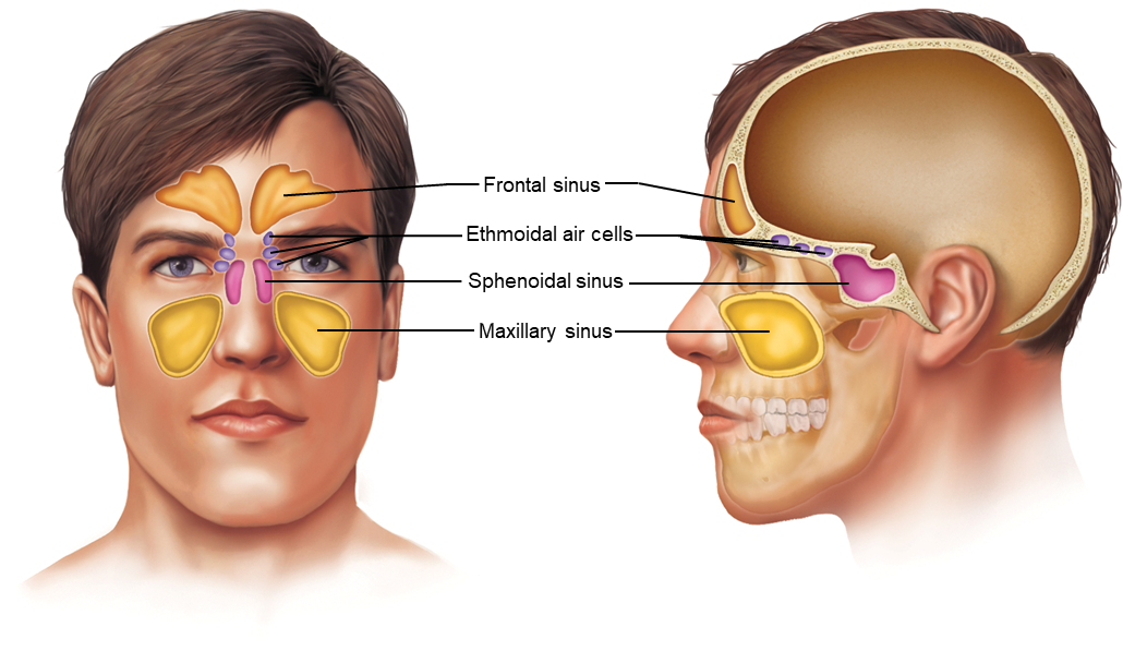
•Paranasal sinuses:
◦Air-filled cavities found in some cranial and facial bones
◦Reduce weight of skull and increase intensity of voice
Questions to consider for the lecture exam:
•Which skull bones are cranial bones, and which bones are facial bones.
— Cranial:
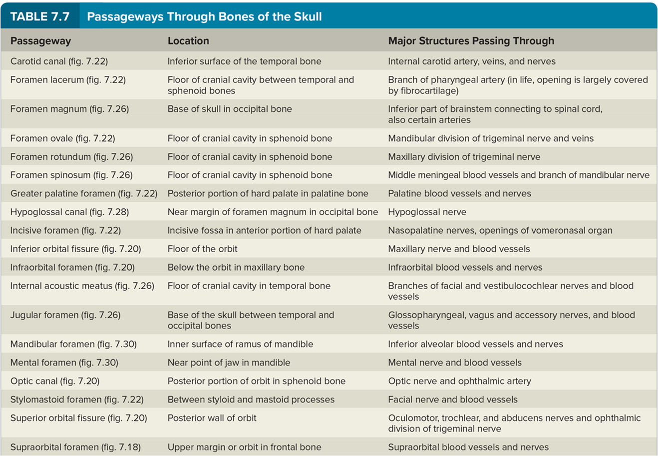
- Frontal bone: 1, Supraorbital foramen)
- parietal bone : 2, connected to frontal bone by coronal suture
- anterior/posterior fontanel (only for newborns)
- occipital bone: 1, connected by Lambdoid suture. External occipital protuberance (large bump on back of head), Foreman Magnum (large hole in skull, boundary between end of brain stem and beginning of spinal cord), On both sides of the Foramen Magnum there are 2 round processes; Occipital condyle where first vertebral bone articulates with (ATLAS,C1),
- Inside of skull (superior view), on each side of the foreman magnum, there are 1 small Hypoglossal Canal. There is a cranial Hypoglossal nerve that passes through the Hypoglossal canal.
- Temporal Bone: 2, separated from parietal bone by squamous suture. Has large bump by ear, Mastoid Process. Anterior of process, there’s an External acoustic (auditory) meatus is a canal that leads to the internal organs of ear for hearing, forms ear canal. Found on the other side of the temporal bone, the Internal Acoustic Meatus is a small opening that ha nerves that leave ear and pass through opening, nerves for hearing, balance, facial nerves. Next to the occipital condyle, there’s a few openings called the Carotid canal (within temporal bone) that has a branch that carotid artery passes through it. Another opening, between temporal bone and occipital bone; Jugular Foreman which contains nerves and internal jugular vein.
- Anterior and inferior to the Internal Acoustic meatus, there’s a pointy projection called the Styloid Process
- Sphenoid bone: 1, “keystone of entire skull (helps hold different parts of skull together)”. In he middle, there’s a Sella Turcica that contains the pituitary Gland. Has several openings, a short canal; Optic Canal (has optic nerve), below optic canal is the Superior orbital Fissure WITHIN, An Inferior Fissure is found BETWEEN bones (Sphenoid and Maxilla, and Palatine Bone). 3 openings lined up together, Foremen Rotundum, right below is the oval shape Foreman Ovale, and small pinpoint opening called the Foramen Spinosum. These 3 openings line up.
- Ethmoid Bone: 1, best found in superior view. Little projection called Crista Galli, layers of connective tissues are the meninges that surround the brain and spinal cord and attach to Critsta galli. The Crista Galli sits on top of cribriform Plate and it has microscopic openings called the Cribriform Foramina. The nerve fibers from nose pass through Cribriform foramina to enter the skull.
- To find the Ethmoid Bones, look within nasal cavity. there’s a Perpendicular plate that forms a top part of the nasal septum. ad folded piece of bone called the Middle Nasal Concha.
— suture Wormian bones- irregular pattern sutures. Creates isolated fragments of bone
—- Facial:
- Zygomatic Bone, forms cheek bone ( 2 bones). Mandible (lower jaw) includes a Mandibular Fossa; a shallow indentation where the mandible articulates with the temporal bone and it has the TMJ (what allows Mandible to be the freely moveable bone of skull)
- Vomer: forms the bottom part of nasal septum
-inferior nasal concha: 2, independent facial bones.
- Nasal Bones: 2, forms bridge of nose
- Lacrimal Bone: 2, small piece of bone
- Maxilla: 2, Forms upper jaw. Contains small opening called the infraorbital foreman.
- Alveolar process: Next to teeth, small projections, Paletine Processes form part of our hard palate.
- Mandible: Freely moveable bone. Contains body of chin, Mental foeman, Ramus and branches 2 rejections called (anterior) Coronoid process and (posterior)mandibular Condyle. It articulates to the temporal bones and created TMJ. Mandibular foreman. Alveolar processes are tooth sockets.
•Which two bones form the zygomatic arch?
- Temporal bone and zygomatic bone
•Which two bones form the nasal septum?
-perpendicular Plate of Ethmoid bone and Vomer
•Which two pairs of bones form the hard palate?
- 2 sets of bones; Paletine processes of maxillary bones and median Paletine suture and Incisive Foremen that contain sensory nerve. Maxillary Bones ad Paletine bones
•Which three cranial bones have a sinus?
- Frontal bones,Ethmoid bone, Sphenoid bone
•Which facial bone has a sinus?
- Maxillary Bones (largest sinus)
•Describe a fontanel.
- “soft spot” between an infants cranial bones which are membrane-covered , fibrous gap where sutures intersect but have not yet fused.
•Describe cleft palate.
- Where Paletine processes do not fuse together as a birth defect but can be fixed by surgery.
The most common bone disease is osteoporosis, which involves a severe loss of bone density.
•Bones become brittle and highly subject to pathological fractures.
◦Fractures occur especially in the hip, wrist, and vertebral column.
•Occurs in both sexes and at all ages, but postmenopausal women are at the greatest risk.
◦Women start with less bone mass than men and lose it faster.
◦After menopause, the ovaries stop producing estrogen, which decreases osteoclast activity.
◦After menopause, osteoclast activity begins to outpace osteoblasts activity, leading to accelerated bone loss.
•The optimal means of preventing osteoporosis is a good diet and exercise habits.
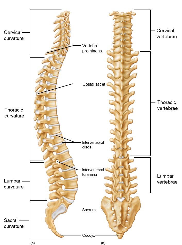
The vertebral column forms vertical axis of skeleton.
•Consists of many vertebrae separated by cartilaginous intervertebral discs, and connected by ligaments
◦Supports head and trunk, permits several types of movements
◦Protects spinal cord in the vertebral canal
•26 separate bones in an adult, and 33 in an infant.
◦7 cervical vertebrae
◦12 thoracic vertebrae
◦5 lumbar vertebrae
◦5 fused sacral vertebrae form the sacrum
◦4 fused coccygeal vertebrae form the coccyx
Questions to consider for the lecture exam:
•How many cervical vertebrae are in the vertebral column?
- 7 in the neck region
•Which cervical vertebra is also known as vertebral prominens?
- C7
•How many thoracic vertebrae are in the vertebral column?
- 12 or the ribs
•How many lumbar vertebrae are in the vertebral column?
- 5
•How many bones fuse to form the sacrum?
- 5
•How many bones fuse to form the coccyx?
- 4
•Which spinal curves are primary?
- Thoracic Curvature and Sacral Curvature; we are born with these
•Which spinal curves are secondary?
- Cervical curvature and lumbar curvature; after birth when we tart crawling and hold our heads upright, when we start walking.
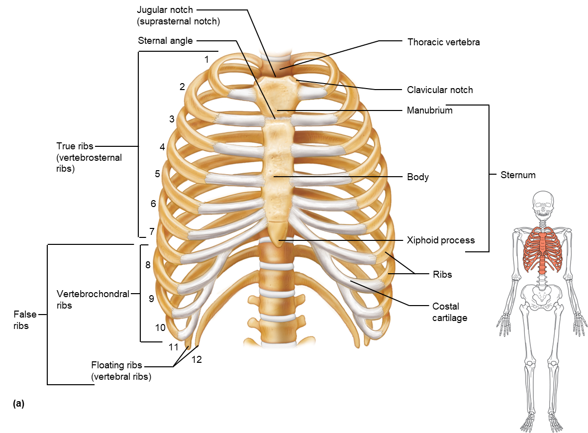
•Which ribs are true (vertebrosternal) ribs?
- 1-7, have direct contact with the sternum with Costal cartilage. They attach ribs to sternum. AKA “vertebrosternal ribs”
•Which ribs are false ribs?
- 8-12, 8-10 have pieces of costal cartilage that have an INDIRECT attachment to sternum. Their cartilage attach to another pice of costal cartilage. AKA “vertebrochondral ribs”. 11-12 don’t have any anterior attachment, AKA “Floating ribs” or “Vertebral ribs”
•Which false ribs are vertebrochondral ribs?
- 8-10
•Which false ribs are floating ribs?
- 11-12
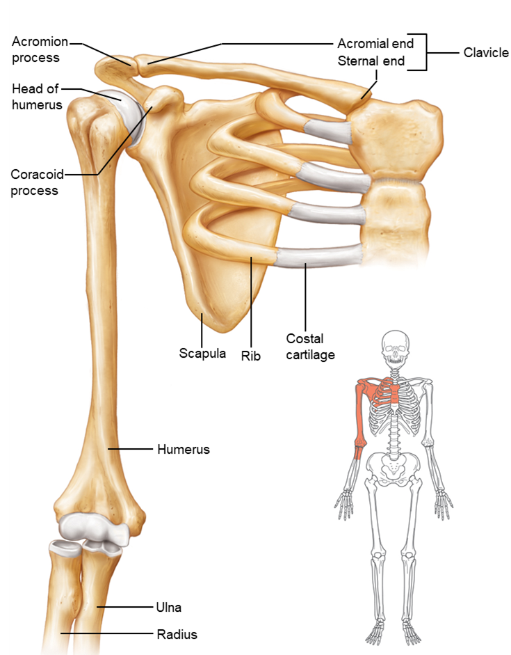
Pectoral (shoulder) girdle:
•Consists of 2 clavicles and 2 scapulae:
◦Clavicles = collarbones; anterior portions
◦Scapulae = shoulder blades; posterior portions
•Forms incomplete ring; open in back
•Supports upper limbs
•Attachment site for muscles that move upper limbs
help attach upper extremity to the trunk of the body, stabilize shoulder joints when arm move, serve as attachment sights for muscles that move the upper extremity.
Pelvic Girdle
•Consists of 2 hip bones (coxal bones, ossa coxae, or innominate bones)
•Pelvic Girdle:
◦Supports trunk of body
◦Protects viscera, such as urinary bladder
◦Transmits weight to lower limbs
◦Provides attachment for lower limbs
•Pelvis = pelvic girdle + sacrum + coccyx
Pelvic brim- pelvic inlet and pelvic outlet. Separates 2 part of the pelvis
Greater pelvis (false) - organs found in pelvis area are found in abdominal cavity
Lesser pelvis- found below the pelvic brim. where the true pelvic cavity i ound and the organs of pelvic cavity are found including urinary bladder and internal reproductive organs,
Male pelvis: more curvature to sacrum which decreases diameter of pelvic outlet. Pubic arch is more narrow than female pelvis.
Female pelvis: iliac crest/ilium are wider and sit lower. Pelvic inlet and outlet have a greater diameter because sacrum sits more vertical. when compared to male pelvis