B3.3 Muscle and motility (HL) Notes
B3.3.1 adaptations for movement as a universal feature of living organisms
There Are Two Types of Movement in Living Organisms
Internal Movement occurs within the body of all living organisms, even stationary ones.
It is essential for nutrient distribution, waste removal, and maintaining cellular functions.
Locomotion on the other hand, involves moving from one location to another.
It enables organisms to find resources, evade predators, and adapt to changing environments.
Example
Internal movement
Plants: Transport water and nutrients through xylem and phloem.
Animals: Processes like peristalsis and blood circulation.
Unicellular Organisms: Cytoplasmic streaming moves nutrients and organelles.
Locomotion
Cheetahs: Sprinting to catch prey.
Birds: Migrating long distances.
Bacteria: Moving via flagella.
Motile vs. Sessile Organisms: A Comparison
Motile Organisms
Actively move through their environment to find food, escape predators, or reproduce.
Adaptations:
Skeletal and Muscular Systems: E.g., elongated limbs in cheetahs for speed.
Energy Storage: Migratory birds store fat reserves before long flights.
Behavioral Strategies: Moving in response to seasonal cues or predator threats.
Sessile Organisms
Remain fixed in one location for most of their lives (e.g., barnacles, corals).
Still exhibit movement at smaller scales:
Feeding: Barnacles use cirri (modified legs) to filter water. Corals use tentacles to capture food.
Larval Motility: Many sessile species have motile larval stages for dispersal.
Anchorage Mechanisms: Strong attachments (e.g., adhesive cement in barnacles, calcium carbonate skeletons in corals) to remain in place.
Tip
Even sessile organisms rely on subtle movements for feeding, reproduction, and survival.
Adaptations for Movement Across Taxonomic Groups
Movement in Unicellular Organisms
Flagella and Cilia: Whip-like or hair-like structures powered by motor proteins.
Cytoplasmic Streaming: In Amoeba, cytoplasm flows to form pseudopodia, aiding movement and feeding.
Movement in Vertebrates
Skeletal Systems: Endoskeletons provide support and anchor muscles.
Muscular Systems: Skeletal muscles contract via the sliding filament model(interaction of actin and myosin).
Joint Structures: Synovial joints (e.g., ball-and-socket) allow a wide range of motion.
Movement in Marine Mammals
Streamlined Bodies: Reduce drag when swimming.
Modified Limbs: Flippers and flukes for propulsion and maneuvering.
Blubber: Provides buoyancy and insulation in cold water.
Hint
Movement adaptations vary widely based on an organism's habitat and lifestyle, highlighting the diversity of evolutionary solutions.
Why Movement Matters
Foraging: Searching for or capturing food (predators chasing prey; herbivores seeking plants).
Escaping Predators: Rapid locomotion reduces the risk of being caught.
Reproduction: Movement facilitates finding mates or dispersing offspring.
Migration: Seasonal journeys (e.g., snow geese) ensure access to food and breeding sites.
Self Review
Internal vs. External Movement: All organisms exhibit internal movement, but only motile organisms show locomotion.
Energetic Trade-Offs: Locomotion requires energy, but the survival and reproductive benefits can outweigh these costs.
Inspiration for Technology: Studying biological movement can inform innovations in robotics and engineering(e.g., mimicking streamlined designs).
B3.3.2 sliding filament model of muscle contraction
Sarcomeres Shorten as Actin and Myosin Slide Past Each Other
Definition
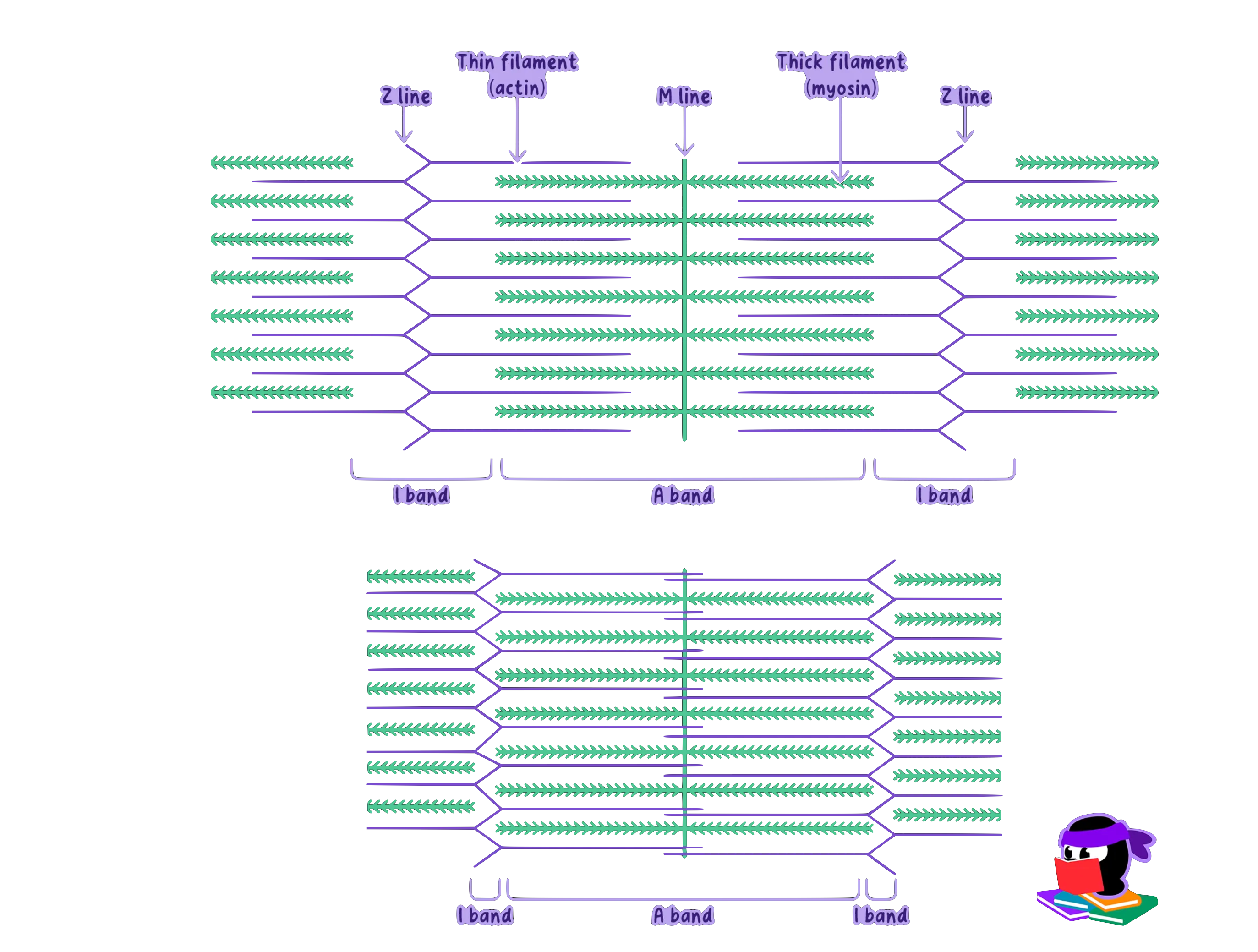
SarcomereA sarcomere is the basic contractile unit of muscle fiber. Each sarcomere consists of two main protein filaments, actin and myosin,which are the active structures responsible for muscular contraction.
The Sarcomere consists of:
Z-Discs (Z-Lines): Define sarcomere boundaries, anchor thin filaments (actin).
Thin Filaments (Actin): Extend from the Z-discs toward the sarcomere center.
Thick Filaments (Myosin): Occupy the central region, interlock with actin filaments.
I-Bands (Light Bands): Contain only actin.
A-Bands (Dark Bands): Overlap region of actin and myosin.
H-Zone: Central part of the A-band where only myosin is present when muscle is relaxed.
Z-Discs are pulled closer together to shortening the sarcomere.
Note
During muscle contraction, the sarcomeres shorten as the Z-discs are pulled closer together.
This shortening occurs because the actin and myosin filaments slide past each other; the filaments themselves do not change length.
Tip
Think of the sarcomere like a spring that compresses during contraction and stretches back out during relaxation.
This elasticity is critical for muscle function and recovery.
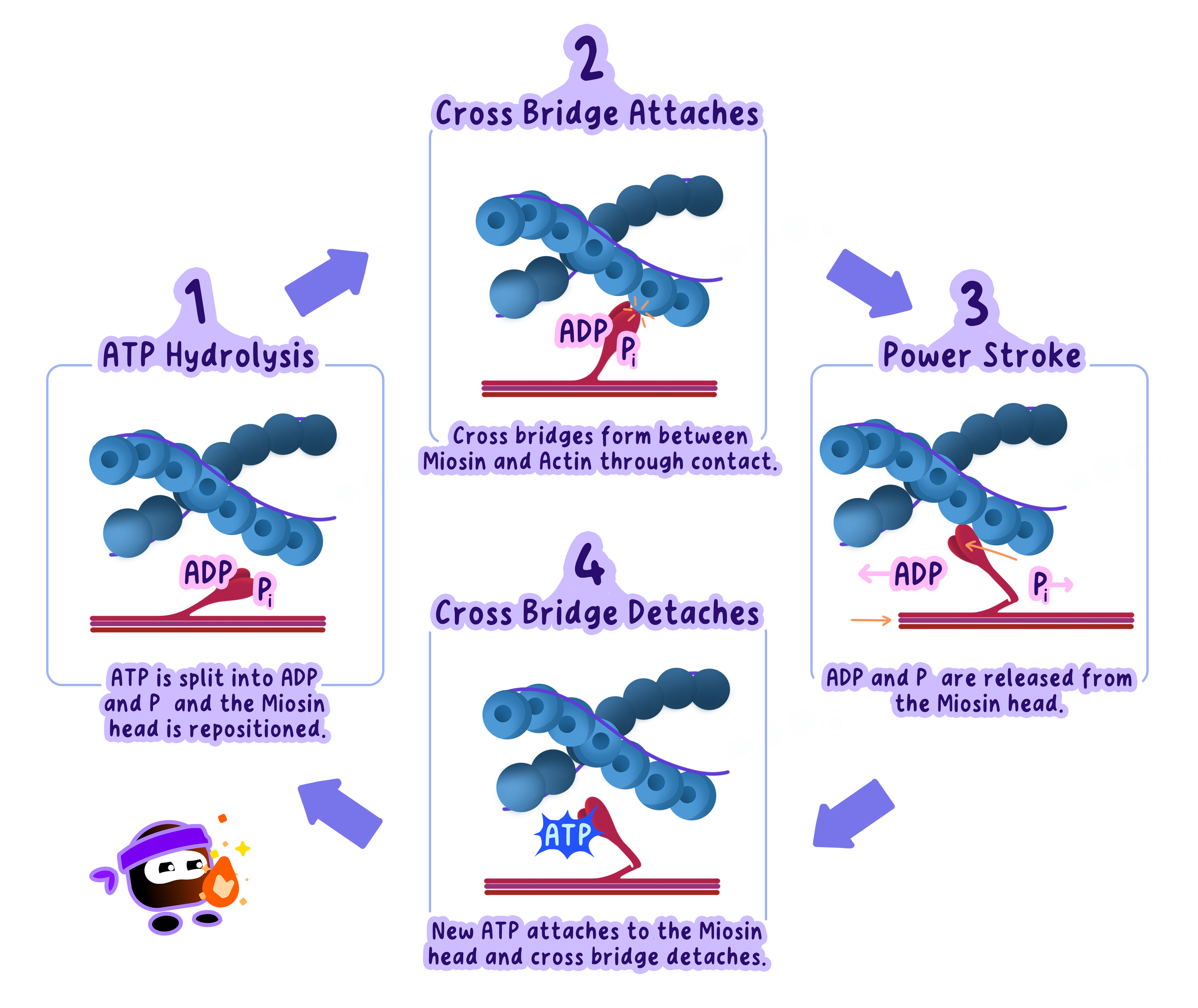
The Sliding Filament Mechanism
Cross-Bridge Formation
Myosin heads attach to binding sites on actin, forming cross-bridges.
Initiated by Ca²⁺ release (triggered by a nerve impulse).
Power Stroke
Myosin heads pivot, pulling actin toward the sarcomere center.
ATP hydrolysis provides the energy for this movement.
Detachment
A new ATP binds to the myosin head, causing it to release actin.
Without ATP, the myosin head would remain locked in place (rigor).
Resetting the Myosin Head
The myosin head hydrolyzes ATP, returning to its cocked position.
Ready to form another cross-bridge for continuous sliding.
Example
Imagine pulling a rope hand-over-hand.
Each time you grip and pull the rope, it's like a myosin head attaching and pulling on an actin filament.
Letting go to re-grip the rope mimics the detachment and resetting of the myosin head.
The Role of ATP and Calcium in Muscle Contraction
The Role of ATP and Calcium (Ca²⁺)
ATP
Energy source for the power stroke and detachment of myosin heads.
In its absence, myosin remains attached (leading to rigor mortis after death).
Warning
Students often assume ATP is only needed for contraction.
In reality, ATP is equally critical for muscle relaxation, as it allows myosin heads to detach from actin.
Calcium Ions (Ca²⁺)
Stored in the sarcoplasmic reticulum and released upon nerve stimulation.
Binds to troponin, shifting tropomyosin to expose actin binding sites.
Allows myosin heads to attach and initiate contraction.
Tip
Think of calcium as the key that unlocks the binding sites on actin, enabling the myosin heads to begin their work.
Coordinated Muscle Contraction
Simultaneous Sarcomere Shortening
All sarcomeres in a myofibril shorten at once.
Multiple myofibrils in a muscle fiber contract together.
Force Generation
Large-scale force is produced by millions of sarcomeres working in unison.
Enables both strong movements (e.g., lifting) and fine movements (e.g., typing).
Hint
This coordination allows muscles to produce both powerful and precise movements, whether you're lifting a heavy object or typing on a keyboard.
Applications and Implications
Athletics: Knowledge of muscle contraction enhances training methods to optimizestrength and endurance.
Medical Context
Diseases like muscular dystrophy and myasthenia gravis involve disrupted actin-myosin interaction.
Treatments focus on restoring proper sliding filament function.
Engineering & Robotics: Biomimetic designs use principles of muscle contraction to create artificial muscles for prosthetics or robots.
Self Review
What roles do actin, myosin, ATP, and calcium play in the sliding filament model?
How does the structure of a sarcomere change during contraction?
Why is the sliding filament model often compared to a "ratchet mechanism"?
What would happen if ATP or calcium were unavailable during muscle contraction?
B3.3.3 role of the protein titin and antagonistic muscles in muscle relaxation
Titin Restores Sarcomere Length and Prevents Overstretching
Definition
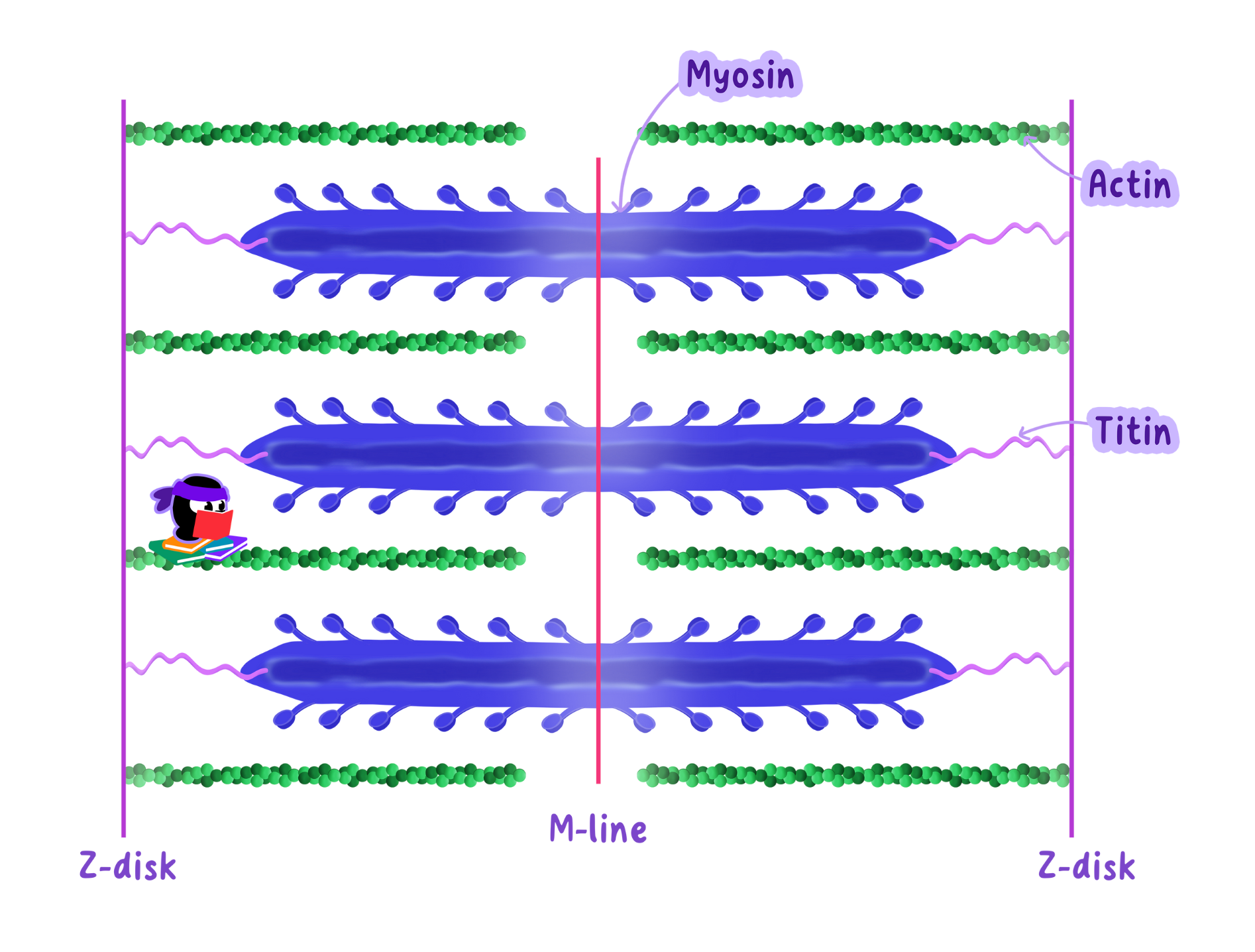
Titin Titin is the largest protein in the human body, spanning from the Z-discs to the myosin filaments in the sarcomere.
It runs parallel to actin, functioning as a molecular spring that stretches and recoils.
Its primary roles include:
Elasticity: Titin stretches during sarcomere elongation and recoils to restore the resting length.
Prevent Overstretching: It limits excessive extension, reducing the risk of muscle damage.
Energy Storage: When stretched, titin stores potential energy that aids both contraction and relaxation.
During muscle relaxation, titin's recoil action pulls the Z-discs back.
This process relies on passive force, as titin's elasticity restores the sarcomere to its resting length without requiring ATP.
Tip
Titin's elasticity is especially important during repetitive movements, such as running or jumping, where muscles are constantly stretched and contracted.
Why Muscles Need Antagonistic Pairs
Definition
Antagonistic musclesAntagonistic muscles are pairs that work in opposition--when one contracts, the other relaxes.
In relaxation, muscles can only pull, not push themselves back to length.
The opposing muscle provides external force to stretch the contracting muscle back to its resting state.
Warning
It's a common misconception that muscles can push themselves back to their original length. Always remember: muscles can only generate force through contraction, not by pushing.
Example
Consider picking up a book from a table.
Your biceps contract to lift your forearm, while your triceps relax.
When you put the book back down, your triceps contract to straighten your arm, and your biceps relax.
The Combined Action of Titin and Antagonistic Muscles
Titin pulls sarcomeres back to resting length, offering passive resistance.
Antagonistic muscle contracts, stretching the relaxed muscle and extending its titinsprings.
This ensures a smooth, coordinated movement and efficient return to resting length.
Analogy
Imagine two people rowing a boat. One rower pulls the oar forward (contraction), while the other pushes it back (relaxation). Titin acts like an elastic cord attached to the oar, ensuring it snaps back to its resting position after each stroke.
Self Review
What are the three main functions of titin in a sarcomere?
Why can't muscles push themselves back to their original length?
Provide an example of an antagonistic muscle pair and explain how they work together during movement.
B3.3.4 structure and function of motor units in skeletal muscle
Structure and Function of Motor Units in Skeletal Muscle
Definition
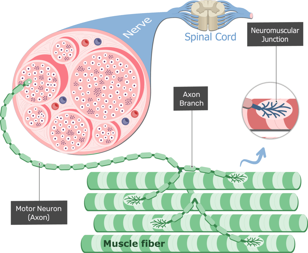
Motor unitA motor unit comprises a single motor neuron and all the skeletal muscle fibers it innervates.
The motor neuron sends electrical signals (action potentials) that simultaneously activate all its muscle fibers.
Neuromuscular junctions (NMJs) are specialized synapses where the neurotransmitter acetylcholine (ACh)initiates muscle fiber contraction.
Components of a Motor Unit
Motor Neuron
Cell body in the spinal cord or brainstem, axon extends to the target muscle.
Transmits the nerve impulses needed for muscle contraction.
Muscle Fibers
Cylindrical cells that respond to stimulation by contracting.
A single motor unit may contain few or hundreds of fibers, depending on precision vs. strength tasks.
Neuromuscular Junction (NMJ)
Synaptic Terminal: End of the motor neuron's axon storing ACh in vesicles.
Synaptic Cleft: Gap between neuron and muscle fiber.
Motor End Plate: Region of muscle me
How Motor Units Work
Nerve Impulse Transmission
The action potential travels along the motor neuron to the synaptic terminal.
Neurotransmitter Release
ACh is released into the synaptic cleft upon arrival of the action potential.
Muscle Fiber Activation
ACh binds to receptors on the motor end plate, opening ion channels.
This triggers an action potential in the muscle fiber, leading to contraction via the sliding filament mechanism.
Tip
Think of a motor unit as a "team": the motor neuron is the coach, and the muscle fibers are the players.
When the coach gives the signal, the entire team acts together.
Motor Units and Muscle Control
Small Motor Units: Precision Tasks
Found in muscles needing fine control (e.g., eye or finger movements).
Each motor neuron innervates few fibers, allowing delicate adjustments in force.
Example
When you thread a needle, small motor units in your fingers and hands allow for the fine adjustments needed to guide the thread through the eye of the needle.
Large Motor Units: Strength Tasks
Found in muscles for powerful movements (e.g., thigh, back).
Each motor neuron innervates hundreds or thousands of fibers, maximizing force output.
Example
When you lift a heavy box, large motor units in your quadriceps and back muscles are activated to generate the necessary strength.
Recruitment of Motor Units
Size Principle
Smaller motor units (fewer fibers) are recruited first for low-force activities.
Larger motor units (more fibers) are added as force requirements increase.
Energy Efficiency: The muscle uses only the energy it needs, conserving resourcesduring less intense tasks.
Warning
Many students assume all motor units in a muscle are activated simultaneously.
In reality, motor units are recruited progressively based on the force required.
Self Review
What are the components of a motor unit?
How do small and large motor units differ in function?
What role does the neuromuscular junction play in muscle contraction?
B3.3.5 roles of skeletons as anchorage for muscles and as levers
Skeletons Act As Anchorage And Levers for Muscles
Definition
SkeletonA skeleton is a hard framework that protects the animal's body.
There are two types of skeletons:
Exoskeletons
Endoskeletons
Note
Exoskeletons must be periodically shed and replaced during growth, a process called molting.
During this time, the organism is temporarily vulnerable to predators and environmental stressors.
Exoskeletons
Found in arthropods (e.g., insects, crustaceans).
External structure made of chitin, providing protection and muscle attachment.
Example
In a grasshopper, leg muscles attach to the exoskeleton, enabling powerful leaps.
Endoskeletons
Found in vertebrates (e.g., humans, cheetahs).
Internal framework of bones, which grows with the organism.
Example
The long bones in a cheetah's legs anchor muscles, allowing speeds of up to 80 km/h.
Skeletons as Anchorage Points for Muscles
Origins and Insertions
Origin: The fixed attachment point (bone does not move during contraction).
Insertion: The movable attachment point (bone does move when the muscle contracts).
Why Anchorage Matters
Muscles only generate force by shortening.
A rigid anchor (bone) ensures force is converted into movement rather than just muscle shortening in place.
Example
The masseter muscle originates on the cheekbone and inserts on the jawbone, elevating the jaw for chewing.
Tip
To remember the difference: the "origin" stays stationary, while the "insertion" moves during contraction.
Skeletons as Levers
Lever Basics
A lever is a rigid structure (bone) that pivots around a fulcrum (joint).
Effort (muscle force) acts against a load (resistance) to produce movement.
Lever Function
Increase Force: Placing the effort farther from the fulcrum amplifies force (but reduces speed/distance).
Increase Speed or Distance: Placing the effort closer to the fulcrum increases speed or distance of movement (but reduces force).
Example
When performing a bicep curl, your forearm acts as a lever.
The elbow joint serves as the fulcrum, the biceps muscle applies the effort, and the weight in your hand experiences the resultant force.
Classes of Levers in the Body
First-Class Lever
The fulcrum lies between the effort and the load.
Example: Nodding the head (neck joint = fulcrum).
Second-Class Lever
The load lies between the fulcrum and the effort.
Example: Standing on tiptoes, where the ball of the foot is the fulcrum.
Third-Class Lever
The effort lies between the fulcrum and the load.
Example: Bicep curl, where the elbow joint is the fulcrum.
Warning
Students often confuse the classes of levers.
To avoid this, focus on the relative positions of the fulcrum, effort, and resultant force in each case.
Practical Implications of Skeletons as Levers
Specialized Locomotion
Digging: Moles have short, sturdy bones to maximize force.
Running: Cheetahs have long, slender bones for greater speed.
Flying: Birds have lightweight, hollow bones for reduced energy cost.
Biomechanical Versatility
Different lever arrangements offer trade-offs between force and speed/distance of movement.
Evolutionary adaptations align bone structure with an organism's environmental and survival needs.
Analogy
A lever works like a seesaw.
Moving the fulcrum changes whether you need more force to lift a heavy object or less force for faster movement
Tok
How might the principles of skeletons and levers inspire innovations in robotics or prosthetics?
Additionally, how do cultural perceptions of movement, such as dance or athletic performance, shape our understanding of biomechanics?
Self Review
Can you identify the origin and insertion points of a muscle in your own body?
How do these points determine the movement caused by the muscle's contraction?
B3.3.6 movement at a synovial joint
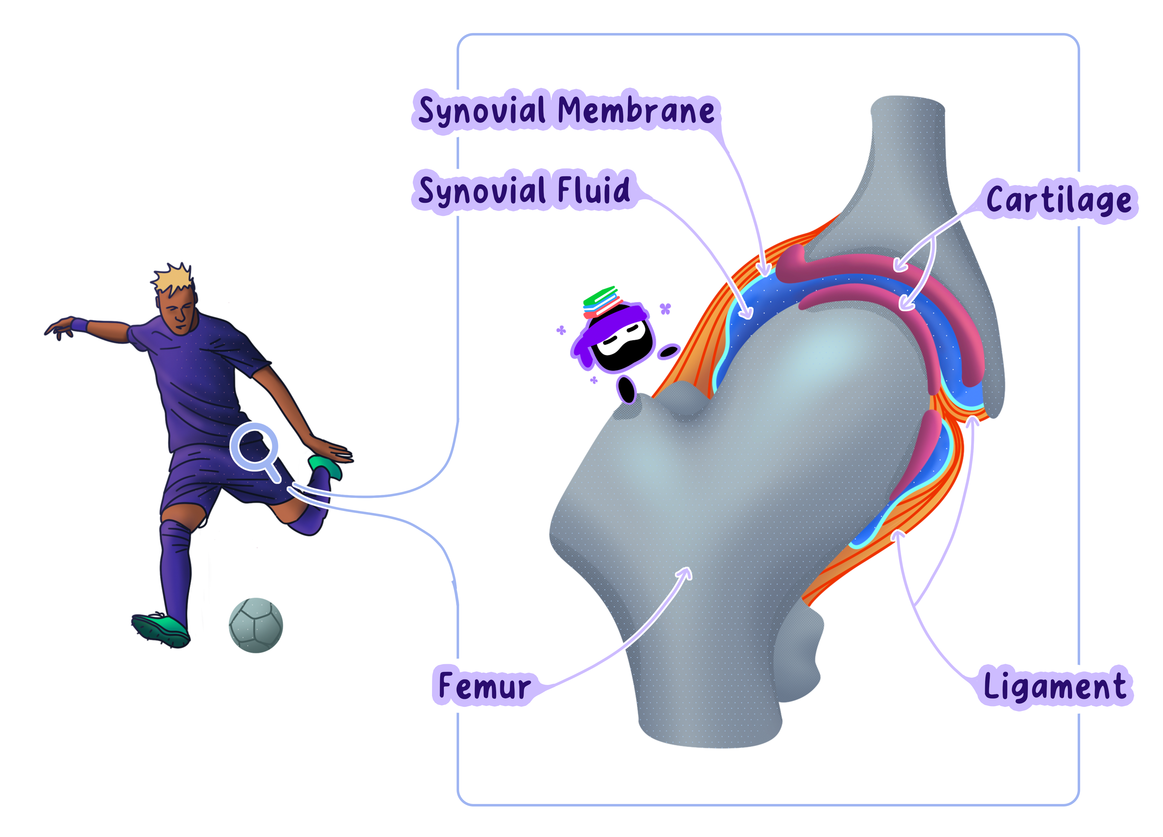
Movement at a Synovial Joint: The Human Hip Joint as an Example
Definition
Synovial jointHighly mobile joints that allow a wide range of movements.
The hip is a ball-and-socket synovial joint connecting the femur (thigh bone) and the pelvis.
This enables flexion, extension, abduction, adduction, and rotation.
Bones AreThe Framework for Movement
Femur: Has a rounded head ("ball") fitting into the acetabulum of the pelvis.
Pelvis: Forms a deep socket ("acetabulum") that holds the femoral head.
Shape & Fit: Determine range of motion and stability.
Tip
Bones act as levers, amplifying the force generated by muscles to produce movement. This principle is essential for efficient locomotion.
Cartilage Reduces Friction and Absorbs Shock
Articular Cartilage: Smooth, tough tissue at bone ends.
Functions:
Reduces friction between moving bones.
Absorbs impact, preventing damage during high-impact activities.
Warning
Students often confuse cartilage with bone. Remember, cartilage is softer, lacks calcium deposits, and is designed for flexibility and shock absorption.
Synovial Fluid Lubricates the Joint
Location: Fills the joint cavity between bones.
Secretion: Produced by the synovial membrane.
Functions:
Lubricates cartilage surfaces, reducing friction.
Nourishes cartilage (since cartilage lacks its own blood supply).
Distributes force, acting as a shock absorber.
Example
Imagine oiling the hinges of a door to prevent squeaking and ensure smooth operation. Synovial fluid performs a similar role in your joints, enabling frictionless movement.
Ligaments Stabilize the Joint
Definition
LigamentsStrong, fibrous bands of connective tissue linking bone to bone.
Hip Ligaments restrict abnormal movements to prevent dislocation.
The joint capsule encloses the hip, helping seal synovial fluid inside.
This provides stability even under heavy stress (e.g., running, jumping).
Note
Ligaments are tough but inelastic. Overstretching them can lead to permanent damage, reducing joint stability.
Muscles Generate Force for Movement
Skeletal Muscles: Attach to bones and contract to produce movement.
Origin & Insertion:
Origin: Usually the stationary bone (e.g., part of the pelvis).
Insertion: The movable bone (e.g., femur).
Contraction: When muscles shorten, they pull the insertion bone, causing joint movement.
Analogy
Think of muscles as engines that convert chemical energy (from ATP) into mechanical work, enabling movement.
Tendons Transmit Muscle Force to Bones
Definition
TendonsFibrous connective tissue that attaches muscle to bone.
Tendons transmit pulling force from muscle to bone.
This allows controlled, efficient movement across the joint.
Tip
Unlike ligaments, which connect bones to bones, tendons connect muscles to bones.
This distinction is key when studying joint mechanics!
Coordinated Action: How the Hip Joint Enables Movement
Muscle Contraction: A muscle shortens and pulls on its tendon.
Force Transmission: The tendon pulls on the femur or pelvis, depending on the movement.
Joint Movement: The ball-and-socket design allows the femur to rotate or move in multiple planes.
Friction Reduction: Cartilage and synovial fluid ensure smooth motion by minimizing resistance.
The Hip Joint Balances Between Stability and Flexibility
Stability
Deep Socket (Acetabulum): Holds the femoral head tightly.
Strong Ligaments & Capsule: Prevents excessive or abnormal movement.
Musculature: Surrounding hip muscles also enhance joint stability.
Flexibility
Ball-and-Socket Design: Permits rotation and movement in multiple directions.
Multiple Planes of Motion: Flexion, extension, abduction, adduction, and circumduction.
Tok
How do engineers design prosthetic hip joints to mimic the natural balance of stability and flexibility?
Self Review
How do cartilage and synovial fluid work together to prevent joint damage?
Why is the hip joint classified as a ball-and-socket joint, and how does this structure support a wide range of motion?
B3.3.7 range of motion of a joint
Range of Motion: Factors That Determine How Joints Move
Definition
Range of motionThe range of motion (ROM) refers to how far the bones of a joint can move relative to each other.
Multiple Factors Influence a Joint's ROM
1. Joint Structure
Hinge Joints (e.g., elbow, knee): Allow movement in one plane, flexion (bending) and extension(straightening).
Ball-and-Socket Joints (e.g., hip, shoulder): Permit movement in multiple planes, including abduction, adduction, flexion, extension, and rotation.
These joints have the greatest ROM.
Example
Your shoulder joint, a ball-and-socket joint, allows you to throw a ball, lift your arm sideways, or rotate it in a circular motion.
Meanwhile, your knee joint, a hinge joint, is limited to bending and straightening, which is ideal for walking and running.
Analogy
Think of a hinge joint as a swinging door, it moves forward and backward but not side to side.
In contrast, a ball-and-socket joint is like a joystick, enabling movement in almost any direction.
2. Ligaments and Joint Capsule
Ligaments: Strong, fibrous tissues that connect bones, limiting excessive or abnormalmovements.
Joint Capsule: A protective envelope around the joint that contributes to stability and restricts excessivemotion.
Note
Ligaments act like seat belts, they keep the joint stable but limit how far it can move.
Overstretching them through injury can compromise joint stability and increase the risk of dislocation.
3. Muscles and Tendons
Muscles generate force, tendons connect muscle to bone.
The flexibility of these tissues heavily influences ROM.
Stretching can lengthen muscles and tendons, improving joint mobility over time.
Tip
To enhance your range of motion safely, focus on dynamic stretches before physical activity and static stretches afterward.
4. Other Factors
Age and Gender: Younger individuals and females often have greater flexibility.
Injuries and Health Conditions: Ligament tears, arthritis, or cartilage damage can reduce ROM.
Activity Level: Regular exercise and stretching help maintain or enhance flexibility.
Warning
It's easy to confuse flexibility with the range of motion.
Flexibility refers to the ability of muscles and tendons to stretch, while the range of motion describes the movement possible at a joint.
Both are interconnected but not identical.
Measuring the Range of Motion
Researchers, therapists, and athletes measure ROM to evaluate joint function or trackrehabilitation progress.
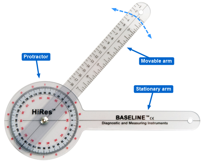
1. Goniometer
A protractor-like device with two arms to measure joint angles.
Center the goniometer on the joint, align one arm with the stationary bone, and the other with the movingbone.
Read the angle for the joint's range of motion.
2. Computer Image Analysis
Uses video or photographic data to measure joint angles digitally.
Valuable for dynamic movements in sports or rehabilitation programs.
Tip
Digital goniometers and mobile apps can simplify measurements and provide additional data, such as tracking progress over time.
Comparing the Range of Motion Across Joints
Elbow Joint (Hinge)
Movements: Flexion and Extension
Range: Approximately 0° (extended) to 140° (fully flexed)
Function: Provides stability for lifting and pushing tasks
Hip Joint (Ball-and-Socket)
Movements: Flexion, Extension, Abduction, Adduction, Rotation
Range (approximate):
Flexion: up to 120°
Extension: up to 30°
Abduction: up to 45°
Adduction: up to 30°
Rotation: up to 45°
Function: Enables diverse movements like walking, running, and sitting cross-legged
Tok
Should athletes prioritize flexibility or stability in their joints? Can increasing the range of motion in some joints lead to a higher risk of injury?
Case_study
Practical Activity: Investigating Range of Motion
Choose Two Joints (e.g., elbow and hip).
Measure ROM for different movements (flexion, extension, abduction, rotation).
Record data and compare results.
Reflect on how joint structure influences ROM.
Self Review
What factors might explain differences in the range of motion between individuals?
How could age, gender, or activity level influence your findings?
B3.3.8 internal and external intercostal muscles
ntercostal Muscles Coordinate Inhalation and Exhalation
The intercostal muscles are located between the ribs and are essential for breathing.
They are divided into two layers: external intercostal muscles and internal intercostal muscles, with fibers oriented in different directions.
The alternating contraction of these layers moves the ribcage in opposite directions, enabling the mechanics of breathing.
Coordinated Contraction and Relaxation
The alternating contraction of these layers moves the ribcage in opposite directions, enabling the mechanics of breathing.
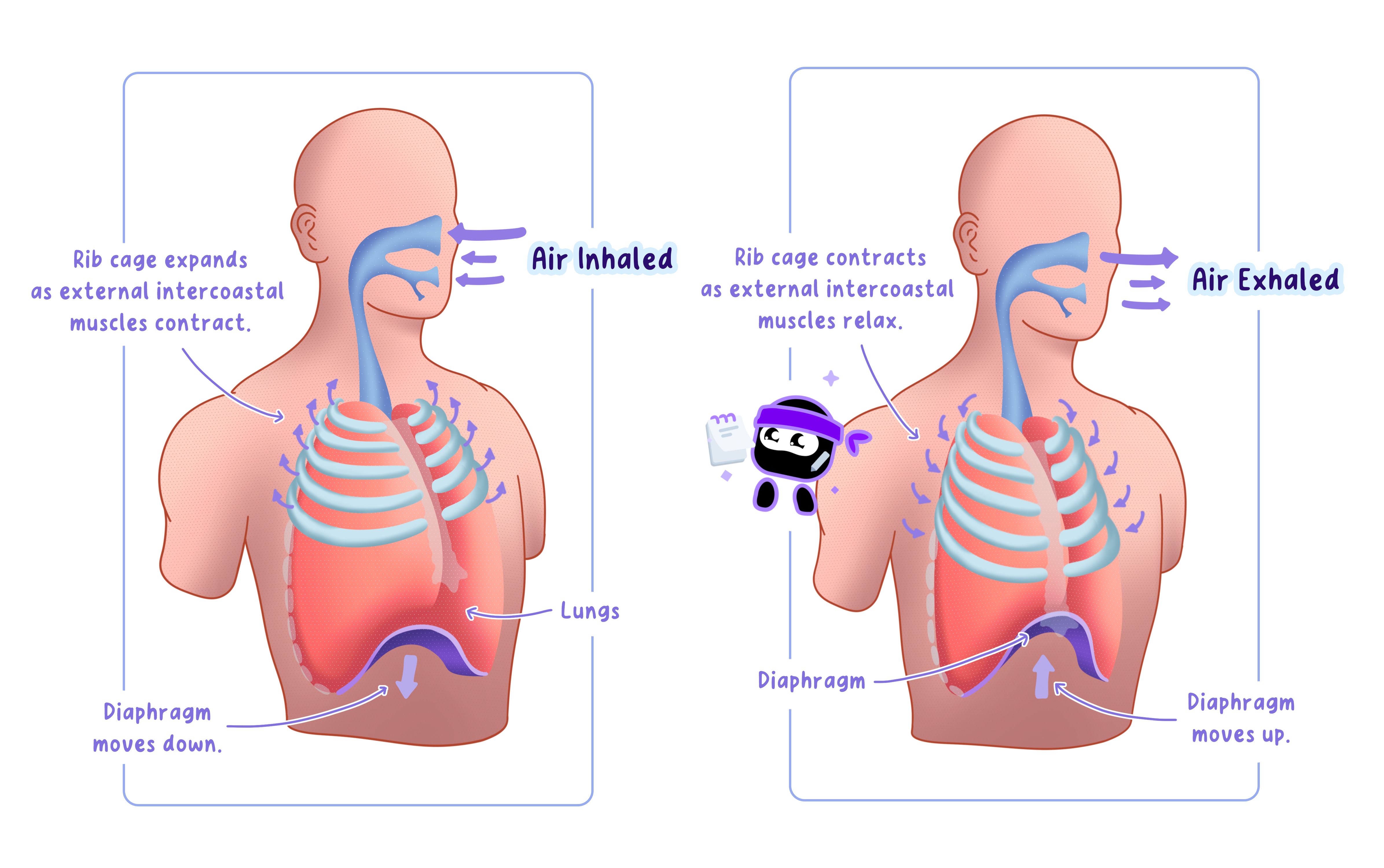
During Inhalation
External intercostal muscles contract, expanding the ribcage outward and upward.
This expansion stretches the internal intercostal muscles, storing potential energy in the protein titin within these muscles.
Analogy
Imagine pulling on a rubber band.
As you stretch it, energy is stored in the band.
Similarly, when the external intercostal muscles contract, they stretch the internal intercostal muscles, storing energy in the titin molecules within their fibers.
During Exhalation
The internal intercostal muscles contract, compressing the ribcage and stretching the external intercostal muscles.
This process efficiently uses the stored energy to return the ribcage to its resting position.
Note
Titin in the intercostal muscles acts like a spring, storing and releasing energy during each breath cycle.
This mechanism reduces the energy cost of breathing by making movements more efficient.
Self Review
Can you describe how the orientation of muscle fibers in the intercostal muscles contributes to their opposing actions?
How does titin enhance the efficiency of these movements?
B3.3.9 reasons for locomotion
1. Foraging for Food: Movement to Satisfy Hunger
Essentials of Feeding
Animals move to find nutrients not immediately available in their current habitat.
Predators stalk or chase prey, herbivores wander or migrate in search of fresh vegetation.
Example
Bees flying from flower to flower to collect nectar and pollen (critical for pollination).
Wildebeests migrating across the African savannah to access lush grasslands.
Hint
Remember that foraging strategies often depend on the type of food an organism consumes, predators move to hunt, while herbivores move to graze or browse.
2. Escaping from Danger: Locomotion as a Survival Mechanism
Avoiding Predators and Hazards
Locomotion enables rapid evasion of threats, including predators or natural disasters.
Speed and agility can confuse predators or allow prey to reach safety.
Example
Springboks in southern Africa rely on high-speed running (up to 80 km/h) and pronking(leaping) to elude cheetahs and alert nearby springboks.
Warning
Many students assume that only prey animals use locomotion to escape danger.
Predators also move to avoid larger predators or to retreat from unsuccessful hunts.
3. Searching for a Mate: Locomotion for Reproduction
Ensuring Genetic Diversity
Animals travel to locate mates, especially in widely dispersed populations.
This movement prevents inbreeding and maintains a robust gene pool.
Example
Male American moon moths fly toward pheromones released by distant females.
Young male lions leave their birth pride and travel to other prides, challenging the dominant male for breeding rights.
Note
Locomotion for mating is not limited to terrestrial animals. In aquatic species like salmon, individuals migrate vast distances to reach spawning grounds where they can reproduce.
Tip
Locomotion for mating often involves energy-intensive journeys, but the reproductive success it enables makes the effort worthwhile.
4. Migration: Long-Distance Movement for Survival
Seasonal or Periodic Relocation
Organisms relocate to avoid food scarcity or harsh climates and, in some cases, to reproduce.
Often entails extended journeys over great distances.
Example
Salmon migrate from the ocean to freshwater rivers to spawn, then return (or die after spawning), ensuring species continuity.
Hint
Migration often involves preparation, such as building fat reserves for energy or timing the journey to coincide with favorable weather conditions.
B3.3.10 adaptations for swimming in marine mammals
Adaptations of Marine Mammals for Aquatic Life
Marine mammals, which evolved from terrestrial ancestors about 50 million years ago, exhibit specialized adaptations for swimming in water, a medium approximately 1,000times denser and far more viscous than air.
These adaptations minimize resistance and enhance locomotion and breathing efficiency in an aquatic environment.
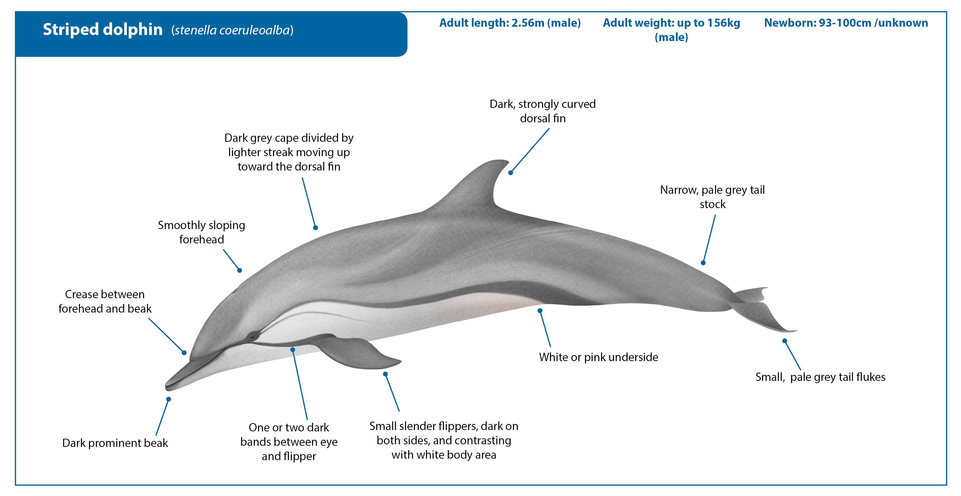
Streamlining Reduces Resistance
To overcome water resistance, marine mammals have evolved a streamlined body shape that reduces drag during motion:
Tapered Body Shape: Bodies are widest near the front and taper towards the rear, minimizing drag compared to other shapes.
Elongated Teardrop Profiles: Flippers, flukes, and dorsal fins have a teardrop-shaped cross-section, reducing water resistance during movement.
Smooth Body Surface: The blubber distribution and the absence of hind limbs and external ear flaps create a smooth contour.
Hairless Skin: Skin lacks hair, further reducing friction with water.
Example
The hydrodynamic design of dolphins enables them to swim at speeds up to 60 km/h, conserving energy by minimizing drag.
Adaptations for Locomotion
Marine mammals rely on specialized structures for efficient and controlled movement:
Flippers: Function as steering devices, replacing the role of front legs.
Flukes: Lobes of the tail move up and down, generating powerful thrust.
Dorsal Fin: Provides stability by preventing rolling during swimming.
Blubber: Offers buoyancy, enabling the animal to float just below the water's surface, even while resting or sleeping.
Tip
The dorsal fin stabilizes dolphins much like a rudder stabilizes a ship, ensuring balance and smooth movement.
Breathing Adaptations: Efficient Airway Design
Marine mammals have developed mechanisms to optimize lung ventilation while avoiding water entering the respiratory system:
Blowhole: Positioned on the top of the head, allowing the animal to breathe at the water's surface without fully lifting its head.
Separated Airways: No connection exists between the mouth and lungs, ensuring water does not enter the lungs during feeding or diving.
Warning
A common misconception is that marine mammals breathe through their mouths.
In fact, their lungs are ventilated exclusively through the blowhole.
Integrated Design for Aquatic Life
The combination of streamlining, locomotion adaptations, and breathing efficiencyenables marine mammals to thrive in aquatic environments.
These adaptations allow them to:
Swim with minimal energy expenditure.
Steer and stabilize effectively during high-speed movement.
Ventilate their lungs efficiently, even in rough seas.
Hint
Aquatic animals are quite literally "built different," just as any specialized function is in order to best adapt to their environment.
Self Review
How does body shape minimize drag?
What distinct roles do flippers and flukes play in swimming?
Why is it crucial that breathing and feeding paths remain separate in marine mammals?