Bio-Lab
The scientific method → process that involves the formation and testing of hypotheses.
called an iterative process
Steps:
Hypothesis
Experiment
Analysis
Conclusion
ask questions → make a hypothesis → test hypothesis → analyze results → draw conclusions → communicate results
if results do not support hypothesis, you do not communicate results to broader community.
Hypothesis → formulated statement of predicted outcomes.
Controlled experiment → allows scientists to test hypothesis and to draw conclusions based on reproducible results.
allows comparison between a control group and a test (experimental) group that has been exposed to an experimental factor (independent variable).
In the analysis phase → the results (dependent variable) of the experiment are examined for agreement with the hypothesis. Any differences that are observed between the two groups can be attributed to the effects of the experimental factor (independent variable).
The conclusion → includes a representation of the results and a discussion regarding the confirmation or rejection of the original hypothesis.
Independent variable → what the scientist intentionally changes.
Dependent variable → what the scientist measures as the result.
Constants → conditions kept same during experiment.
Control group → provides a comparison to determine the effect of the independent variable
Writing a hypothesis → If the (independent variable) changes, then the (dependent variable) will change in a predicted way, because of a scientific reason.
Controls give scientists something to compare their results against. Constants prevent other factors from changing the results.
Macromolecules → large biological molecules important for cell structure
Monomer → a substance that is the smallest unit of a category of substances.
Polymer → a substance made up of many units of a common chemical attached to each other.
Lipid
Lipid → a macromolecule made up of dozens to hundreds of molecules of mostly carbon and hydrogen.
Triglycerides → made up of a three-carbon glycerol molecule with three fatty acid chains attached to it.
Fatty acids → long chains of carbon with hydrogen attached, making them nonpolar molecules.
Saturated fatty acid → a chain of carbon atoms using only single carbon-to-carbon bonds with hydrogen atoms attached to the carbon atoms. → NO double bonds.
Unsaturated fatty acid → a chain of carbon atoms that contain one (monounsaturated) or more (polyunsaturated) double bonds between carbons with hydrogen atoms attached to the carbon atoms. → double bonds.
Positive result → evenly distributed oily stain, water completely evaporates, and nonlipid substances mixed with water will leave a circular stain.
Protein
Protein → polymers consisting of long chains of amino acid monomers.
Two amino acids = dipeptide
Multiple amino acids folded to form a functional structure make up a protein.
The “R” in the amino acid structure represents any one of 20 possible functional groups found in the amino acids used in biological systems.
Biuret reagent → a light blue reagent that tests for protein.
Positive result → anytime the reagent changes from its original blue color to a pink or purple color. Any tube that is blue, no matter which shade of blue, does NOT contain protein.
The Biuret reagent turns from light blue to pink if a low concentration of protein is present.
The Biuret reagent turns from light blue to dark purple indicates a high protein concentration.
light blue → pink = low concentration
light blue → dark purple = high concentartion
Carbohydrates
Carbohydrates → organic molecules that contain carbon, hydrogen, and oxygen. The monomer units of carbs are monosaccharides, while the polymer unit of carbs are known as polysaccharides.
molecules that are not readily tested with any of the four utilized reagents are: RNA, DNA
Starch → rice
Simple sugar → sugar
Protein → egg
Lipid → vegetable oil
Starch → a polysaccharide consisting of long chains of glucose.
storage form of carbohydrates in plants. As a food source, starch is vital to survive.
(Plant based)
carbohydrate polymer made of many smaller monomers, glucose.
Iodine → yellowish-brown colored liquid that turns blue or black (purple) when it contacts starch.
tests for polysaccharides, not monosaccharides.
Glucose → a small, six-carbon sugar molecule found in starch and glycogen.
monosaccharide.
Monosaccharide → one individual molecule of sugar; the building blocks of carbohydrates.
Disaccharide → a carbohydrate made up of two sugar molecules linked together.
Maltose → a disaccharide consisting of two glucose molecules bound together.
Simple sugar → monosaccharides and disaccharides.
Oligosaccharide → a carbohydrate made up of more than two sugar molecules linked together.
Polysaccharide → a carbohydrate made up of hundreds to thousands of sugar molecules linked together.
Glycogen → an animal-based polysaccharide.
Positive control → any procedure that is known to produce the desired result. What you are looking for is present.
Negative control → any procedure that is known not to produce the desired result. What you are looking for is not present.
How to interpret the starch test:
positive test result occurs when iodine turns color.
negative test result occurs when iodine stays yellow.
medium blue → small amount of starch
dark blue → medium amount of starch
blue-black → large amount of starch.
Sugar
Benedict reagent → identify simple sugars.
reaction requires heat to take place.
Benedict test for sugar:
positive result occurs anytime the reagent changes from its original blue color.
Blue → lack of simple sugars
Green (low) → Orange (medium) → Red (high)
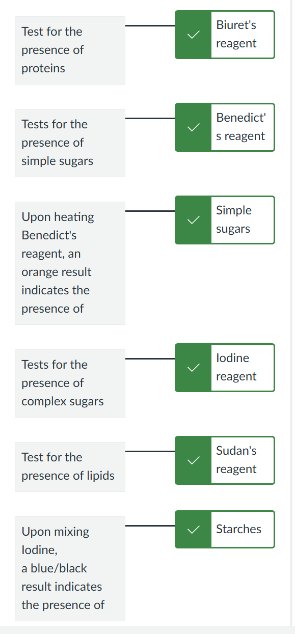
Proteins → Biuret’s reagent
Simple Sugars → Benedict’s reagent
Complex Sugars → Iodine reagent
Lipids → Sudan’s reagent
Accuracy → how closely the measured values agree with the true value.
Precision → how closely repeated measures of the same thing agree with each other.
repeatability of a measurment.
Calculated volume (mL) = mass (g) / density (g/mL)
Deviation → absolute value of the differences between the trial calculated volume and average calculated volume.
Standard deviation → square root of the variance; indicates how far data vary from the mean.
Variance → sum of squared deviations / (number of trials - 1)
Measurement precision can be increased by → increasing fineness of measure.
Accuracy can be increased by → calibrating instruments correctly.
Increasing the precision of the measuring instrument → decreases the variance and standard deviation.

Pipette → narrow tube with attached pump used to measure and transfer specific volumes of liquid.
Pipette pump → device attached to pipette tube and has a plunger that is raised and lowered to draw fluid into tube and dispense liquid out of tube.
Pipette tip → pointed end of a pipette that is placed into a liquid. Tip is a separate piece that must be attached to the micropipette before each use.
Micropipette → measuring and transferring very small volumes of liquid, usually in microliters, has a build-in pipette pump where a desired volume is set using a dial.
a new disposable micropipette tip must be attached for each use.
micropipettes measure in one millionth of a liter units
plunger is depressed to the first stop prior to drawing solution into micropipettor.
In a 200 micropipette → top digit represents hundreds, next is tens, then ones.
In a 20 micropipette → top digit represents tens, then ones, then tenths.
Transfer (bulb) pipette → small pipettes used for transferring relatively small liquid volumes when precise measurement is not critical.
Volumetric pipette → pipettes with precise, graduated markings used to accurately measure and move specific volumes of liquid.
Proper Technique For Pipetting
never place by mouth.
when drawing liquid into pipette, do not submerge entire pipette, only the tip.
make sure tip stays immersed in liquid and does not touch bottom of tube or beaker.
never fill up pipette or micropipette all the way.
never touch end of pipette tip when transferring liquid.
carry pipette or micropipette vertically.
when dispensing liquid, use thumbwheel for slow and controlled dispensing, dispense only part of liquid.
for micropipettes, never re-use tips.
Different models of micropipettes:
0.5-10 → volume of 2.6
5-50 → volume of 20.0
20-200 → volume of 50
200 → volume of 98

Elodea (plant cell)
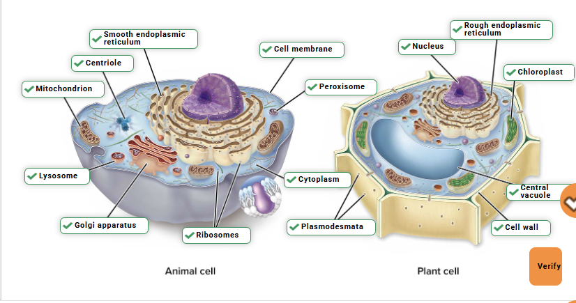
chloroplasts
central vacuole
cell wall
Cheek cell (animal cell)
nucleus
cell membrane
cytoplasm
All cells contain
DNA
RNA
Ribosomes
Proteins
Cytoplasm
Cell membrane
Archaea → one of two domains of prokaryotes; single-celled and metabolically diverse; closest prokaryotic relatives of eukaryotes.
Bacilli (singular, bacillus) → rod-shaped bacteria.
Bacteria → one of two domains of prokaryotes; single-celled and metabolically diverse; oldest and most numerous organisms on Earth.
Calibration slide (stage micrometer) → microscope slide etched with precise scale lines of true length.
Cell → small membrane-bound compartment that is the basic unit of living organisms that performs all the processes of life.
Cell membrane → lipid rich barrier that encloses all cells and regulates the passage of substances into and out of cells.
Cell wall → rigid, protective layer surrounding cells of many prokaryotes, protists, fungi, and plants.
Cellular respiration → group of cellular reactions that extracts energy from food molecules.
Central vacuole → membrane-bounded organelle in plant cells that stores water, ions, sugars, and other molecules.
Chloroplasts → site of photosynthesis in eukaryotes; found in cells of plants and some protists.
Cocci (singular, coccus) → spherical-shaped bacteria.
Cyanobacteria → group of aquatics and photosynthetic bacteria, include Anabaena, and played an important role in contributing oxygen to the ancient earth’s atmosphere.
Cytoplasm → semifluid matrix that fills interior of cell and contains a cell’s molecules, structures, and in eukaryotes, organelles.
Eukaryotes → organisms composed of one or more cells containing a membrane-bounded nucleus and other membrane-bounded organelles; includes protists, fungi, plants, and animals.
larger than prokaryotes.
Eukaryotic cells → complex cells that contain a nucleus and other membrane-bounded organelles.
make up both unicellular and multicellular organisms.
Mitochondria → organelles that are the sites of cellular respiration reactions in eukaryotes.
Nucleoid → area in prokaryotic cell where DNA is located.
Nucleus → membrane-bounded compartment that contains the DNA in a eukaryotic cell.
Ocular micrometer → see ocular scale.
Ocular scale (ocular micrometer) → row of lines etched into the eyepiece of a compound microscope; can be calibrated and used to measure size of specimens being viewed.
Organelle → membrane-bounded compartment of a eukaryotic cell that performs a specialized function.
Photosynthesis → group of cellular reactions that capture light energy and use it to power the production of organic molecules; oxygen is a by-product.
Plasmodesmata (singular, plasmodesma) → channels in the cell walls of adjacent plant cells that act as tunnels for the exchange of materials between cells.
Plasmolysis → process in which plant cells lose water to their surroundings, causing the cell to shrink and the cell membrane to pull away from the cell wall.
Prokaryotes → organisms composed of a cell that lacks a nucleus and other membrane bounded organelles; includes bacteria and archaea.
Prokaryotic cells
cells that have a nucleoid area containing the DNA but lack a membrane-bounded nucleus.
Spirilla (singular, spirillum) → spiral or corkscrew-shaped bacteria.



Magnification → increase in image size.
Resolution → ability to distinguish fine detail.
Contrast → ability to distinguish objects, such as cells, from the background.

Field of view → amount of the slide you are able to see through the eyepiece.
as magnification increases, the field of view decreases.
Depth of Field → vertical distance that is in focus.
how much thickness stays in focus.
power increases → depth of field decreases.
Low power → depth of field greater
High power → depth of field smaller
Low power → width of view larger
High power → width of view smaller
Low power → less detail
High power → more detail
Parfocal → object viewed should stay in focus when changing magnification.
Total magnification → overall enlargement of the image of a specimen.
to calculate total, multiply magnification of the ocular lens (10X) with the magnification of the objective lens.
Low magnification → see more specimen, less detail
High magnification → less specimen, more detail
as magnification of image increases, light intensity decreases and will have to be adjusted.
as magnification increases, both width and depth of field decrease.
Condenser lens → focuses the light on the slide.
Ocular lens → always 10X
Sliding bar → on the condenser lens is the aperture → as you move the aperture to decrease the light to the slide, the contrast increases. As you move the aperture to increase the light, the contrast decreases.


Light Microscope
increases ability to see detail by 1000x.
objects as small as 0.1 micrometer can be seen.
Ocular lens → eyepiece you look through (10x)
Objective lenses → magnify the specimen (4x,10x,40x)
Revolving nosepiece → rotates to change objective lenses.
Stage → holds the slide.
Stage clips → keep the slide in place.
Coarse adjustment knob → moves the stage a lot for rough focusing.
Fine adjustment knob → makes small adjustments to sharpen image.
high power
Diaphragm → controls how much light passes through.
Condenser → focuses light onto the specimen.
Light source → provides light to see the specimen.
Arm → supports the microscope; used when carrying.
Base → bottom support of the microscope.
Macromolecule Metabolism
Natural Cellular processes are very slow
most chemical reactions would normally occur only at high temperatures.
much higher than those inside a cell
Reactions require major boost in chemical reactivity
allows reactions to proceed rapidly in cell.
Enzymes
Catalytic proteins → speed up the chemical reactions that occur in the body and are unchanged by reaction.
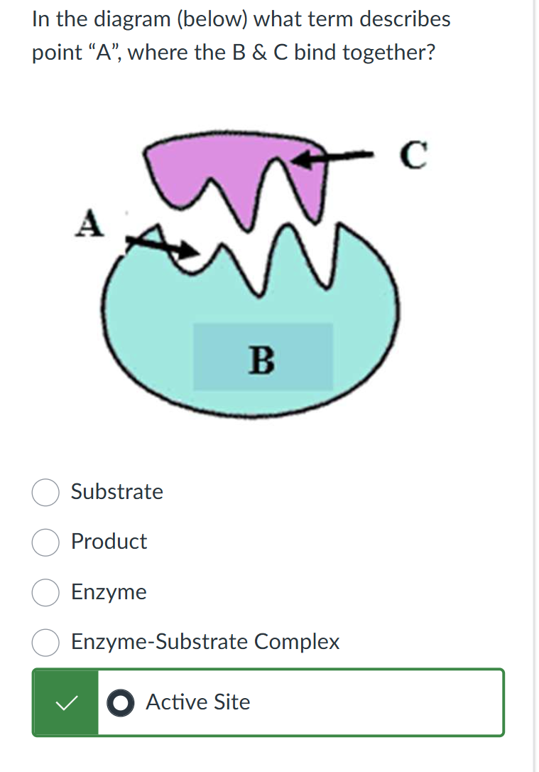
bind to a substrate at active site.
protein structure → enzyme-substrate specificity
reusable
Enzyme changes shape → induced fit
holds substrate in a way that reduces activation energy.
Activation energy → amount of energy required for a reaction to proceed.
enzymes reduce → increased rate of reaction.
Enzyme activity can be affected by:
temperature
pH
enzyme & substrate concentration
inhibitors → blocks enzymes & reduce function
temp and pH affect the rate of diffusion.
Temperature
Increase in temperature
increase kinetic energy
increase collisions
too high → denatures enzyme
Decrease in temperature
lower kinetic energy
less frequent collision
too low → deactivation of enzyme
pH
change in pH disrupts the enzymes shape and structure.
Substrate and Enzyme Concentration
initially, more substrate = faster reactions = more frequent collisions.
will reach a saturation point
all enzymes are bound to substrate.
will need to add more enzymes to increase reaction rate.
Catalysis → conversion of substrate to product.
decrease as temperature lowers.
Active site → location on an enzyme to which a substrate bind
Denaturation → loss of an enzyme’s functional shape.
Induced fit → alteration of the enzyme binding site once a molecule binds.

Diffusion Across a Selectively Permeable Membrane
Diffusion → net movement of a solute from an area of higher concentration to an area of lower concentration.
result of the random movement of molecules.
Concentration gradient → present when there is a different concentration of solutes within a solution.
Rate of diffusion → may be influenced by density of media, concentration gradient, temperature, molecular size, and membrane permeability.
Media → substance through which diffusion occurs, such as water and air.
Solution → a mixture of two substances with one dissolved in the other.
Solute → the substance dissolved in a solution.
Solvent → substance that there is most of in a solution.
Selectively permeable membrane → allows some substances to pass through and blocks others based on size.
Movement of Water Across a Selectively Permeable Membrane
Selectively permeable (semipermeable) membrane → only allows certain molecules through and blocks others.
Osmotic pressure → influenced by changes in nonpermeable solute concentration.
Higher nonpermeable solute concentration → increases amount of water that will cross the membrane.
Hypertonic → a solution with a higher concentration of nonpermeable solutes in comparison to another solution.
higher solute concentration outside a bag.
Isotonic → a solution has the same solute concentration inside and outside the cell.
animal cells
Hypotonic → a solution with a lower concentration of nonpermeable solutes in comparison to another solution.
plant cells
Osmosis → movement of water across a selectively permeable membrane from a hypotonic solution into a hypertonic solution.
movement of water across a membrane.
Active transport → moves substances from low to high concentration, requiring ATP.
Osmotic pressure → the pressure applied to a selectively permeable membrane to prevent osmosis.
Thistle tube → a long tube of glassware with an open round reservoir at one end.
Hypertonic: water leaves the cell → cell shrinks
Hypotonic: water enters the cell → cell swells
Isotonic → no net water movement → cell stays the same size.
Active transport: low → high (requires energy)
Passive transport: high → low (no energy required)
Blood cells placed in a hypotonic solution experience hemolysis.
water enters the red blood cells
cells swell and burst
hemolysis → bursting of red blood cells
Cellular Respiration - Measuring Energy Production in Plants
Primary goal: to make ATP
Stages of respiration: Glycolysis → breakdown of pyruvate → citric acid cycle → oxidative phosphorylation
Aerobic cellular respiration:
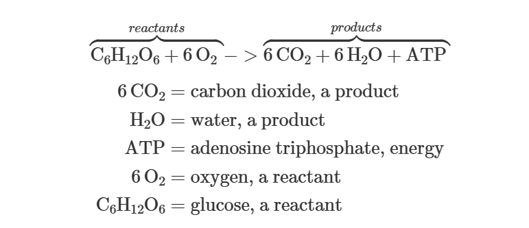
One glucose molecule reacts with 6 oxygen molecules to produce 6 carbon dioxide, 6 water, and energy to make between 28-38 ATP molecules.
Aerobic cellular respiration → multistep process that requires oxygen.
First step → glycolysis → takes place in cytoplasm.
Next steps → Krebs cycle and electron transport chain → occurs in the mitochonrdria.
At the end of ETC → oxygen is consumed when it accepts electrons and is reduced to form water.
Volume of oxygen consumed -→ estimates amount of cellular respiration.
Monosaccharide → one individual molecule of sugar; building blocks of carbohydrates.
glucose, fructose
Disaccharide → carbohydrate made up of two sugar molecules linked together.
maltose, sucrose
Polysaccharide → carbohydrate made up of hundreds to thousands of sugar molecules linked together.
cellulose, glycogen, starch
Germinating → beginning to grow.
Polyester → will not soak up KOH, can be used as a barrier between KOH and the seeds.
Oxygen consumption → measured using a respirometer.
How to read respirometer → the graduated side arm contains a drop of red fluid to mark the amount of oxygen consumed from inside the experimental container. Read left edge of red marker for consistency.
Photosynthesis - Photosynthetic Pigments
Plants use carbon dioxide + energy from sun during photosynthesis to produce glucose.
White light → made up of a range of wavelengths from red with the longest wavelength to violet with the shortest wavelength.
combination of wavelengths.
each group of wavelengths has a characteristic color.
Plants use multiple photosynthetic pigments to absorb light energy used in photosynthesis. The wavelengths of light absorbed can be used in photosynthesis, and the wavelengths of light reflected are what we observe.

Chlorophyll extract → appears green, hiding presence of multiple pigments.
possible to visualize different pigments found in chlorophyll by separating them - one method is paper chromatography.
Photosynthetic Pigments:
Chlorophyll a → absorbs red-violet light, appears blue green.
Chlorophyll b → absorbs red-violet light, appears yellow green.
Carotenes → absorb blue-violet light, appears orange.
Xanthophylls → absorb blue light, appears yellow.
Why does chlorophyll b appear yellow green → it reflects light in those wavelengths that absorbs violet and red.
Why did the different photosynthetic pigments migrate to different points on the chromatography paper → the pigments differ in their solubility in the solvent and affinity for the paper.
Paper Chromatography
chloroplast extract is placed at end of chromatography paper, and the end is placed in a solvent.
the solvent carries the molecules as it migrates up the paper → separating the molecules.
molecules are separated based on their chemical composition.
readily soluble molecules move the farthest compared to molecules more strongly attracted to the paper.
paper chromatography works by separating the different photosynthetic pigments into bands.
Purpose of chromatography solvent → the pigments dissolve in the solvent, which allows them to be separated.
Purpose of using extract from chloroplasts → contain photosynthetic pigments.

Quiz Questions


highest Rf = most nonpolar
Dark reaction → stroma
Light reaction → thylakoid membrane
DNA Biology and Technology - Transcription, Translation, and Mutation
DNA (deoxyribonucleic acid) → a molecule found in all cells that stores the genetic information of the cell.
Eukaryotic cells → found in nucleus.
Prokaryotic cells → found in nucleoid region of the cytoplasm.
RNA (ribonucleic acid) → exists in all cells, synthesized using DNA as a template and functions in the stages of protein synthesis.
Molecule that consists of a strand of nucleotides and carries the genetic instructions of DNA to other parts of the cell for protein synthesis.
RNA nucleotides contain the sugar ribose, a phosphate group, and one of four nitrogenous bases, adenine (A), guanine (G), cytosine (C), and uracil (U).
Nucleotide → subunit of DNA and RNA that consists of a five-carbon sugar bonded to a nitrogenous base and a phosphate group.
5 kinds of nitrogenous bases: adenine (A), guanine (G), cytosine (C), thymine (T; DNA only), uracil (U; RNA only).
Genetic information in DNA and RNA is encoded in the sequence of bases in the nucleotides.
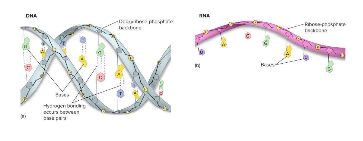
DNA contains the instructions for making all of the RNA and proteins required by a cell. These instructions are encoded in segments of DNA called genes.
Gene → sequence of DNA nucleotides that codes for a specific protein or RNA molecule.
Gene expression → describes the process of how information in a gene is used to produce proteins.
DNA is used as a template for the synthesis of RNA, which then directs the production of proteins from amino acid subunits.
The process in which an mRNA copy of a gene is transcribed and then translated at the ribosome to produce a protein.
Three main types of RNA are synthesized from DNA and used in gene expression:
messenger RNA (mRNA)
ribosomal RNA (rRNA)
transfer RNA (tRNA)
Messenger RNA (mRNA) → a molecule of RNA that encodes a protein; consists of codons and binds to ribosomes during translation.
Carries the genetic information that specifies a protein.
Ribosomal RNA (rRNA) → a molecule of RNA that combines with proteins to form a ribosome.
Combines with proteins to form ribosomes, which are the sites of protein synthesis.
Transfer RNA (tRNA) → a molecule of RNA that binds an amino acid at one end and has an anticodon at the other end; brings amino acids to the ribosome during protein synthesis.
Brings amino acids to the ribosomes.
Two Stages of Gene Expression
Transcription
Translation
Transcription → process that produces RNA using DNA as a template.
An mRNA copy of a DNA sequence is made.
Translation → process that produces a polypeptide at a ribosome according to the sequence of nucleotides in mRNA.
The information in mRNA is used to assemble a polypeptide.
Ribosome → structure in cells that is made of ribosomal RNA (rRNA) and proteins and that binds mRNA and tRNA to synthesize polypeptides and proteins.
Polypeptide → a long chain of amino acids linked together; once it folds into its functional shape, a polypeptide is called a protein.
Process of Transcription
An enzyme called RNA polymerase attaches to one strand of the DNA called the template strand, at a start sequence for a gene called the promoter.
RNA polymerase then moves along the DNA template strand and synthesizes and RNA copy of the DNA by adding RNA nucleotides.
Complementary base-pairing rules determine what RNA nucleotides are added to the growing RNA strand, with uracil replacing thymine in RNA.
When the end of the gene is reached at a sequence called the terminator on the template DNA, RNA polymerase and the new strand of RNA are released from the DNA.
RNA Polymerase → enzyme that uses a DNA template to synthesize a molecule of RNA during transcription.
Template Strand → the strand in a DNA molecule that is used to build a new DNA strand in DNA replication or an RNA strand in transcription.
Terminator → a nucleotide sequence at the end of a gene that signals where a gene’s coding region ends.
Promoter → a nucleotide sequence at the beginning of a gene where RNA polymerase binds to begin transcription.
Amino Acid → organic molecule, monomer of proteins.
composed of a central carbon atom attached to an amino group (-NH2) a carboxyl group (-COOH), a hydrogen, and a variable side group (R group).
Process of Translation
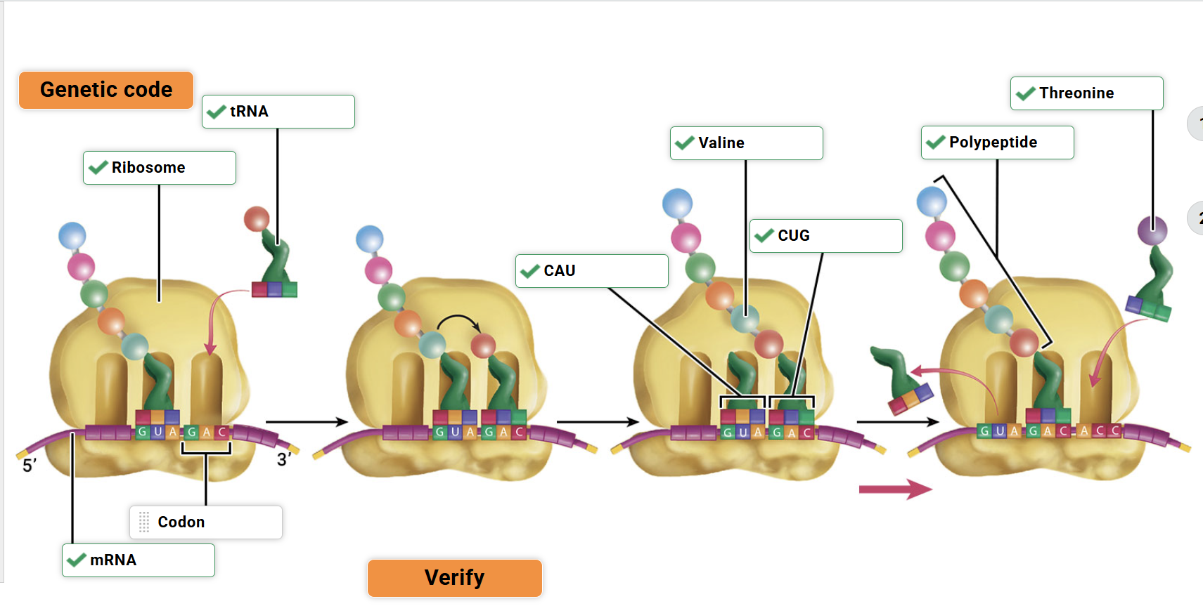
Ribosomal subunits attach to mRNA.
The ribosome moves down the mRNA three nucleotides at a time; each group of three nucleotides on mRNA is called a codon and corresponds to one amino acid, as specified by the genetic code.
Amino acids are brought to the ribosome by tRNA molecules, which have an anticodon that binds to a complementary codon on the mRNA.
Amino acids are added to a growing polypeptide chain one at a time in the order dictated by the sequence of nucleotides on the mRNA.
When the end of a gene sequence on the mRNA is reached, the components of translation dissemble, and the completed polypeptide is released.
The polypeptide can fold or combine with other polypeptides to form a functional protein.
Codon → a sequence of three mRNA bases that specifies an amino acid or the termination of translation
Anticodon → a sequence of three tRNA bases that is complementary to and base-pairs with an mRNA codon.
Genetic Code → the correspondence between nucleotide triplets and specific amino acids.

Mutation → a change in the nucleotide sequence of DNA, which can affect the amino acid sequence of a protein after transcription and translation take place.
Ultimate source of genetic variation among organisms.
Can be caused by DNA replication errors, environmental chemicals or radiation → changes can affect one base pair, a few base pairs, or large parts of chromosomes.
Point Mutations → change o one base pair and can be substitutions, insertions, or deletions.
Substitution → one DNA base is replaced with another.
Insertion → a nucleotide is added.
Deletion → a nucleotide is removed.
Types of Mutations
Silent Mutation → the altered codon specifies the same amino acid; there is no change to the amino acid sequence of the protein.
Missense Mutation → the altered codon specifies a different amino acid; one amino acid is replaced with another, which can drastically affect the protein product.
Nonsense Mutation → the altered codon is a “stop” codon; the protein product is shortened, which can severely affect the proteins’ function.
Frameshift Mutation → the insertion or deletion of a nucleotide shifts the codon reading frame for translation.
Can change the amino acid sequence from the site of the mutation, with devastating effects on protein function.
These type of changes in the DNA sequence affect transcription and can alter the final protein product of translation.
Nucleic Acids
DNA and RNA are both nucleic acids composed of nucleotide subunits linked into a chain.
A nucleotide consists of a five-carbon sugar, a phosphate group, and nitrogenous base.
In DNA, the five-carbon sugar is deoxyribose.
In RNA, the five-carbon sugar is ribose.

A DNA molecule is a double helix composed of two strands of nucleotides whereas an RNA molecule is single-stranded.
In DNA and RNA, the nitrogenous bases of the nucleotides protrude to one side of the backbones.
In the DNA double helix, complementary base pairs on the two strands are joined by hydrogen bonds, such that A pairs with T and C pairs with G.
Transcription complementary base-pairing → used to produce RNA, with the exception that adenine (A) on the template DNA is paired with uracil (U) on the RNA strand.
Translation → amino acid specified by each mRNA codon is dictated by the genetic code.
Some amino acids are encoded by more than one codon.
Three codons (UAA, UAG, UGA) do not code for amino acids and are called “stop” codons because they signal the end of translation.
The codon AUG is typically the first codon in mRNA and is called the “start” codon.
The order of the codons in mRNA determines the order that tRNAs bring amino acids to the ribosome, and therefore, determines the final sequence of amino acids in a polypeptide.


DNA Biology and Technology - DNA and RNA Structure
DNA → molecule that stores genetic material and consists of a double strand of nucleotides
DNA nucleotides contain the sugar deoxyribose, a phosphate group, and one of four nitrogenous bases → adenine (A), guanine (G), cytosine (C), and thymine (T).
2 strands of nucleotides twisted together - double helix shape.
RNA → molecule that consists of a strand of nucleotides and carries the genetic instructions of DNA to other parts of the cell for protein synthesis.
RNA nucleotides contain the sugar ribose, a phosphate group, and one of four nitrogenous bases → adenine (A), guanine (G), cytosine (C), and uracil (U).
Nucleotide → subunit of DNA and RNA that consists of a five-carbon sugar bonded to a nitrogenous base and a phosphate group.
Deoxyribose → five-carbon sugar found in the nucleotides of DNA; lacks an -OH group bonded to the 2’ carbon.
Ribose → five-carbon sugar found in the nucleotides of RNA.
Nitrogenous base → a nitrogen-containing organic molecule that forms part of a nucleotide.
The nitrogenous bases adenine (A) and guanine (G) consist of two carbon-nitrogen rings, while the nitrogenous bases cytosine (C), thymine (T), and uracil (U) consist of one carbon-nitrogen ring.
Phosphate group → functional group containing a phosphorus atom and four oxygen atoms (PO4) that is a component of nucleotides.\
Double Helix → shape of a DNA molecule consisting of two strands twisted together, similar to a spiral staircase but with alternating major and minor grooves along the outside of the double helix.
DNA replication → process by which DNA makes a copy of itself.
Replication fork → site on a DNA molecule where the double helix separates and unwinds to begin DNA replication.
Transcription → process that produces RNA using DNA as a template.
RNA polymerase → enzyme that uses a DNA template to synthesize a molecule of RNA during transcription.
Template Strand → the strand in a DNA molecule that is used to build a new DNA strand in DNA replication or an RNA strand in transcription.
Coding Strand → the strand in a DNA molecule that is not used as a template.


DNA Biology and Technology - Gel Electrophoresis
Individuals with different genetic phenotypes may have different sized DNA fragments when the DNA is cut by enzymes.
DNA fragments can be separated by size using an electrophoresis gel.
DNA fragments are negatively charged and when placed into an electrophoresis gel will travel through an electrical field towards the positive pole.
Electrophoresis buffer floods the gel and allows for the creation of an electric field.
Different DNA fragment sizes will be separated as smaller pieces will move more rapidly.
Loading dye allows for the visualization of the location of the DNA fragments in the gel.
Electrophoresis bromide binds to the DNA and will fluoresce under UV light showing the location of DNA fragments.
TNF alpha → important protein that helps the body resist disease.

Cell Division - Examining Meiosis
Two types of cell division occur in eukaryotic cells → mitosis and meiosis.
In multicellular eukaryotes, most cells of the body reproduce by mitosis.
Some cells in multicellular eukaryotes often called germ-line cells undergo → meiosis.
Meiosis → a cell division process in certain eukaryotic cells that partitions the genetic material into four genetically different daughter nuclei, each with half the number of chromosomes as the parent cell.
Produce gametes.
Shuffle genetic material.
After the chromosomes are divided, the cytoplasm is partitioned to form daughter cells in a process called cytokinesis.
Meiosis includes two divisions of the genetic material → meiosis I and meiosis II. The genetic material is replicated once, before meiosis I, chromosome segregation and cytokinesis occur twice.
The result is that one nucleus divides to produce four daughter nuclei with half the number of chromosomes.
Two processes occur during meiosis that generate genetic variability:
Crossing over → homologous chromosomes exchange genetic material.
Independent assortment → chromosome pairs randomly align along the metaphase plate and assort independently into daughter nuclei.
Meiosis I Four Phases:
Prophase I
Spindle fibers form.
Homologous chromosomes pair up.
Crossing over occurs in which homologous chromosomes may exchange segments.
Nuclear envelope breaks down.
Metaphase I
Homologous chromosome pairs align along equator of cell.
The alignment of homologs toward each pole of the cell is random.
Anaphase I
Homologous chromosomes separate and move to opposite poles of the cell.
Telophase I
Nuclear envelopes form around two daughter nuclei.
Spindle disappears.
Meiosis II Four Phases
Prophase II
Spindle fibers form.
Nuclear envelopes break down.
Metaphase II
Chromosomes align singly along equator of cell.
Anaphase II
Sister chromatids separate and move to opposite poles of the cell.
Telophase II
Nuclear envelopes form around four daughter nuclei.
Spindle disappears.

In animals, germ line cells are found in the ovaries and testes and undergo meiosis to produce gametes.
Gametes → haploid cells produced by meiosis that unite during fertilization as part of sexual reproduction.
Contain half the number of chromosomes as body cells.
Often called eggs and sperm. → zygote
In human females, meiosis begins in the ovaries to produce gametes called eggs.
Germ-line cells called oocytes mature through early stages of meiosis, progress from being contained within small primary follicles → larger secondary follicles → Graafian follicles, have a large fluid-filled antrum.
After ovulation and fertilization, an oocyte becomes an egg cell.
In human males, meiosis takes place in the testes to produce gametes called sperm.
Germ-line cells called spermatocytes undergo meiosis as they move from the outer wall of seminiferous tubules toward the inner lumen of the tubules, where they mature into sperm cells.
Each testis contains many coiled seminiferous tubules.
Micrograph → a photograph taken through a microscope of a magnified specimen.
Mitosis → a cell division process in eukaryotes that divides the genetic material into two identical daughter nuclei.
Homologous chromosomes → two chromosomes that share same size, centromere position, and sequence of genes.
Sister chromatids → one of two identical copies of a chromosome produced by DNA replication and attached to each other at the centromere.
Centromere → constriction where sister chromatids of a duplicated chromosome are held together.
Diploid → cell condition in which two of each type of chromosome are present.
Haploid → cell condition in which only one of each type of chromosome is present.
Gametes, such as sperm and eggs, are haploid.
Cytokinesis → division of the cytoplasm into daughter cells.
Germ-line cells → specialized cells that undergo meiosis to give rise to gametes.
Zygote → diploid cell formed by the union of two gametes during fertilization.
Ovary → paired gonad organ in females containing germ-line cells that give rise by meiosis to eggs.
Oocyte → cell in an ovary that will undergo meiosis to form an ovum (egg).
Follicle → an oocyte and its surrounding tissue sac.
Testis → paired gonad organ in males containing germ-line cells that give rise by meiosis to sperm.
Spermatocyte → cell in a testis that will undergo meiosis to form a sperm cell.


Cell Division - Examining Mitosis
Eukaryotes, the genetic material consists of multiple chromosomes contained in the nucleus of the cell.
Eukaryotic cell division thus consists of a sequence of steps to properly divide the multiple chromosomes into daughter cells.
Mitosis → cell division process in eukaryotes that divides the genetic material into two identical daughter nuclei.
A cell stage called interphase precedes mitosis.
Interphase → each chromosome replicates to become two identical sister chromatids attached at centromere.
Cell growth and protein synthesis also occur during interphase in preparation for cell division.
Mitosis Four Phases
Prophase
Chromosomes condense.
Spindle fibers form.
Nuclear envelope breaks down.
Metaphase
Chromosomes align along equator of cell.
Anaphase
Sister chromatids separate and move to opposite poles of the cell.
Telophase
Nuclear envelopes re-from.
Chromosomes condense.
After chromosomes divide, the cytoplasm is partitioned to form two daughter cells → cytokinesis.
In animals, plants and other multicellular eukaryotes, most of the body cells divide using mitosis.
In eukaryotes, mitosis functions to produce more cells for growth, replace damaged cells, and to repair tissues.
Mitosis also plays a role in producing identical daughter cells during asexual reproduction in some organisms.
Chromosome → a molecule of DNA wrapped around proteins and contained in the nucleus of eukaryotic cells.
Humans have 23 pairs of these in each somatic cell.
Spindle → a set of microtubule proteins that coordinate the movement and division of chromosomes during mitosis.
Sister chromatids → one of two identical copies of a chromosome produced by DNA replication and attached to each other at the centromere.
Centrosome → microtubule-organizing center in cells; in animals cells, it contains two, short cylinders of microtubules called centrioles.
Chromatid → one strand of DNA that makes up a chromosome.
Cleavage Furrow → in animal cells, this location pinches in to separate two new daughter cells.
