Bone and Bone Tissue Anatomy and Physiology
Anatomy and Physiology Overview
Orthopedics and Osteology
Orthopedics: A branch of medicine focused on the prevention and correction of disorders and injuries related to bones, joints, and muscles.
Osteology: The scientific study of bones.
Functions of Bone
Shape: Provides structure to the body, giving it form and shape.
Support: Bones in the legs, pelvis, and spinal column support the body and maintain an upright posture.
Protection: Encloses and protects delicate internal organs.
Movement: Facilitates movement through interaction with muscles. Essential for limb movement and breathing.
Electrolyte Balance: Stores and releases essential minerals such as calcium and phosphorus, crucial for various body functions.
Blood Production: Houses bone marrow, which is a primary site for blood cell formation.
Acid-Base Balance: Bone tissue absorbs and releases alkaline substances to help maintain pH balance in the body.
Classification of Bones
Long Bones: Longer than they are wide; function as levers to facilitate movement. Examples include:
Femur (thigh bone)
Humerus (arm bone)
Short Bones: Approximately equal in length and width; cube-shaped. Examples include:
Carpal bones (wrist)
Tarsal bones (ankle)
Flat Bones: Thin, flat, or slightly curved; protective in nature. Examples include:
Frontal bone (skull)
Ribs
Sternum
Scapula
Irregular Bones: Do not fit into other categories; various shapes and sizes. Example include:
Vertebrae
Parts of a Long Bone
Diaphysis: The main shaft of the bone, characterized by a hollow cylinder composed of compact bone; provides strength.
Articular Cartilage: Smooth tissue covering the epiphysis (end of the bone) that facilitates joint movement.
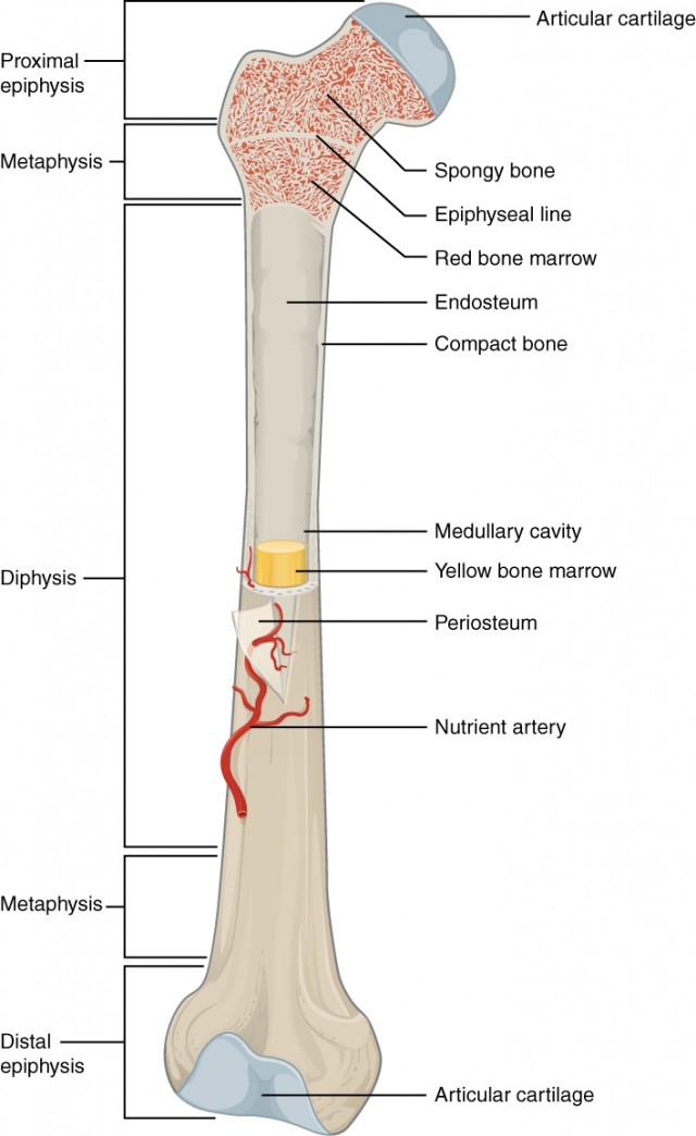
Medullary Cavity: Central, hollow section of the bone; houses bone marrow.
Endosteum: A thin epithelial membrane lining the medullary cavity.
Red Bone Marrow: Fills the medullary cavity in children; in adults, much of this marrow is replaced by yellow marrow.
Periosteum: A dense, fibrous membrane that covers the diaphysis; contains bone-forming cells and blood vessels; important for bone health and nutrient delivery.
Composition of Bone Tissue
Osseous Tissue: Another term for bone; consists of a matrix made up of collagen fibers and crystalline salts (mainly calcium and phosphate).
Strength: Bone has a compressive and tensile strength comparable to steel and reinforced concrete:
Tensile Strength: High resistance to stretching forces, attributable to collagen fibers.
Compressive Strength: Capable of withstanding strong squeezing forces due to calcium salts.
Torsional Strength: Bone is weak against twisting forces; fractures often result from torsional stress.
Bone Cells
Osteoblasts: Cells responsible for bone formation; secrete components of the bone matrix.
Osteoclasts: Responsible for bone resorption; dissolve unhealthy or unwanted bone tissue.
Osteocytes: Mature osteoblasts trapped within the hard matrix of the bone; they function to maintain bone density and regulate bone metabolism.
Factors Affecting Bone Growth and Maintenance
Heredity: Genetic factors determine maximum potential height and bone structure.
Nutrition: Essential nutrients include calcium, phosphorus, and vitamins D, C, and A:
Calcium: Crucial for bone mineralization.
Phosphorus: Works in conjunction with calcium.
Vitamins: Support bone formation.
Hormones: Various hormones including growth hormone, thyroxine, parathyroid hormone, insulin, and sex hormones (estrogen and testosterone) play roles in bone growth.
Types of Bone
Spongy Bone
Consists of a latticework of bone structure called trabeculae, which provides strength and adaptability to stress.
The spaces between trabeculae are filled with bone marrow.
Compact Bone
Structured in layers called lamellae forming around a central canal (haversian canal).
The basic structural unit is the osteon, which facilitates the transport of blood vessels and nutrients through tiny gaps called lacunae and connections known as canaliculi.
Volkmann’s canals provide transverse connections for blood vessel distribution.
Bone Marrow
A soft tissue residing in the medullary cavity and within spongy bone:
Red Bone Marrow: Produces red blood cells, plentiful in children; in adults, found in certain bones (ribs, sternum, vertebrae, skull, pelvis, as well as upper parts of the humerus and femur).
Yellow Bone Marrow: Replaces red marrow with age, mainly composed of fat cells.
Bone Development
Intramembranous Ossification
Early formation of skull and facial bones starting as connective tissue where stem cells differentiate into osteoblasts; mineralization occurs to form actual bone tissue.
Endochondral Ossification
Involves formation of bones from existing cartilage models, beginning around three months into fetal development.
As gestation proceeds, osteoblasts replace cartilage with new bone formation, initiating ossification.
Bone Growth Mechanisms
Epiphyseal Plate: In growing children, this layer of hyaline cartilage allows for lengthening of bones. Chondrocyte multiplication leads to calcification of cartilage.
Growth continues until mature age (16-25) when cartilage is replaced leading to the epiphyseal line, marking growth cessation.
Widening occurs through deposition by osteoblasts in the periosteum.
Bone Remodeling
The dynamic process of renewing bone structure through:
Resorption: Removal of old bone.
Ossification: Addition of new bone.
Osteoclasts and osteoblasts work in balance, adapting bone density as necessary based on physical activity.
Bone adapts to stresses; increased exercise promotes bone density, while inactivity can lead to loss of bone mass.
Osteoporosis
Defined as “porous bones,” a condition wherein bone density decreases to a critical level, making them fragile and prone to fractures from minor stresses.
Common among approximately 10 million Americans with another 44 million showing low bone density (osteopenia).
Risk Factors:
Gender: Women, especially postmenopausal, are at a heightened risk due to hormonal changes affecting bone density.
Lifestyle: Smoking, poor dietary intake of calcium and vitamins, and conditions like diabetes increase susceptibility.
Fractures
Pathologic Fracture: Breakage in a bone weakened by disease that would not occur in a healthy bone.
Simple Fracture: Bone remains aligned without damage to surrounding tissue.
Compound Fracture: The bone pierces through the skin, presenting risk of infection and severe tissue damage.
Greenstick Fracture: An incomplete fracture often seen in children; the structure is still intact on one side.
Comminuted Fracture: Bone fragments into multiple pieces, typically from high-impact trauma.
Spiral Fracture: Fracture occurring in a twisting manner, complicating realignment.
Fracture Repair Process
Hematoma Formation: Blood vessel rupture leads to clot formation, creating inflammation and a temporary structure.
Soft Callus Formation: Fibrocartilage and collagen fill in, acting as a soft immobilization structure.
Hard Callus Formation: Osteoblasts create a bony callus around the fracture site, stabilizing it for repair.
Remodeling: The callus is replaced with mature bone through continuous matrix remodeling.
Functions of Flat Bones
Flat Bones: Serve protective roles for internal organs and provide a base for muscle attachment. They are typically thin and broad. Examples include:
Frontal bone (skull)
Ribs
Bones of the Face and Skull
Bones forming the face include:
Nasal bones
Maxilla
Zygomatic bones
Mandible
Bones forming the skull include:
Frontal bone
Parietal bones
Temporal bones
Occipital bone
Total Bones in Adult Body
An average adult human has 206 bones.
Fusion of Skull
The skull bones fuse completely by the age of 2 years, but the sutures may continue to ossify into early adulthood.
Fracture Healing Duration
A fracture typically takes about 6 to 8 weeks to heal, depending on the fracture type and location.
Effects of Physical Activity on Bones
Physical activity increases bone density and strength through mechanical stress that triggers bone remodeling, promoting the deposition of new bone.
Gender Differences in Pelvis
The female pelvis is usually wider and has a more circular shape compared to the male pelvis, facilitating childbirth. Males have a narrower pelvis adapted for bipedal locomotion.
Bone Lengthening
Bone lengthening stops after the epiphyseal plates close, typically occurring between ages 16 and 25.
Purpose of Sinuses
Sinuses reduce the weight of the skull, provide resonance for the voice, and produce mucus to keep the nasal passages moist.
Age and Bone Marrow Composition
At birth, bones are filled mostly with red bone marrow, which decreases and is replaced by yellow bone marrow in adulthood, with significant portions being yellow by age 30.
Bones in the Arms
The primary bones in the arms include:
Humerus (upper arm)
Radius (forearm, laterally)
Ulna (forearm, medially)
Differences Between Bones and Other Tissues
Bones differ from other tissues in being mineralized, providing a rigid structure; they also contain a matrix that includes collagen fibers, contributing to their strength.
Bone Formation in Utero
Bone formation begins in utero at around 6 weeks of gestation.
Function of Intervertebral Disks
Intervertebral disks act as shock absorbers for the spine and allow for flexibility and movement between vertebrae.
Purpose of Bone Surface Markings
Bone surface markings provide sites for muscle attachment, joint articulation, and the passage of nerves and blood vessels.
Structural Parts of the Vertebra
Each vertebra consists of:
Body: Weight-bearing part
Vertebral arch: Forms the vertebral foramen
Spinous process: Provides attachment for muscles
Transverse processes: Provide lateral stability
Vertebral Sections
The vertebral column is divided into five sections:
Cervical (7 vertebrae, neck region)
Thoracic (12 vertebrae, upper back)
Lumbar (5 vertebrae, lower back)
Sacral (5 fused vertebrae, pelvic region)
Coccygeal (4 fused vertebrae, tailbone)