Transport & Trafficking
Key Terms
Compartmentalisation - The separation of the cell into different sections / organelles based on their functions.
Peroxisomes - Break down lipids etc
Autophagosomes - Autophagy which means self eating - so the cell eat its self and then recycles these.
Endosomes - Sorts and transports substances taken in from the outside of the cell to their correct destinations
Microtubules - Supports shape and separates chromosomes during division.
Actin filaments - Protein strands that support shape and cell movement
Leptocyte - A metabolic active liver cell
Amphiphilic - have hydrophobic and hydrophobic properties.
Exocytosis - The process by which a cell releases substances by transporting them in vesicles that fuse with the cell membrane.
Constitutive Pathway: Continuous secretion of soluble proteins and plasma membrane lipids. - makes it and then immediately secretes it
Regulated Pathway: Secretion of proteins triggered by specific signals (e.g., hormones or neurotransmitters).
Endocytosis - where the cell takes substances into cytoplasm by surrounding them in its cell membrane.
Phagocytosis: Ingestion of large particles
Pinocytosis: Ingestion of fluids and small molecules.
Myosin: Acts along actin microfilaments.
Kinesin: Moves along microtubules in one direction.
Dynein: Moves in the opposite direction along microtubules.
Dividing cells Compartments
Compartmentalisation - The separation of the cell into different sections / organelles based on their functions.
It is important for organising and increasing the speed of cellular activities and functions.
Based on organelles:
size
Cellular Compartments include
Nucleus - Contains the genetic material.
Plasma membrane - Control what enters and leaves the cell
Endosomes - Sorts and transports substances taken in from the outside of the cell to their correct destinations
Actin filaments - Protein strands that support shape and cell movement
Microtubules - Supports shape and separates chromosomes during division.
Mitochondria - Creates ATP and is the site of respiration
Lysosomes - Contains digestive enzymes
Golgi apparatus - Modifies, sorts and packages proteins and lipids for transport.
Autophagosomes - Autophagy which means self eating - so the cell eat its self and then recycles these.
Endoplasmic Reticulum (E.R.) - RER synthesis and transports proteins. SER makes lipids and removes toxins
Peroxisomes - Break down lipids
Membranes - Enclose organelles and are important for cellular functions.
Membranes
Functions:
Controlling transport across the cell
Concentrating enzyme activity in the membranes
To speed up the process
Controlling cell communication
Connecting cells
Recognising cells
Self and non Self
Structure & Composition
Lipid Bilayer - Thin double-layered sheet of lipids that are amphiphilic
They self assemble
2 sides of the bilayer are asymmetrical
to 10 nm in thickness.
Types of lipids in lipid membranes
Phosphoglycerides - Phosphate + Glycerol + 2 fatty acids, is it is specific type of phospholipid
Sphingolipids - Contains a long chain of amino acids called sphingosine
Sterols - Regulates membrane fluidity and stability. (Cholesterol)
Glycolipids
Membrane Proteins
Transmembrane - cross the bilayer and are usually α-helical
Channel proteins are made up of large channels called β barrels
Attach to one side
Anchored by a lipid molecule
Membrane Protein Functions
Transporter and Channels for movement
Receptors fro receiving external signals
The cytoskeleton (the cell cortex of proteins) binds to the membrane, providing strength.
Transport between compartments
Three primary methods of protein transport include:
Gated Transport - Movement of molecules between the nucleus and cytoplasm through nuclear pores complexes that allow certain substances to pass.
Transmembrane Transport: Is the movement of molecules across the cell membrane through the lipid bilayer or transport proteins.
Vesicular Transport: Exocytosis & Endocytosis
Transmembrane Transport Mechanisms
Types of Transport
Passive Transport
Active Transport
Simple diffusion
Facilitated diffusion
Signal sequences
Passive Transport - The movement of substance from a high concentration to a low concentration with out the use of energy in the from of ATP
Simple Diffusion - The Movement of small lipid solubles across the lipid bilayer, along their concentration gradient.
Facilitated Diffusion - The Movement of water soluble and other molecules, across the lipid bilayer, along their concentration gradients. Using a carrier or channel protein.
Active Transport - The movement of substances against their concentration gradient, via ATP.
E.g. The Na+/K+ pump maintains ionic balance by pumping Na+ out of the cell.
Counteracting intracellular solute concentrations and thereby preventing cellular lysis due to osmotic pressure.
Signalling Processes - The specific amino aids sequences that direct proteins to their correct destinations within the cell.
Vesicular Transport
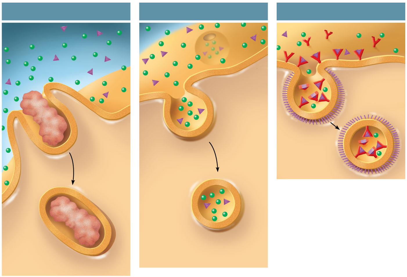
Endocytosis - where the cell takes substances into cytoplasm by surrounding them in its cell membrane.
Types
Phagocytosis: Ingestion of large particles (e.g., food vacuoles). - cell eating
Pinocytosis: Ingestion of fluids and small molecules. - cell drinking
Receptor-Mediated Endocytosis: Specific uptake of molecules via receptor-ligand interactions.
Exocytosis - The process by which a cell releases substances by transporting them in vesicles that fuse with the cell membrane.
Process
proteins are secreted
they are transported in vesicles to the plasma membrane
they fuse and release their contents externally
Proteins can either be soluble in the vesicle or integrated within the vesicle membrane.
Constitutive Pathway: Continuous secretion of soluble proteins and plasma membrane lipids. - makes it and then immediately secretes it
Regulated Pathway: Secretion of proteins triggered by specific signals (e.g., hormones or neurotransmitters).
Nuclear Pores
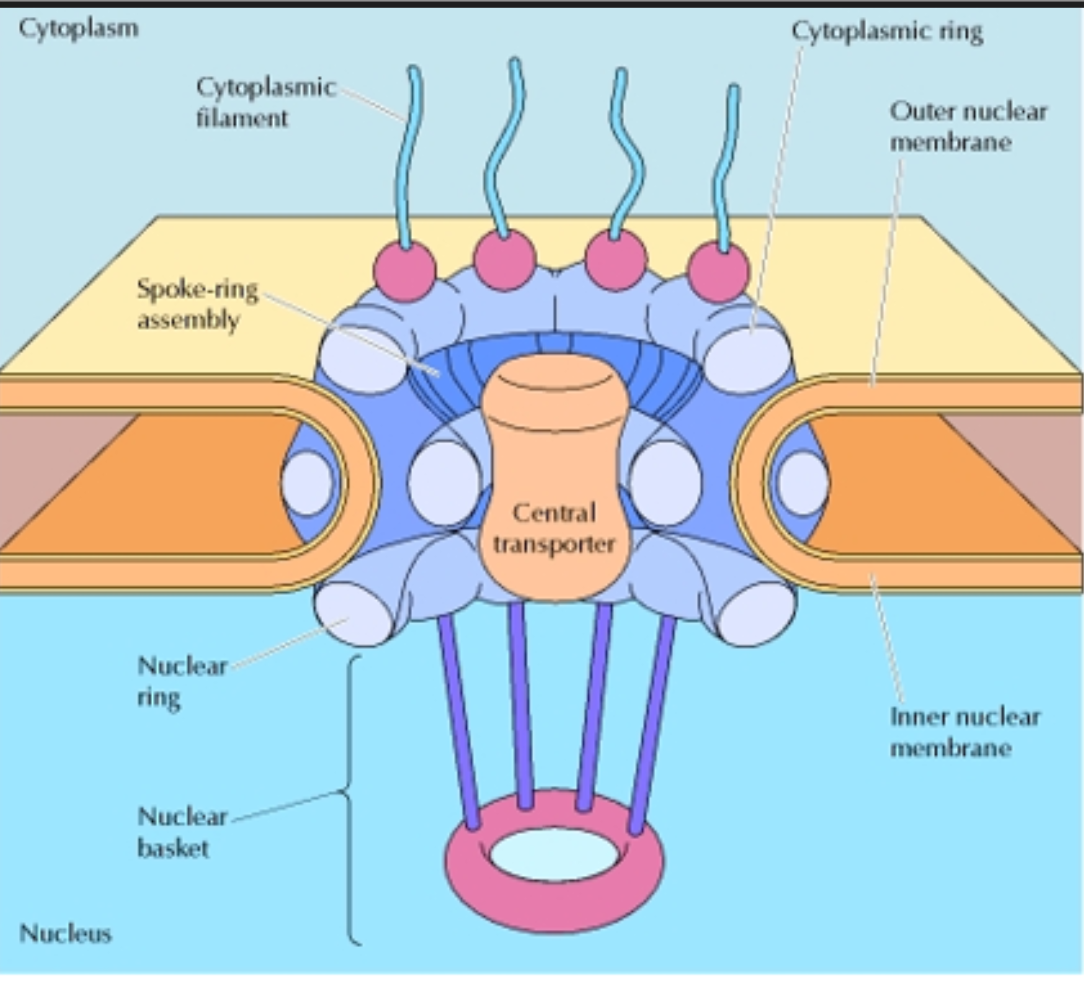
Nuclear pore complexes (NPCs) - specialised structures that act as the primary gateway for the transport of molecules between the cell’s nucleus and the cytosol.
Structure & location
embedded in the nuclear envelope
3000-4000 NPCs
Cytoplasmic filaments
Cytoplasmic ring
Spoke-ring assembly (scaffold)
Central transporter (central channel)
Nuclear ring
Nuclear basket
Cytoplasmic filaments
Project into the cytoplasm.
Function: First point of contact for proteins entering the nucleus.
Bind transport receptors (importins/exportins) carrying cargo.
Cytoplasmic ring
Ring on the cytoplasmic side.
Anchors the cytoplasmic filaments and helps stabilize the pore.
Spoke-ring assembly (scaffold)
Forms the main structural framework of the NPC.
Gives the pore its shape and anchors it in the nuclear envelope.
Central transporter (central channel)
The actual passageway through the pore.
Lined with FG nucleoporins (rich in phenylalanine-glycine repeats).
Acts as a selective barrier:
Small molecules (< ~40–60 kDa) diffuse freely.
Larger proteins and RNAs require active transport via importins/exportins and Ran-GTP.
Nuclear ring
Ring on the nuclear side.
Supports the nuclear basket.
Nuclear basket
Basket-like filaments extending into the nucleus.
Functions:
Helps export mRNA.
Participates in gene regulation.
Quality control of RNA before export.
Motor proteins
Motor Proteins - A protein that uses energy (ATP) to move materials around the cell along cytoskeletal filaments.
3 Types of motor proteins
Myosin: Acts along actin microfilaments.
Kinesin: Moves along microtubules in one direction.
Dynein: Moves in the opposite direction along microtubules.
Myosin
Acts along actin microfilaments.
Transports vesicles and organelles
Structure
Head
Binds to actin filaments.
Contains an ATP-binding site that provides energy for movement.
Also called the motor domain.
Neck/stalk
Connects the head to the tail.
Acts as a lever arm to generate movement.
Tail
Attaches to cargo (such as vesicles or organelles) or to other myosin molecules.
Determines the specific function of the myosin.
How Myosin Motor Proteins Moves
One head binds the microtubule.
ATP binds to the attached head.
The neck linker swings forward.
The second head steps ~8 nm to the next binding site.
ATP is hydrolysed and the rear head detaches.
The cycle repeats.
Direction of Kinesin Transport
Direction - Plus (+) end
Movement - From the centre of the cell (near the centrosome/MTOC) towards the cell periphery (plasma membrane).
This is called anterograde transport.
Kinesin
moves organelles and vesicles throughout the cytoplasm
Structure
2 motor (globular) head
Each containing ATP-binding sites (ATPase)
Microtubule-binding sites
Neck Linker
Short flexible region behind each head
changes position when ATP binds
Coiled-coil stalk
Long α-helical region where the two heavy chains wrap around each other.
Provides structural support.
Connects the motor heads to the cargo-binding region
Tail domain
Located at the opposite end from the heads.
Often associated with light chains.
Binds cargo such as:
Vesicles
Mitochondria
Lysosomes
Protein complexes
How Kinesin Moves
One head binds the microtubule.
ATP binds to the attached head.
The neck linker swings forward.
The second head steps ~8 nm to the next binding site.
ATP is hydrolysed and the rear head detaches.
The cycle repeats.
Direction of Kinesin Transport
Direction - Plus (+) end
Movement - From the centre of the cell (near the centrosome/MTOC) towards the cell periphery (plasma membrane).
This is called anterograde transport.
Colour Changes in Animals
Zebra Fish
Motor proteins transport pigment molecules along microtubules to change the colour of some organisms.
light - dark in this case
Cephalopods
contains pigments called chromatophores that change shape due to action of muscle fibers