lymphatic system
2. Lymphatic System
Definition
The lymphatic system consists of:
Groups of cells
Tissues
Organs
Function:
Monitor body surfaces and internal fluid compartments
React to harmful substances
3. Main Cells of the Lymphatic System
Lymphocytes
Definitive cell type of the lymphatic system
Effector cells in immune responses against harmful substances
4. Components of the Lymphatic System
Important lymphatic tissues and organs:
Diffuse lymphatic tissue
Lymphatic nodules
Lymph nodes
Spleen
Bone marrow
Thymus
These lymphatic organs and tissues are collectively called the immune system.
Lymphatic Vessels
Connect parts of the lymphatic system to the blood vascular system
5. Sites of Lymphocyte Development
Lymphatic tissues are sites where lymphocytes:
Proliferate
Differentiate
Mature
Lymphocyte “Education”
Occurs in:
Thymus
Bone marrow
Gut-associated lymphatic tissue (GALT)
Purpose:
Lymphocytes become immunocompetent
They learn to distinguish:
Self → molecules normally present in the body
Nonself → foreign molecules
6. Antigen
Definition
An antigen is:
Any substance that can induce a specific immune response.
Types of Antigens
Antigens may be:
Soluble substances
Foreign proteins
Polysaccharides
Toxins
Infectious organisms
Foreign tissues
Transformed (cancerous) cells
Antigen Processing
Most antigens must be processed by immune cells before other immune cells can respond.
7. Exposure to Pathogens
The body is constantly exposed to:
Pathogenic organisms
Infectious microorganisms
Toxins
Foreign cells
Foreign tissues
Additionally:
Normal cells may transform into cancer cells, appearing foreign to the immune system.
8. Types of Immune Defenses
Immune responses are divided into:
Nonspecific (Innate) immunity
Specific (Adaptive) immunity
These represent the two lines of immune defense against foreign invaders and transformed cells.
9. Nonspecific (Innate) Immunity
Characteristics
Preexisting
Nonspecific defenses
Represents the first line of defense against microbial invasion.
Components of Innate Immunity
1. Physical Barriers
Prevent entry of organisms:
Skin
Mucous membranes
2. Chemical Defenses
Destroy invading microorganisms:
Low pH
3. Secretory Substances
Examples:
Thiocyanate in saliva
Lysozymes
Interferons
Fibronectin
Complement in serum
Function:
Neutralize foreign cells
4. Phagocytic Cells
Examples:
Macrophages
Neutrophils
Monocytes
5. Natural Killer (NK) Cells
10. Specific (Adaptive) Immunity
When It Occurs
Activated when nonspecific defenses fail
Characteristics
Targets specific invaders
Initiated by initial contact with a specific antigen
Immune Memory
Adaptive immunity often leads to immune memory.
Genetic Basis
Adaptive immunity develops through:
Random somatic rearrangements of genes encoding:
Immunoglobulins
T-cell receptors (TCRs)
11. Activation of Lymphocytes
During adaptive immune responses:
B lymphocytes
T lymphocytes
become activated to destroy invading organisms.
12. Types of Specific Immune Responses
1. Humoral Immune Response
Produces antibodies
Antibodies are proteins
Function:
Mark invaders for destruction by other immune cells
2. Cellular Immune Response
Targets:
Transformed cells
Virus-infected cells
Destroyed by:
Specific “killer” cells
13. Immune System Activation
After invasion by bacteria or other pathogens:
Immune system becomes activated
Inflammatory response occurs
Pathogens are destroyed
Long-term immune memory is generated
Cells of the Lymphatic System
1. Overview
Cells of the immune system consist of:
Lymphocytes
Supporting cells
These cells together make up the cells of the immune system.
2. Types of Lymphocytes
There are three major types of lymphocytes:
B cells
T cells
Natural Killer (NK) cells
3. Supporting Cells of the Immune System
Supporting cells interact with lymphocytes and have important roles in:
Presentation of antigens to lymphocytes
Regulation of immune responses
Types of Supporting Cells
Monocytes
Macrophages
Neutrophils
Basophils
Eosinophils
Reticular cells
Dendritic cells
Follicular dendritic cells
Langerhans’ cells
Epithelioreticular cells
4. Role of Epithelial and Stromal Cells
Specialized epithelial and stromal cells provide the environment for immune reactions by:
Secreting specific substances
Regulating:
Cell growth
Cell migration
Activation of effector cells
Activation of supporting cells
5. Organization of Supporting Cells
Structure
Supporting cells in lymphatic organs are organized into loose meshworks.
Reticular Cells and Reticular Fibers
In:
Lymph nodules
Lymph nodes
Spleen
Reticular cells and reticular fibers produced by these cells form elaborate meshworks.
Cells Located in These Meshworks
Lymphocytes
Macrophages
Dendritic cells
Follicular dendritic cells
Other immune system cells
These cells also exist in loose connective tissue of the body.
6. Langerhans’ Cells
Location
Found only in the middle layers of the epidermis
Function
Carry out surveillance and defense
7. Epithelioreticular Cells
Location
Found in the thymus
Function
Form the structural meshwork of thymic tissue
Important Feature (Exam Point)
Despite their name:
They do NOT produce reticular fibers
They are NOT related to reticular fibers
8. Identification of Cells in Lymphatic Tissue
Different lymphatic tissue cells are identified by surface markers called:
Cluster of Differentiation (CD) Markers
General Characteristics of Lymphocytes
Definition
Circulating lymphocytes are the chief cellular constituents of lymphatic tissue.
Circulating Pool
Most lymphocytes (≈70%) in blood or lymph form a circulating pool of immunocompetent cells.
Circulation Cycle
Lymphocytes:
Exit systemic circulation
Enter lymphatic tissues
Perform immunologic surveillance
Return to systemic circulation
Main Circulating Population
The circulating population mainly consists of:
Long-lived mature lymphocytes
Mainly T cells
Capable of:
Recognizing foreign antigens
Responding to foreign antigens
These cells move from one lymphatic tissue site to another.
2. Non-Circulating Lymphocyte Population
About 30% of lymphocytes:
Do not circulate between blood and lymphatic tissues.
These include:
Short-lived immature cells
Activated lymphocytes destined for specific tissues
Migration
These cells:
Leave capillaries
Migrate into tissues, especially:
Connective tissue under the respiratory epithelium
Connective tissue under the gastrointestinal epithelium
Connective tissue under the urogenital epithelium
Intercellular spaces of epithelia
3. Major Types of Lymphocytes
Three functional types:
T lymphocytes
B lymphocytes
Natural Killer (NK) cells
Important Exam Point
The classification of lymphocytes is independent of morphological (size) characteristics.
4. T Lymphocytes (T Cells)
Differentiation
Differentiate in the thymus
Proportion
60–80% of circulating lymphocytes
Function
Involved in cell-mediated immunity
Lifespan
Long life span
Surface Markers
T cells express:
CD2
CD3
CD5
CD7
T-cell receptors (TCRs)
Subclassification
Based on presence or absence of:
CD4
CD8
5. Helper T Cells (CD4⁺ T Cells)
Characteristics
Express CD4 marker
Subtypes
Divided based on cytokine secretion:
TH1 Cells
Produce:
Interleukin-2 (IL-2)
Interferon-γ (IFN-γ)
Tumor necrosis factor-α (TNF-α)
Function
Interact with:
Cytotoxic CD8⁺ T cells
NK cells
Macrophages
Role
Important for controlling intracellular pathogens, including:
Viruses
Certain microorganisms
TH2 Cells
Produce:
IL-4
IL-5
IL-10
IL-13
Function
Interact with:
B lymphocytes
Role
Initiate antibody-mediated immune responses
Target extracellular pathogens
6. Cytotoxic T Lymphocytes (CD8⁺ T Cells)
Marker
Express CD8
Function
Kill target cells such as:
Virus-infected cells
Cancer-transformed cells
Cells infected with intracellular microorganisms
Parasite-infected cells
Transplanted cells
7. Regulatory (Suppressor) T Cells
Function
Suppress immune responses to:
Foreign antigens
Self antigens
Mechanism
They influence activity of other immune cells.
Example Regulatory T Cell Markers
CD4
CD25
FOXP3
FOXP3 indicates expression of forkhead family transcription factors.
Effects
Regulatory T cells can:
Reduce ability of T lymphocytes to initiate immune responses
Suppress B-cell differentiation
Regulate erythroid cell maturation in bone marrow
Another suppressor T-cell type:
CD8⁺ CD45RO⁺ T lymphocyte
9. B Lymphocytes (B Cells)
Differentiation
B cells differentiate in bursa-equivalent organs in mammals:
Bone marrow
GALT
Function
Participate in humoral immunity by producing:
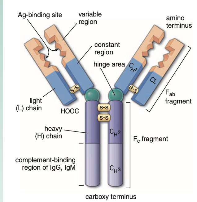
Antibodies (immunoglobulins, Ig)
These are immune proteins circulating in the blood.
Proportion
20–30% of circulating lymphocytes
B-Cell Receptors (BCRs)
B cells express:
Membrane-bound immunoglobulins
Called B-cell receptors (BCRs)
Function:
Serve as antigen-specific binding sites
Immunoglobulin Changes During Maturation
During differentiation:
Immature B cells → express IgM
Mature B cells → express IgD
MHC Expression
B cells express:
Major histocompatibility complex II (MHC II) molecules
CD Markers of B Cells
CD9
CD19
CD20
Natural Killer (NK) Cells
1. Definition
Natural killer (NK) cells:
Are neither T cells nor B cells
Are specialized to kill certain types of target cells
2. Type of Immunity
NK cells are part of:
Nonspecific (innate) immunity
3. Origin
NK cells develop from:
Common lymphoid progenitor (CLP) cell
This is the same progenitor that gives rise to B cells and T cells.
4. Proportion in Blood
NK cells constitute:
5–10% of circulating lymphocytes
5. Thymus and TCR Expression
NK cells:
Do NOT mature in the thymus
Do NOT express T-cell receptors (TCRs)
6. Target Recognition
NK cells are genetically programmed to recognize:
Virus-infected cells
Tumor (transformed) cells
7. Mechanism of Killing
NK cells kill target cells similar to cytotoxic CD8⁺ T lymphocytes.
After recognizing a transformed cell, NK cells:
Become activated
Release:
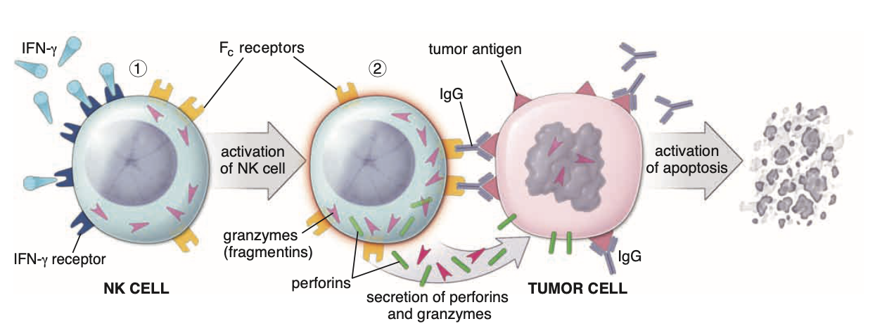
Perforins
Granzymes (fragmentins)
Role of Perforins and Granzymes
Perforins
Create channels (pores) in the target cell plasma membrane
Granzymes
Cause fragmentation of DNA
8. Result of NK Cell Action
These processes lead to:
Apoptosis
orLysis of the target cell
9. Regulation of NK Cells
NK cell activity is regulated by:
Natural Cytotoxicity Receptors (NCRs)
Regulation occurs through:
Activating NCRs
Inhibiting NCRs
These receptors are located on the surface of NK cells.
10. NK Cell Markers (CD Markers)
Specific markers of NK cells include:
CD16a
CD56
CD94
Lymphocyte Development and Differentiation
1. Antigen-Independent Differentiation (Primary Lymphatic Organs)
Lymphocytes first undergo antigen-independent differentiation in primary lymphatic organs.
Primary (Central) Lymphatic Organs
In humans and other mammals:
Bone marrow
GALT (gut-associated lymphatic tissue)
→ together called bursa-equivalent organsThymus
Function of Primary Lymphatic Organs
In these organs lymphocytes:
Differentiate into immunocompetent cells
Become genetically programmed to recognize a single antigen
This process is called:
Antigen-independent proliferation and differentiation
After becoming immunocompetent, lymphocytes:
Enter blood or lymph
Are transported throughout the body
Become dispersed in connective tissues
2. Antigen-Dependent Activation (Secondary Lymphatic Organs)
Lymphocytes undergo antigen-dependent activation in secondary lymphatic organs.
Secondary (Peripheral) Lymphatic Organs
These include:
Lymphatic nodules
Lymph nodes
Tonsils
Spleen
Structure of Effector Lymphatic Tissue
In these tissues:
Immunocompetent lymphocytes
Plasma cells (derived from B cells)
Macrophages
organize around:
Reticular cells
Reticular fibers
to form effector lymphatic tissues and organs.
Result of Antigen-Dependent Activation
T and B lymphocytes differentiate into:
Effector lymphocytes
Memory cells
Immune Responses to Antigens
1. Initial Response: Inflammation
The initial response to an antigen is:
Inflammation
The inflammatory response is a nonspecific defense.
Antigen may be:
A foreign molecule
A pathogenic organism
Actions During Inflammation
The inflammatory response may:
Sequester the antigen
Digest it with enzymes from neutrophils
Phagocytose and degrade it in macrophages
Macrophage degradation may lead to:
➡ Presentation of antigen fragments to immunocompetent lymphocytes
This triggers a specific immune response.
Specific Immune Responses
Specific immune responses are classified into:
Primary immune response
Secondary immune response
2. Primary Immune Response
Definition:
The first encounter of the body with an antigen.
Characteristics
Lag period of several days
Antibodies detected are mostly IgM
Initial response initiated by one or a few B lymphocytes
After this response:
Some antigen-specific B lymphocytes remain as memory cells
3. Secondary Immune Response
Characteristics:
More rapid
More intense
Higher levels of antibodies (usually IgG)
Reason:
Presence of memory B lymphocytes already programmed for that antigen.
Types of Specific Immune Responses
There are two major types:
Humoral (antibody-mediated) immunity
Cell-mediated immunity
Humoral Immunity
Definition
Humoral immunity is mediated by:
Antibodies
Source of Antibodies
Produced by:
B lymphocytes
Plasma cells derived from B lymphocytes
Cell-Mediated Immunity
Mediated By
Specific T lymphocytes
Function
T cells attack and destroy:
Virus-infected cells
Foreign cells
TCR–CD3 Complex
The TCR–CD3 complex:
Contains TCR α and β chains
Associated with CD3 molecules
Function
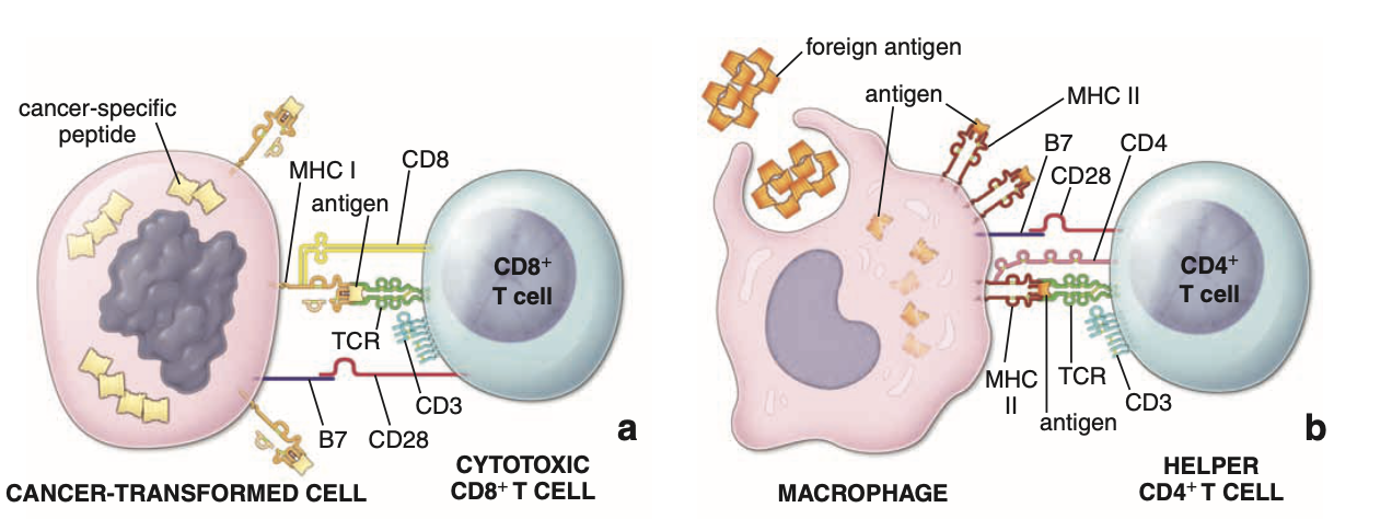
When TCR binds antigen presented on MHC:
Signal transmitted through CD3
T cell becomes activated
T cell secretes interleukins
T cells divide and differentiate
Activation of T and B Cells — Exam Notes
1. Activation of T Cells
Requirement for Activation
Activation of T cells requires costimulatory signals.
Both:
Helper T cells (CD4⁺)
Cytotoxic T cells (CD8⁺)
require two stimulatory signals for:
Full activation
Differentiation
Proliferation
2. Two Signals Required for T-Cell Activation
First Signal
The first signal is produced by:
Interaction of:
T-cell receptor (TCR)
CD4 or CD8 molecules
with the:
Antigen–MHC complex
Second Signal (Costimulatory Signal)
The second signal is the costimulatory signal.
It results from interaction between:
Membrane molecules on T cells
Molecules on antigen-presenting cells (APCs)
Important Costimulatory Interactions
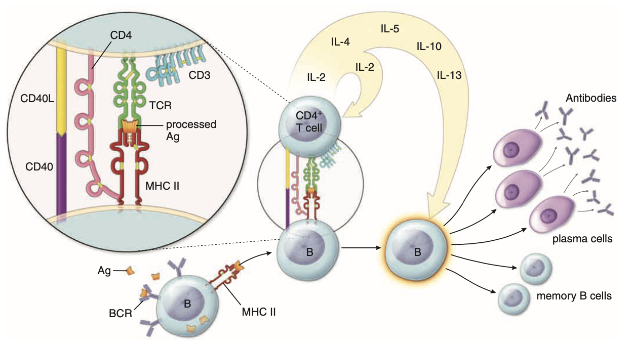
1⃣ CD28 (T cell) ↔ B7 / CD86 (APC)
2⃣ CD40 (APC) ↔ CD40L / CD154 (T cell)
3. Activation of Helper T Cells (CD4⁺)
When a helper CD4⁺ T lymphocyte recognizes an antigen:
The TCR binds to antigen–MHC II complex
If the costimulatory signal is present:
➡ The helper T cell becomes activated
Result
Activated helper T cells release:
Cytokines
4. Cytokines
Definition
Cytokines are:
Immune signaling proteins
Biologic modulators of immune responses
Cytokines Produced by CD4⁺ T Cells
The cytokines secreted by helper T cells are called:
Interleukins (ILs)
Function of Interleukins
Interleukins stimulate:
T cells
B cells
NK cells
to:
Differentiate
Proliferate
5. Activation of Cytotoxic T Cells (CD8⁺)
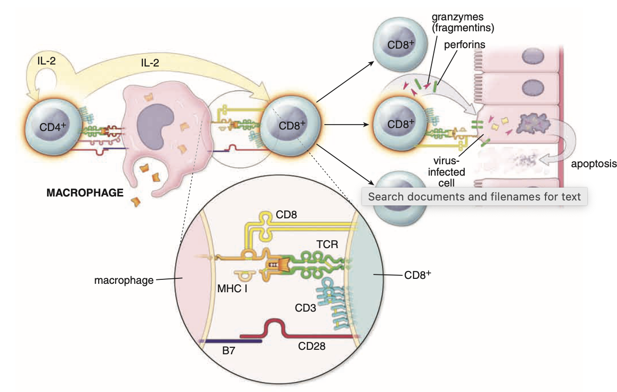
When a cytotoxic CD8⁺ T lymphocyte (CTL) recognizes:
Antigen–MHC I complex
the TCR binds to it.
If a costimulatory signal is present (CD40–CD40L interaction):
➡ The CTL becomes activated.
Activated CTL Functions
Activated CTLs:
Release cytokines
Stimulate cell proliferation
Destroy abnormal host cells
Examples of target cells:
Virus-infected cells
Transformed (cancer) cells
Some CTLs may destroy target cells without costimulatory signals.
6. MHC Restriction of T Cells
Cytotoxic T Cells
CD8⁺ T cells are MHC I restricted
Meaning:
They recognize antigens presented by MHC I molecules.
Helper T Cells
CD4⁺ T cells are MHC II restricted
Meaning:
They recognize antigens presented by MHC II molecules.
7. MHC Molecules and Antigen Recognition
MHC I
Interacts with:
TCR
CD8 molecules
Present on:
Target cells
Function:
Allows cytotoxic T cells to recognize:
Virus-infected cells
Transformed cells
MHC II
Interacts with:
TCR
CD4 molecules
Found on:
Antigen-presenting cells (APCs) such as:
Macrophages
Function:
Present antigens to helper T cells.
8. Activation of B Cells
For B lymphocytes to become activated and differentiate into plasma cells:
➡ They must interact with helper T lymphocytes.
Antigen Recognition by B Cells
Each B lymphocyte recognizes:
One specific antigen
Two Signals Required for B-Cell Activation
Signal 1
Interaction between:
B-cell receptor (BCR)
Antigen
The antigen is then:
Internalized by receptor-mediated endocytosis
Antigen fragments are displayed on MHC II molecules
Signal 2
Provided by helper T cells.
Interaction involves:
CD40 (B cell)
CD40L / CD154 (helper T cell)
This interaction completes B-cell activation.
9. Differentiation of Activated B Cells
Activated B lymphocytes differentiate into:
1⃣ Plasma cells
2⃣ Memory B cells
10. Plasma Cells
Function
Plasma cells:
Synthesize and secrete antibodies
During differentiation:
B cells switch from producing membrane BCRs to soluble antibodies.
11. Memory B Cells
Memory B cells:
Respond more rapidly to future encounters with the same antigen.
12. Antigen–Antibody Complex
The antibody produced by plasma cells binds the antigen forming:
Antigen–antibody complex
These complexes can be eliminated by:
NK cells
Macrophages
Eosinophils
13. Antibody-Dependent Cell-Mediated Cytotoxicity (ADCC)
Definition
In ADCC, antibodies direct immune cells to kill target cells.
Mechanism
IgG antibodies:
Bind to target cells
Immune cells recognize the Fc region of IgG using:
Fc receptors
Cells possessing Fc receptors include:
NK cells
Macrophages
Neutrophils
Eosinophils
Result
Binding of antibody-coated cells leads to:
Apoptosis
Lysis of the target cell
14. Complement System
If the antigen is a bacterium:
The antigen–antibody complex can activate:
Complement system
Complement proteins bind bacteria and promote:
Phagocytosis by macrophages
Complement-bound cells can also be targets of ADCC.
15. Cell-Mediated Immune Response
Main Cells
Cytotoxic CD8⁺ T lymphocytes (CTLs)
Function
Target and destroy:
Virus-infected cells
Transformed cells
16. CTL Killing Mechanism
After antigen recognition:
1⃣ CTLs undergo clonal expansion
2⃣ Differentiate into effector killer cells
Cytotoxic Proteins Released
CTLs release:
Perforins
Granzymes
Perforins
Form pores in the target cell membrane
Increase membrane permeability
Granzymes
Enter target cell through perforin pores
Activate caspases
Induce apoptosis
17. Fate of CTLs After Killing
After killing the target cell:
Most CTLs undergo apoptosis
Some become memory cells
18. Regulatory (Suppressor) T Cells
Markers
Regulatory T cells are:
CD4⁺ CD25⁺ FOXP3⁺ T lymphocytes
Origin
Originate in the thymus
Proportion
About 5% of total T-cell population
Functions
Regulatory T cells:
Suppress immune responses of other lymphocytes
Maintain immunological self-tolerance
Prevent autoimmune diseases
Cytokines Produced
Regulatory T cells secrete:
IL-10
Transforming growth factor-β (TGF-β)
TGF-β strongly suppresses proliferation of:
T effector cells
B effector cells
Cytokines
Definition
Cytokines are:
Soluble polypeptide molecules
Produced mainly by activated T lymphocytes
Function
Cytokines affect:
T cells
B cells
Monocytes
Macrophages
Antigen-presenting cells
Cytokines vs Growth Factors
Both are similar, but differ in:
➡ Target cell populations
21. Cytokines as Chemical Messengers
Cytokines act as:
Chemical messengers between immune cells
They can act:
Autocrine → on the same cell
Paracrine → on neighboring cells
Communication with Other Systems
Cytokines also communicate with:
Central nervous system
Endocrine system
Hemopoietic system
Cytokine Receptors
Cytokines act through specific receptors.
Cells regulated by cytokines therefore possess:
Cytokine receptors
22. Interleukins
Source
Interleukins are synthesized mainly by:
Helper CD4⁺ T lymphocytes
To a lesser extent by:
Monocytes
Macrophages
Endothelial cells
Function
Interleukins promote:
Growth
Differentiation
of:
T cells
B cells
Hematopoietic cells
Antigen-Presenting Cells (APCs)
APCs interact with helper CD4⁺ T lymphocytes to facilitate immune responses.
The interaction between most antigens and antibodies alone is not sufficient to stimulate immune responses.
The antigen must be:
• Broken into small peptide fragments
• Presented with MHC II molecules
• Displayed to helper CD4⁺ T lymphocytes
Antigen can also be processed as part of the B-cell activation pathway.
APCs and the Mononuclear Phagocytic System (MPS)
Most APCs belong to the mononuclear phagocytic system (MPS).
APCs include:
• Macrophages
• Perisinusoidal macrophages (Kupffer cells) of the liver
• Langerhans’ cells in the epidermis
• Dendritic cells of spleen and lymph nodes
Two APCs that do not belong to the MPS:
• B lymphocytes
• Type II and Type III epithelioreticular cells of the thymus
Antigen Processing by APCs
To present antigen to helper T cells:
The APC endocytoses the antigen
The antigen is degraded into peptides (18–20 amino acids)
In the endosomal compartment, peptides bind to MHC II molecules
The antigen–MHC II complex is transported to the cell membrane
The complex is displayed on the APC surface
Additional Functions of Macrophages
Besides acting as APCs, macrophages have other important immune functions.
They:
• Endocytose and partially degrade protein and polysaccharide antigens before presenting them with MHC II to CD4⁺ T cells
• Digest pathogenic microorganisms through lysosomal activity together with helper CD4⁺ T lymphocytes
• Secrete multiple cytokines, including:
– Lymphokines
– Complement components
– Interleukins
– Acid hydrolases
– Proteases
– Lipases
Activated Macrophages
Activated macrophages destroy phagocytosed bacteria and foreign antigens.
After contact with antigen, macrophages undergo activation processes with functional and morphological changes.
Classically Activated Macrophages (M1 Macrophages)
Activated by:
• Interferon-γ (IFN-γ)
Characteristics:
• Increase in cell size
• Increased lysosomes
• Increased cytoplasmic vacuoles
Functions:
• Highly phagocytic
• Strong ability to lyse ingested pathogens and foreign antigens
They promote:
• Inflammation
• Destruction of extracellular matrix
• Apoptosis
Alternatively Activated Macrophages (M2 Macrophages)
Activated by:
• Interleukins
Functions:
• Downregulate inflammation
• Promote rebuilding of extracellular matrix
• Stimulate cell proliferation
• Promote angiogenesis
Macrophage Role in Removing Foreign Materials
Macrophages help sequester and remove foreign materials and organisms that:
• Do not provoke an immune response
• Are ingested but not digested
Foreign Body Giant Cells
When macrophages cannot eliminate foreign material, they may fuse together.
This forms multinucleated giant cells called:
Langerhans’ giant cells
Function:
• Isolate pathogens or foreign materials from the body.
Diffuse Lymphatic Tissue and Lymphatic Nodules
Diffuse lymphatic tissue and lymphatic nodules:
• Guard the body against pathogenic substances
• Are the site of the initial immune response
Location
Diffuse lymphatic tissue is commonly found in the:
• Alimentary canal
• Respiratory passages
• Genitourinary tract
These areas contain accumulations of lymphatic tissue that are not enclosed by a capsule.
Diffuse Lymphatic Tissue
Lymphocytes and other free immune cells are located in the:
• Lamina propria (subepithelial connective tissue)
This form of lymphatic tissue is called:
• Diffuse lymphatic tissue
or
• Mucosa-associated lymphatic tissue (MALT)
It is called MALT because it is associated with mucous membranes.
Function
Cells of diffuse lymphatic tissue are strategically positioned to:
• Intercept antigens
• Initiate an immune response
After Antigen Exposure
After contact with antigen:
Cells travel to regional lymph nodes
They undergo proliferation and differentiation
The progeny return to the lamina propria
These returning cells function as:
• Effector B lymphocytes
• Effector T lymphocytes
Importance of Diffuse Lymphatic Tissue
Its importance in protecting the body from antigens is shown by two findings:
Presence of Plasma Cells
Large numbers of plasma cells are present, especially in the:
• Lamina propria of the gastrointestinal tract
This indicates local antibody secretion.
Presence of Eosinophils
Large numbers of eosinophils are frequently observed in the:
• Lamina propria of the intestinal tract
• Lamina propria of the respiratory tract
This indicates:
• Chronic inflammation
• Hypersensitivity reactions
Lymphatic Nodules in the Alimentary Canal
1. General Features of Lymphatic Nodules
Lymphatic nodules are usually found in structures associated with the alimentary canal, including:
Tonsils
Ileum
Vermiform appendix
General distribution
Usually dispersed singly in a random manner.
In the alimentary canal, some aggregations of nodules occur in specific locations.
Aggregations of Lymphatic Nodules
1. Tonsils
Location & Arrangement
Tonsils form a ring of lymphatic tissue at the entrance of the oropharynx.
Types of Tonsils
Pharyngeal tonsils (adenoids)
Located in the roof of the pharynx.
Palatine tonsils
Located on either side of the pharynx
Positioned between the palatopharyngeal and palatoglossal arches
Lingual tonsils
Located at the base of the tongue
✔ All tonsils contain aggregates of lymphatic nodules
Histological Structure of Palatine Tonsils
Consist of dense accumulations of lymphatic tissue.
Located in the mucous membrane.
Epithelium
Covered by squamous epithelium.
Tonsillar Crypts
The epithelium dips into underlying connective tissue.
This forms numerous tonsillar crypts.
✔ Walls of crypts contain numerous lymphatic nodules
Lymphatic Drainage of Tonsils
Tonsils do NOT possess afferent lymphatic vessels.
Lymph drains from tonsillar lymphatic tissue via efferent lymphatic vessels.
2. Peyer’s Patches
Location
Found in the ileum (distal portion of the small intestine).
Structure
Consist of numerous aggregations of lymphatic nodules.
Cells Present
Contain:
T lymphocytes
B lymphocytes
Additional Nodules
Numerous isolated single (solitary) lymph nodules occur along:
Large intestine
Small intestine
3. Vermiform Appendix
Origin
Arises from the cecum.
Histology
Lamina propria heavily infiltrated with lymphocytes.
Contains numerous lymphatic nodules.
Mucosa-Associated Lymphatic Tissue (MALT)
Naming Principle
Diffuse lymphatic tissue and lymphatic nodules are named according to the region or organ in which they appear.
Types
1. GALT (Gut-Associated Lymphatic Tissue)
Found in the alimentary canal.
2. BALT (Bronchus-Associated Lymphatic Tissue)
Found in the bronchial tree.
3. MALT (Mucosa-Associated Lymphatic Tissue)
Includes:
GALT
BALT
Distribution of MALT
Diffuse lymphatic tissue and lymphatic nodules occur in many regions of the body, for example:
Female reproductive tract
Present where the mucosa is exposed to the external environment.
Important Exam Point
✔ All lymphatic nodules enlarge after encounters with antigen.
Lymph Nodes
Definition
Lymph nodes are small, encapsulated organs that filter lymph.
Located along the pathway of lymphatic vessels.
General Characteristics
Shape: small bean-shaped organs
Type: encapsulated lymphatic organs
Size
Range from about 1 mm (barely visible with unaided eye)
Up to 1–2 cm in their longest dimension
Function
Serve as filters through which lymph percolates on its way to the blood vascular system.
Distribution
Widely distributed throughout the body
Concentrated in certain regions:
Axilla
Groin
Mesenteries
Lymphatic Vessels of the Lymph Node
Two types of lymphatic vessels serve the lymph node:
1. Afferent Lymphatic Vessels
Convey lymph toward the lymph node
Enter at various points on the convex surface of the capsule
2. Efferent Lymphatic Vessels
Convey lymph away from the lymph node
Leave at the hilum
Hilum
Depression on the concave surface of the node
Serves as entrance and exit for blood vessels and nerves
Transport of Activated Lymphocytes
Activated lymphocytes remain in the lymph node to:
Proliferate
Differentiate
They are carried to the lymph node primarily by blood vessels.
Supporting Elements of the Lymph Node
1. Capsule
Dense connective tissue
Surrounds the lymph node
2. Trabeculae
Also dense connective tissue
Extend from the capsule into the substance of the node
Form a gross supporting framework
3. Reticular Tissue
Composed of:
Reticular cells
Reticular fibers
Forms a fine supporting meshwork throughout the remainder of the organ.
Composition of Reticular Meshwork
Cells of mesenchymal origin
Reticular fibers
Ground substance produced by those cells
Cells of the Reticular Meshwork
The reticular meshwork of the lymph node contains several cell types that perform functions in immune responses.
General Morphology
Cells appear:
Stellate or elongated
Have oval euchromatic nuclei
Contain small amounts of acidophilic cytoplasm
They are capable of taking up dyes and colloidal materials.
Using immunocytochemistry and transmission electron microscopy, several cell populations are identified.
Types of Cells in the Reticular Meshwork
1. Reticular Cells
Indistinguishable from typical fibroblasts
Functions
Synthesize and secrete type III collagen (reticular fibers)
Produce associated ground substance forming the stroma.
Structural Role
Elongated cytoplasmic processes wrap around bundles of reticular fibers
This isolates these structural components from lymphatic tissue parenchyma.
Additional Functions
Express surface molecules
Produce substances that attract:
T cells
B cells
Dendritic cells
2. Dendritic Cells (DCs)
Origin
Bone marrow–derived antigen-presenting cells (APCs)
Function
Monitor the local environment for foreign substances
Process antigens
Present antigens to antigen-specific T cells
Efficiency
More efficient in antigen presentation than other APCs
Antigen Presentation
Can present virtually any protein antigen
Presented on:
MHC I
MHC II
Surface Molecules
Express exceptionally high levels of MHC II
Express costimulatory molecules needed for T cell activation
Location in Lymph Node
Usually found in T lymphocyte–rich areas
3. Macrophages
Functions
Phagocytic cells
Antigen-presenting cells
Molecules Expressed
MHC I
MHC II
Costimulatory molecules
Comparison with Dendritic Cells
Lower levels of MHC II and costimulatory molecules
Therefore less efficient APCs than dendritic cells
Major Capability
Very high capacity for endocytosis and digestion of internalized materials
4. Follicular Dendritic Cells (FDCs)
Morphology
Have multiple thin, hair-like branching cytoplasmic processes
Location
Processes interdigitate between B lymphocytes in germinal centers
Antigen Handling
Antigen–antibody complexes adhere to cytoplasmic processes
Attachment occurs via antibody Fc receptors
Antigen Retention
Antigen can remain on the cell surface for:
Weeks
Months
Years
Important Characteristic
Antigen is not endocytosed (unlike macrophages)
APC Status
Not antigen-presenting cells
Reason: lack MHC II molecules
Thymus
Definition
The thymus is a lymphoepithelial organ located in the superior mediastinum.
General Characteristics
Location
Superior mediastinum
Anterior to the heart and great vessels
Shape
Bilobed organ
Developmental Process
The epithelium invaginates.
The thymic rudiment grows caudally as a tubular projection of endodermal epithelium into the mediastinum.
The advancing tip proliferates.
It eventually becomes disconnected from the branchial epithelium.
Cellular Origin of T Cells
Common lymphoid progenitor (CLP) cells from the bone marrow:
Migrate to the thymus.
Develop into immunocompetent T cells.
Invade the epithelial rudiment.
Occupy spaces between epithelial cells.
✔ As a result, the thymus becomes a lymphoepithelial organ.
General Architecture of the Thymus
Capsule
The thymus has a thin connective tissue capsule.
Trabeculae
Trabeculae extend from the capsule into the parenchyma.
Contents of Capsule and Trabeculae
They contain:
Blood vessels
Efferent lymphatic vessels (but NOT afferent lymphatic vessels)
Nerves
Connective Tissue Components
In addition to collagen fibers and fibroblasts, the connective tissue contains variable numbers of:
Plasma cells
Granulocytes
Lymphocytes
Mast cells
Adipose cells
Macrophages
Thymic Lobules
Trabeculae divide the thymus into thymic lobules.
Important Exam Point
These are NOT true lobules.
Structure
Each lobule consists of:
Cortical caps
Covering portions of the continuous inner medullary tissue
Histologic Appearance
In some sections the cortical cap and medullary tissue resemble a lymphatic nodule with a germinal center, which may confuse identification.
Thymic Parenchyma
Contains developing T cells within an extensive meshwork formed by epithelioreticular cells.
Thymic Cortex
Location
Outer portion of the parenchyma
Staining
Markedly basophilic in H&E preparations
Reason
Presence of closely packed developing T lymphocytes with intensely staining nuclei.
Thymocytes
Developing T lymphocytes are called thymocytes.
Cellular Organization
Thymocytes occupy spaces within a meshwork of epithelioreticular cells.
Other Cells
Macrophages are dispersed among cortical cells.
Origin and Development of T Cells
Developing T cells arise from CLPs originating in bone marrow.
During development in the thymus:
CLP-derived cells pass through several developmental stages.
These stages are identified by expression of different CD molecules.
Epithelioreticular Cells
Characteristics
Have features of both:
Epithelial cells
Reticular cells
Function
Provide the framework for developing T cells.
Comparison with Other Lymphatic Organs
In other lymphatic tissues:
Framework = reticular cells + reticular fibers
In the thymus:
Reticular connective tissue cells and reticular fibers are absent
Epithelial Features
Epithelioreticular cells possess:
Intercellular junctions
Intermediate filaments
Types of Epithelioreticular Cells
✔ Six types exist
Three types in cortex
Three types in medulla
Cortex Epithelioreticular Cells
Type I Epithelioreticular Cells
Location
Boundary of the cortex and connective tissue capsule
Between cortical parenchyma and trabeculae
Surround adventitia of cortical blood vessels
Function
Separate thymic parenchyma from connective tissue of the organ
Structural Feature
Occluding junctions between cells
Result
Form a barrier isolating developing T cells from:
Capsule
Trabeculae
Perivascular connective tissue
Type II Epithelioreticular Cells
Location
Within the cortex
Ultrastructure
Desmosomes connect cytoplasmic processes of adjacent cells.
Cytoplasmic processes contain abundant intermediate filaments.
Morphology
Stellate shape
Large pale nucleus that stains lightly in H&E due to abundant chromatin.
Function
Compartmentalize the cortex into isolated areas for developing T cells.
Immunologic Role
Express:
MHC I
MHC II
✔ Participate in thymic education of T cells.
Type III Epithelioreticular Cells
Location
Boundary between cortex and medulla
Structure
Occluding junctions between cytoplasmic processes
Function
Form a functional barrier between cortex and medulla
Immunologic Molecules
Express:
MHC I
MHC II
Macrophages in the Thymic Cortex
Function
Phagocytose T cells that do not fulfill thymic education requirements.
Apoptosis of T Cells
T cells are programmed to die before leaving the cortex.
✔ Approximately 98% of T cells undergo apoptosis.
Identification
Cortical macrophages are difficult to see in H&E preparations.
PAS Reaction
Their large lysosomes stain with PAS.
✔ Therefore they are called PAS cells.
Interaction Between Cells (Cross-Talk)
Epithelioreticular cells play an important role in the development of immunocompetent T cells.
Developing T cells control the microarchitecture of thymic epithelioreticular cells.
✔ This mutual influence is called cross-talk.
Thymus (Medulla) —
Distinguishing Feature of the Thymic Medulla
Thymic (Hassall’s) corpuscles
Derived from Type VI epithelioreticular cells
Characteristic feature of the thymic medulla
Thymic Medulla
Definition
The thymic medulla is the inner portion of the thymic parenchyma.
Cellular Composition
Contains:
Large numbers of epithelioreticular cells
Loosely packed T cells
Staining Characteristics
Stains less intensely than the cortex.
Reason
Contains mostly large lymphocytes
These lymphocytes have:
Pale-staining nuclei
More cytoplasm than small lymphocytes
Epithelioreticular Cells in the Medulla
Like the cortex, the medulla contains three types of epithelioreticular cells:
Type IV
Type V
Type VI
Type IV Epithelioreticular Cells
Location
Located between the cortex and the medulla
Close to type III cells
Structure
Have sheet-like processes
Possess occluding junctions:
Between adjacent cells
Between type IV and type III cells
Function
Together with type III cells, they create the barrier at the corticomedullary junction.
Type V Epithelioreticular Cells
Location
Found throughout the medulla
Structure
Cytoplasmic processes of adjacent cells are joined by desmosomes
Function
Provide the cellular framework of the medulla
Compartmentalize groups of lymphocytes
Nuclei
Nuclei contrast markedly with densely staining lymphocyte nuclei
Type VI Epithelioreticular Cells
Function
Form thymic (Hassall’s) corpuscles
Thymic (Hassall’s) Corpuscles
Structure
Isolated masses of closely packed type VI epithelioreticular cells
Cells arranged concentrically
Cells have flattened nuclei
Ultrastructural Features (TEM)
Cells contain:
Keratohyalin granules
Bundles of cytoplasmic intermediate filaments
Lipid droplets
Cell Connections
Cells are joined by desmosomes
Central Changes
The center may show keratinization
Characteristics of Thymic Corpuscles
Unique structures of the thymic medulla
Antigenically distinct
Functionally active multicellular components
Function (Proposed)
Thought to produce interleukins:
IL-4
IL-7
These cytokines function in:
Thymic differentiation
Education of T lymphocytes
Blood Supply of the Thymus
Entry of Blood Vessels
Blood vessels pass from trabeculae into the thymic parenchyma
Typical Pathway
Blood vessels enter the medulla from deeper parts of the trabeculae
Perivascular Connective Tissue Sheath
Blood vessels carry a sheath of connective tissue
Thickness
Thicker around large vessels
Gradually thinner around smaller vessels
Composition of the Perivascular Connective Tissue
When Thick
Contains:
Reticular fibers
Fibroblasts
Macrophages
Plasma cells
Other loose connective tissue cells
When Thin
Contains:
Reticular fibers
Occasional fibroblasts
Blood–Thymus Barrier & T-Cell Educatation
Blood–Thymus Barrier
Function
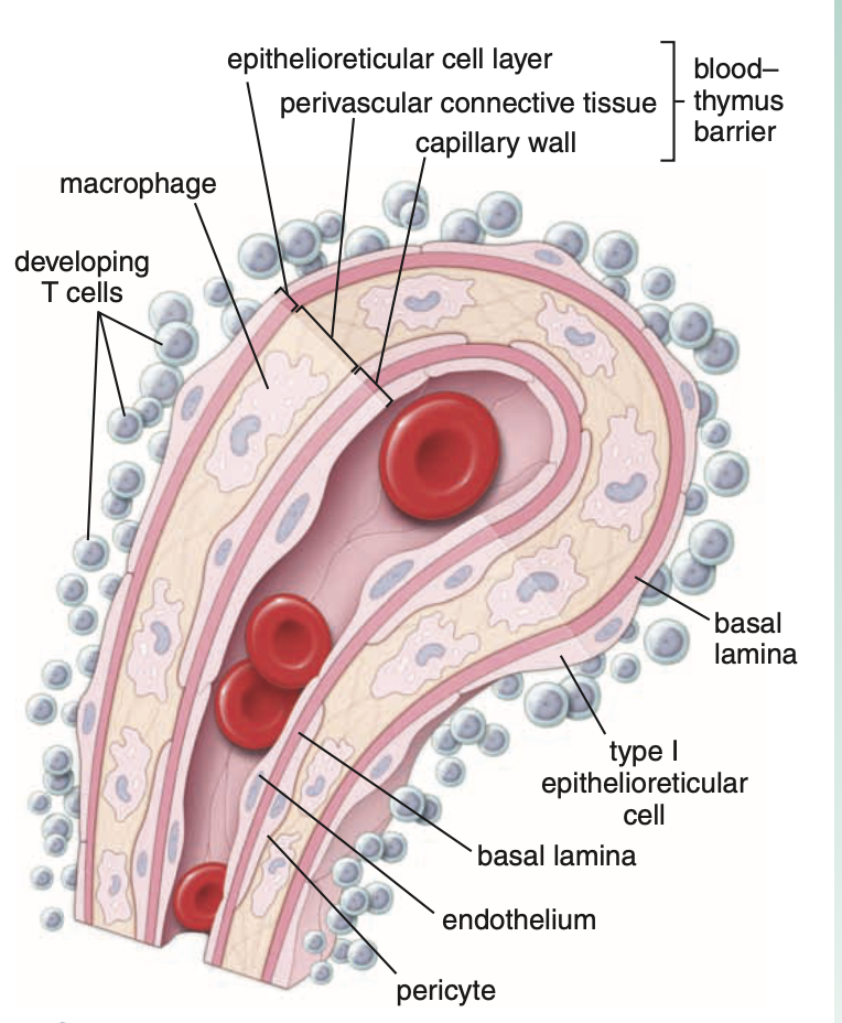
The blood–thymus barrier protects developing lymphocytes in the thymus from exposure to antigens.
Lymphocytes reaching the thymic cortex are prevented from contacting antigens by a physical barrier called the blood–thymus barrier.
Components of the Blood–Thymus Barrier
(From the lumen of cortical blood vessels outward)
1. Endothelium of Capillaries
Capillary endothelium is continuous type with occluding junctions.
Highly impermeable to macromolecules.
Major structural component of the barrier in the cortical parenchyma.
Associated Structures
Basal lamina of endothelial cells
Occasional pericytes
These structures are also part of the capillary wall.
2. Macrophages
Located in the surrounding perivascular connective tissue.
Function:
Phagocytose antigenic molecules that escape from the capillary lumen into the cortical parenchyma.
3. Type I Epithelioreticular Cells
Have occluding junctions.
Provide additional protection to developing T cells.
Arrangement
Surround the capillary wall in the cortex.
Structural Role
Their basal lamina forms another major structural component of the blood–thymus barrier.
T-Cell Education
Site
The thymus is the site of T-cell education.
Origin of Cells
During fetal life:
The thymus becomes populated by multipotential lymphoid stem cells.
These originate from the bone marrow.
These cells will develop into immunocompetent T cells.
Thymic Cell Education
Definition
Stem cell maturation and differentiation into immunocompetent T cells.
Mechanism
Characterized by expression and deletion of specific surface CD molecules.
Stages of T-Cell Differentiation
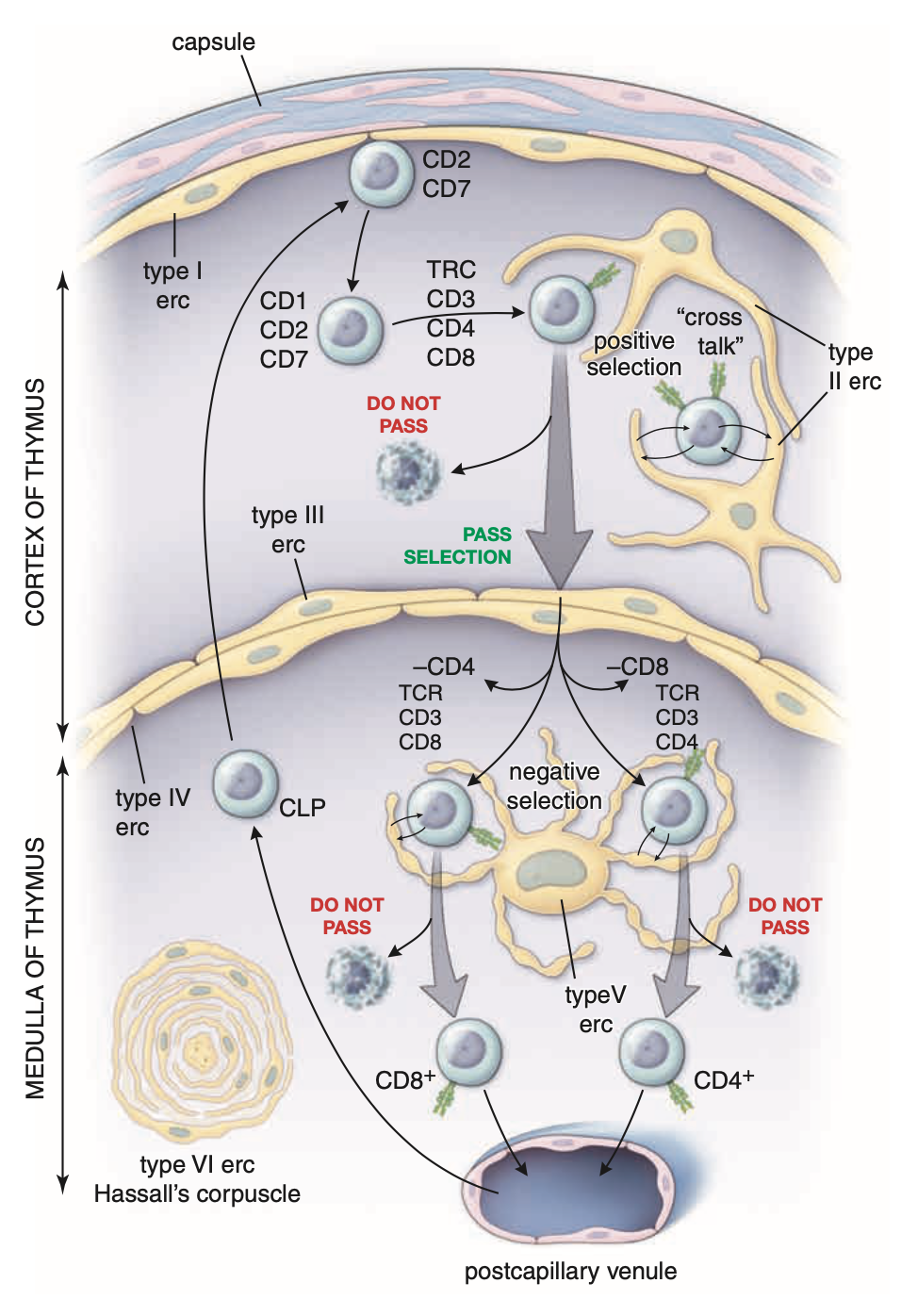
1. Early Stage (Double-Negative Stage)
Markers
Expression of:
CD2
CD7
Meaning of “Double-Negative”
Absence of both CD4 and CD8 molecules.
2. Middle Stage
Marker
Expression of CD1 molecule
Indicates the middle stage of T-cell differentiation.
3. Double-Positive Stage
As maturation progresses, T cells express:
TCRs
CD3
CD4
CD8
This stage is called the double-positive stage of T-cell differentiation.
Positive Selection
T cells are presented with self and foreign antigens by:
Type II epithelioreticular cells
Type III epithelioreticular cells
Outcome
If the lymphocyte recognizes self-MHC molecules and antigen → survives.
This survival process is called positive selection.
If recognition does not occur
The cell dies.
Movement After Positive Selection
Cells that pass positive selection:
Leave the cortex
Enter the medulla
Negative Selection
Occurs in the medulla.
Process
Cells that recognize self-antigen displayed by self-MHC are eliminated.
This process is called negative selection.
Final Differentiation (Single-Positive Stage)
Cells surviving negative selection differentiate into:
1. Cytotoxic T Lymphocytes
CD8⁺ T cells
Lose CD4
Retain CD8
2. Helper T Lymphocytes
CD4⁺ T cells
Lose CD8
Retain CD4
This stage is called the single-positive stage of T-cell differentiation.
Exit from the Thymus
Mature T cells leave the thymus:
From the medulla
Into the blood circulation
Factors Promoting Thymic Cell Education
Substances secreted by epithelioreticular cells promote the process:
Interleukin-4 (IL-4)
Interleukin-7 (IL-7)
Colony-stimulating factors
Interferon-γ
Spleen
1. Basic Overview
The spleen is the largest lymphatic organ.
Size: Approximately the size of a clenched fist.
Location:
Upper left quadrant of the abdominal cavity.
It has a rich blood supply.
Key Function
Filters blood.
Reacts immunologically to blood-borne antigens.
Filtering Functions
The spleen has two types of filtering functions:
Morphologic filtering
Immunologic filtering
Components that allow immune monitoring of blood
The spleen contains:
Large numbers of lymphocytes
Specialized vascular spaces or channels
Meshwork of reticular cells
Reticular fibers
Macrophages
Dendritic cells
These components allow the spleen to monitor blood immunologically, similar to how:
Macrophages and dendritic cells in lymph nodes monitor lymph.
2. Capsule and Trabeculae
Capsule
The spleen is enclosed by a dense connective tissue capsule.
Trabeculae
Trabeculae extend from the capsule into the parenchyma of the organ.
Contents of Capsule and Trabeculae
They contain:
Myofibroblasts
Contractile cells
Produce extracellular connective tissue fibers
Functional Significance
In many mammals:
The spleen stores large volumes of red blood cells.
When capsule and trabeculae contract:
Stored RBCs are discharged into systemic circulation.
Human spleen
Normally retains relatively little blood.
However, it can contract due to contractile cells in capsule and trabeculae.
3. Hilum of the Spleen
Location
The hilum is located on the medial surface of the spleen.
Structures Passing Through the Hilum
Splenic artery
Splenic vein
Nerves
Lymphatic vessels
Lymphatic Vessels
Originate in the white pulp near the trabeculae.
Provide a route for lymphocytes leaving the spleen.
4. Splenic Pulp
Most of the spleen consists of splenic pulp.
It is divided into two regions based on the color of fresh sections:
White pulp
Red pulp
Appearance
White pulp: Circular or elongated whitish-gray areas
Surrounded by red pulp
5. White Pulp
Definition
White pulp consists of lymphatic tissue, mainly lymphocytes.
Key Structural Concept
White pulp is a thick accumulation of lymphocytes surrounding an artery.
Histological Appearance (H&E)
Appears basophilic.
Reason
Due to dense heterochromatin in nuclei of numerous lymphocytes.
6. Arterial Supply of White Pulp
Path of the Splenic Artery
Branches of the splenic artery pass through:
Capsule
Trabeculae
They then enter the white pulp.
Central Artery
Inside white pulp:
The arterial branch is called the central artery.
7. Periarterial Lymphatic Sheath (PALS)
Definition
Lymphocytes aggregating around the central artery form the PALS.
Full name:
Periarterial Lymphatic Sheath
Shape
Has a roughly cylindrical configuration.
Conforms to the course of the central artery.
Appearance in Cross Section
Appears circular.
May resemble a lymphatic nodule.
Distinguishing Feature
Presence of the central artery distinguishes it from typical lymphatic nodules elsewhere.
8. Splenic Nodules
Formation
Nodules are localized expansions of the PALS.
Effect on Central Artery
The central artery becomes eccentrically placed rather than central.
Cellular Composition
B lymphocytes occupy the nodules.
Cells in PALS
Mainly T lymphocytes.
Functional Classification
The PALS is a thymus-dependent zone.
Comparable Structure
Similar to the deep cortex of a lymph node.
9. Germinal Centers
Location
Usually present in splenic nodules.
Development
Form after antigen exposure when B cells proliferate.
Timing
In humans, germinal centers develop within 24 hours after antigen exposure.
Size
Can become extremely large and visible with the naked eye.
10. Splenic Nodules (Malpighian Corpuscles)
Definition
Enlarged nodules with germinal centers.
Other Name
Malpighian corpuscles
Important Note
Not to be confused with renal corpuscles, which have the same name.
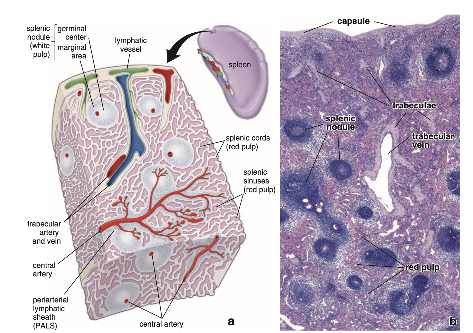
Spleen – Red Pulp
Main Function of Red Pulp
Red pulp contains large numbers of red blood cells that it filters and degrades.
Appearance
Red appearance in:
fresh state
histologic sections
Reason:
Contains large numbers of red blood cells.
Structure of Red Pulp
Red pulp consists of:
Splenic sinuses
Splenic cords (cords of Billroth)
Splenic Cords (Cords of Billroth)
Structure
Loose meshwork of reticular cells
Reticular fibers
Collagen Types
Reticular fibers contain:
Type III collagen
Type V collagen
Cells Present in Splenic Cords
The reticular meshwork contains:
Erythrocytes
Macrophages
Lymphocytes
Dendritic cells
Plasma cells
Granulocytes
Function of Macrophages in Splenic Cords
Phagocytose damaged red blood cells
Begin hemoglobin breakdown
Reclaim iron
Fate of Iron
Used in formation of new red blood cells
Megakaryocytes in Red Pulp
Present in some species:
rodents
cat
Not present in humans
except during fetal life
Splenic (Venous) Sinuses
Definition
Special sinusoidal vessels
Lining
Lined by rod-shaped endothelial cells
Endothelial Cells of Sinuses
Characteristics:
Extremely long cells
Longitudinal axis runs parallel to vessel direction
Few contact points between adjacent cells
Result
Prominent intercellular spaces
Function of These Spaces
Allow blood cells to pass easily:
into sinuses
out of sinuses
Macrophage Processes
Macrophage processes extend:
between endothelial cells
into sinus lumen
Function
Monitor passing blood for foreign antigens
Basal Lamina of Splenic Sinuses
Key Feature
Sinuses do NOT have a continuous basal lamina.
Instead:
Strands of basal lamina loop around the outside of the sinus.
Appearance
Similar to hoops around a barrel
Orientation
At right angles to the long axis of endothelial cells
Staining
This material stains with:
Silver-containing reagents
PAS reaction
Cells NOT Present in Sinus Walls
The walls lack:
Smooth muscle
Pericytes
Additional Structural Feature
Reticular cell processes may extend to the basal side of endothelial cells.
Associated with reticular fibers merging with:
perisinusoidal loops of basal lamina
Histology Section Appearance
Blood fills:
sinuses
splenic cords
This may obscure underlying structures, making it difficult to distinguish sinuses from cords in sections.
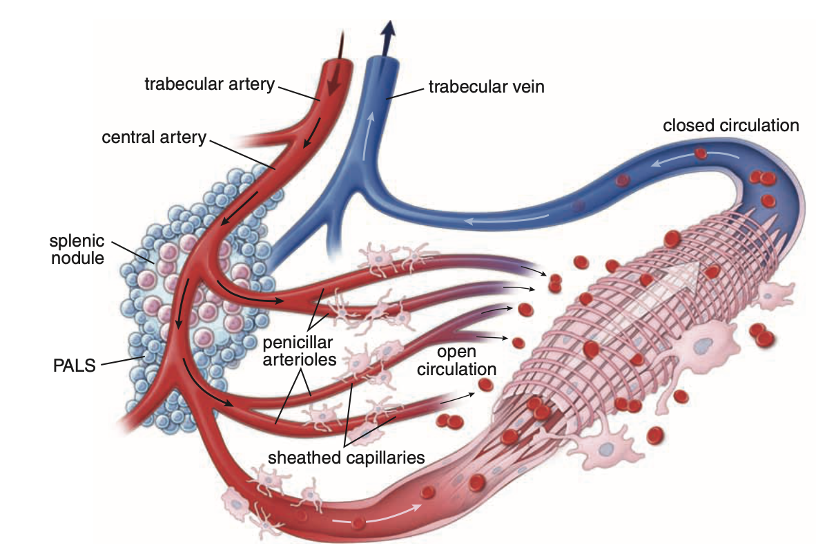
Blood Circulation in the Red Pulp
Arterial Pathway
Branches of splenic artery enter white pulp from trabeculae
Central artery gives branches to:
white pulp
sinuses at white pulp perimeter
Marginal Sinuses
Located at perimeter of white pulp.
Penicillar Arterioles
Central artery continues into red pulp
Branches into straight arterioles called penicillar arterioles
Capillaries
Penicillar arterioles continue as arterial capillaries.
Sheathed Capillaries
Some capillaries are surrounded by aggregations of macrophages.
These are called:
Sheathed capillaries
Open Circulation
Mechanism
Sheathed capillaries empty:
directly into the reticular meshwork of splenic cords
NOT directly into sinuses.
Blood then:
Percolates through splenic cords
Is exposed to macrophages
Reenters circulation by squeezing through sinus walls
Importance
Only route of venous return in humans
Closed Circulation
In some species (rat, dog):
Blood from sheathed capillaries enters splenic sinuses directly
This is called:
Closed circulation
Venous Drainage ( open )
Blood enters splenic sinuses
Drains into tributaries of trabecular veins
Trabecular veins merge into larger veins
Blood leaves spleen via splenic vein
Splenic vein joins intestinal drainage in hepatic portal vein
Functional Significance of Open Circulation
Open circulation:
Exposes blood efficiently to macrophages of red pulp
Allows macrophages to screen antigens
Functions of the Spleen
The spleen performs both:
Immune functions
Hemopoietic functions
Reason:
It filters blood, just as lymph nodes filter lymph.
Immune System Functions of the Spleen
Include:
Antigen presentation
by APCs
mainly dendritic cells and macrophages
Initiation of immune response
Activation and proliferation of
B lymphocytes
T lymphocytes
Production of antibodies
against antigens in circulating blood
Removal of macromolecular antigens from blood
White Pulp Immune Activity
Occurs in white pulp, including:
Activation of T cells
Differentiation of B cells
Formation of plasma cells
Antibody secretion
White pulp functions like other lymphatic organs.
Hemopoietic Functions of the Spleen
Include:
Removal and destruction of
senescent erythrocytes
damaged erythrocytes
abnormal erythrocytes
platelets
Retrieval of iron from erythrocyte hemoglobin
Formation of erythrocytes during early fetal life
Storage of blood
especially RBCs
in some species
Blood Filtration in Red Pulp
Primary role:
Blood filtration
Removes:
particulate material
macromolecular antigens
aged blood cells
abnormal blood cells
damaged blood cells
platelets
Role of Macrophages in RBC Breakdown
Macrophages in red pulp:
break down senescent RBCs
Iron Fate
Iron from hemoglobin is stored as:
Ferritin
Hemosiderin
Heme Breakdown
Heme → bilirubin
Bilirubin transported to:
Liver via portal system
In liver:
conjugated with glucuronic acid
Conjugated bilirubin:
secreted into bile
gives bile characteristic color
Recognition of Senescent RBCs by Macrophages
Macrophages detect aged RBCs through:
1. Nonspecific Mechanisms
Due to morphologic and biochemical changes in aging erythrocytes.
Effects:
RBCs become more rigid
More easily trapped in red pulp mesh
2. Specific Mechanisms
Involve:
Opsonization of RBC membrane
By:
anti-band 3 IgG antibodies
This triggers:
Fc receptor–dependent phagocytosis
Additional Recognition Mechanism
Changes in glycosylation of glycophorins in aging erythrocytes:
act as recognition signals
trigger elimination of senescent erythrocytes by macrophages