INTEGUMENTARY SYSTEM
Skin is the largest organ and is responsible for insulation and protection
Includes Hair + Nails
Nails have keratinised dead cells that make them hard and not easily subjected to wear and tear
Hair helps regulate body temperature and protect the skin from the sunlight
Hair Shaft
Top visible part of the hair
Has 3 layers:
Outer Cuticle: outermost layer that is clear and protective
Medulla Cortex: has the structure for keratin
Inner medulla: Inner core
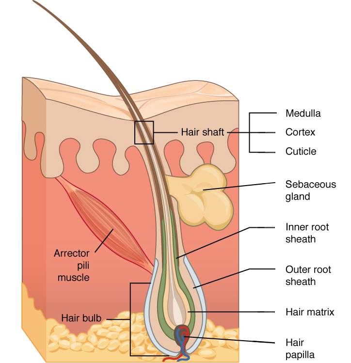
Hair follicle is connected to a muscle called the arrector pili muscle
Arrector pili muscles contract thus pulling hair upright and leading to goosebumps
Types of Exocrine Glands
SEBACEOUS GLANDS
Produce sebum (oily substance ) that helps to lubricate and waterproof the skin and hair.
Overproduction of sebum by the sebaceous glands results in acne
Androgen Hormones: trigger or stimulate the sebaceous glands during puberty to produce more sebum.
SUDORIFEROUS GLANDS
Secrete sweat in order to regulate body temperature
The sweat released, evaporates thus cooling the body
2 Types of Sudoriferous glands
Merocrine(Eccrine Glands ) :
located all over the body especially on the forehead, palms & soles of the feet.
Responsible for thermoregulation, cool the body by releasing sweat which evaporates
Apocrine Glands:
Located in the axilla, groin & areolar (nipples)
Responsible for emotional sweating
Become active during puberty & are associated with a strong odour/ scent
CERUMINOUS GLANDS
Secrete cerumen (earwax)
Cerumen traps dust and bacteria & lubricates ( prevents dryness) the ear canal
4. MAMMARY GLANDS
Part of the female reproductive system
Secrete milk which provides nutrients to the baby
HYPODERMIS
Also known as the subcutaneous layer
It is the deepest layer beneath the skin & it connects the skin to the muscles & bones below
It keeps everything in place
Composed of 2 tissues:
adipose tissue which is responsible for insulation & support
Areolar Connective Tissue which has passageways for blood vessels which carry food & water to the skin to keep it healthy
EPIDERMIS
Has 4 cell types namely:
Keratinocytes
Melanocytes
Langerhan Cells
Merkel Cells
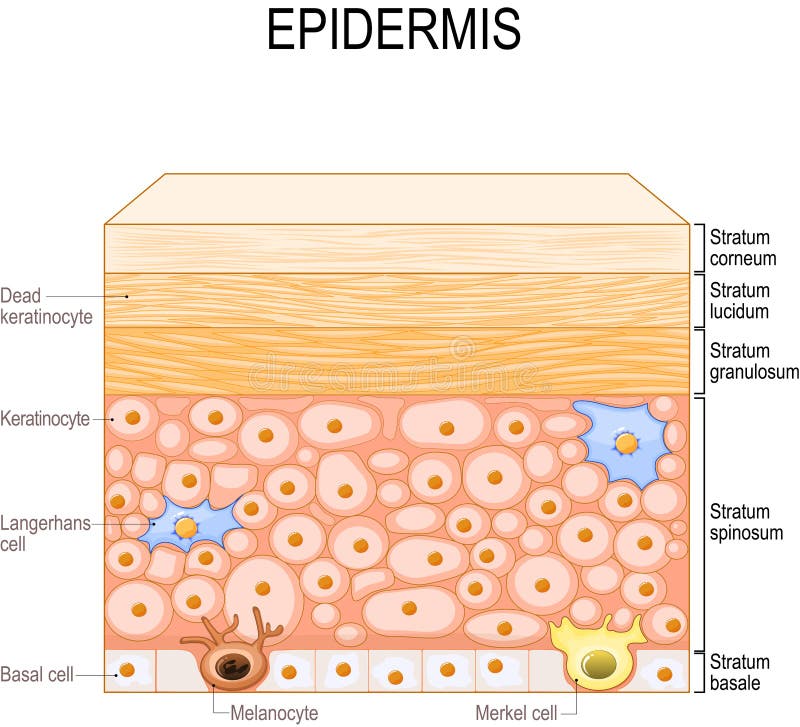
Keratinocytes:
produce keratin for protection
Located through out the epidermis
Make up 90% of the epidermal cells
Melanocytes
Produce melanin for protection against harmful radiation
Located in the Basal Layer
Use an enzyme called tyrosinase to convert tyrosine to melanin
2 Types Of Melanin
Eumelanin: dark brown/ black
Pheomelanin: red/ yellow
Langerhan Cells
Also known as epidermal dendritic cells
Responsible for immune response (locate any foreign substance then alert the immune system which produces a response)
Located in the Stratum Spinosum
Merkel Cells
Responsible for touch sensation
Located in the Basal Layer
THIN SKIN
Has 4 layers
Stratum Basale
Stratum Spinosum
Stratum Granulosum
Stratum Corneum
Located in most parts of the body and is subject to wear & abrasion
STRATUM GRANUNOLOSUM
Keratinocytes undergo apoptosis
Apoptosis ensures that skin remains fresh & healthy
NB: The degree of thickness of the skin is determined by the amount of friction and pressure an area experiences. Increased friction results in rapid & increased cell division resulting in more keratin production and therefore forming thick skin
TONOFILAMENTS
Help skin cells stay strong & provide shape
Connect skin cells to one another
Contain keratin
SKIN GRAFTS
When the basal area is damaged beyond repair, cell division for new skin to form cannot occur and therefore skin grafts will be required
Types Of Skin Grafts
Autografts: removing skin from one area of an individuals body and putting it in another area. There is decreased rejection since the skin is from the same individual
Isograft: graft from an identical twin, involves 2 people
Autologous Graft: An individual’s skin samples are taken & grown in the lab until a large sheet of skin has been formed to transplant on the area of damage.
EPIDERMAL RIDGES
Protrude downward into the epidermis
Interlock with the dermal papillae thus anchoring the epidermis to the dermis
Connection between epidermal ridges & dermal papillae results in skin being intact & reduces sliding
DERMIS
Located below the epidermis
Has 2 regions:
Papillary region: loose connective tissue, has dermal papillae
Reticular Region: Irregular Dense connective tissue, highly vascularised
Both the papillary and reticular region have Free Nerve Endings which are pain receptors