tooth morphology
The dentition (natural teeth in position) are arranged in two arches. The upper arch is the maxillary arch, because the teeth are set in the maxilla bone. The lower teeth are located in the mandible bone, and therefore are located in the mandibular arch (Figure 9-1). The maxillary arch is fixed to the skull and the mandibular arch is movable, bringing the biting force toward the maxillary arch. Each arch has an identical number of teeth, and the teeth are designed so that proper function and positioning can be maintained. The teeth in the maxillary arch slightly overlap the mandibular teeth when in proper alignment. The teeth in each arch touch the teeth adjacent (next) to them, except for the last tooth in each arch. The teeth from the maxillary arch contact the teeth from the mandibular arch each time the mouth is closed. Each tooth supports the teeth beside it and the teeth in the opposing arch so that displacement does not occur.
Each of the dental arches is divided in two halves by an imaginary line called the midline (median line), which creates two sections called quadrants (one-fourth of the dental arches). Thus, there are four quadrants, containing eight permanent teeth each, found in the dentition. The arrangement of the teeth is identical in each quadrant, and each quadrant is named according to its location in the dentition
The quadrants are labeled according to the patient’s right or left. Looking into the oral cavity from the front of the patient makes the directions of right and left reversed to the dental assistant.
The dentition can also be divided into sextants, or sixths. There are two posterior sextants and one anterior sextant in each arch. The anterior sextant is comprised of the six front teeth
Types of Teeth and Their Functions
Humans grow two sets of teeth: primary and permanent. The primary teeth erupt first and are replaced by permanent teeth between the ages of 6 and 17.
The primary (deciduous [di-SI-jeh-wus]) teeth in each quadrant are named similar to the permanent teeth. The deciduous (i.e., first) dentition consists of 20 teeth: 10 in each arch and 5 in each quadrant. The following teeth are found in each quadrant: Starting from the midline, the first tooth is called the central incisor and is used to cut or bite the food that is ingested. The second tooth from the midline, the lateral incisor, is also used for cutting. The third tooth from the midline is the canine (cuspid). This tooth is slightly bulkier in size and aids in tearing food. The next two teeth are molars and are named the first molar, which is the one closest to the midline, and the second molar. Molars are used to chew food.
When compared to the permanent dentition, the deciduous dentition contains an identical number of central incisors, lateral incisors, and canines, but has no premolars and one less molar per quadrant
Permanent Teeth
Permanent teeth are arranged similarly to the deciduous teeth. Adults have 32 permanent teeth: 16 in each arch and 8 in each quadrant. Each quadrant has the permanent central incisor, the lateral incisor, and the canine (cuspid), as did the deciduous quadrant. Directly after the canine (cuspid) in the permanent dentition are the first and second premolars.
The premolars are often called bicuspids because they usually have two (bi) cusps (pointed or rounded mounds on the crown of the tooth). However, two of the eight bicuspids may have three cusps; therefore, the term bicuspid is not technically correct. However, it is important to be aware of the names commonly used for the same teeth (for example, canines or cuspids and premolars or bicuspids).
The premolars are used to pulverize food. In other words, the premolars break the food down into smaller sizes to ready them for the chewing process, which is performed by the molars.
After the premolars, the permanent dentition has the first, second, and third molars. The first molars are closest to the midline, and the third molars—which are farthest from the midline—are commonly termed the “wisdom teeth.”
The teeth in either arch that are toward the front of the mouth from cuspid to cuspid are the anterior teeth. The central incisors, lateral incisors, and canines (cuspids) are termed anterior teeth for both the deciduous and permanent dentition. Anterior teeth have single roots and a cutting or tearing edge called the incisal edge.
The teeth in either arch that are located in the back of the mouth are termed posterior teeth. The molars are posterior teeth in the deciduous dentition, and the premolars (bicuspids) and molars are posterior teeth in the permanent dentition. Posterior teeth normally have more than one root and multiple cusps for pulverizing and chewing.
Eruption Schedule
The primary dentition (deciduous teeth) begins eruption (emerges into the oral cavity) around 6 months of age. All 20 teeth are normally erupted by the age of 3 years (Table 9-1). The period when both primary teeth and permanent teeth are in the dentition is called the mixed dentition period (Figure 9-5). This period lasts from approximately 6 to 12 years of age. After the age of 12, most of the primary teeth have exfoliated (shed from the oral cavity). The permanent dentition begins to erupt from about 6 years of age until around 17 to 21 years of age (Table 9-2).
Table 9-1
Eruption and Exfoliation Dates for Primary Teeth
Tooth | Eruption Date (Months) | Exfoliation Date (Years) | Maxillary Order |
|---|---|---|---|
Central incisor | 6–10 | 6–7 | #1 |
Lateral incisor | 9–12 | 7–8 | #2 |
Canine | 16–22 | 10–12 | #4 |
First molar | 12–18 | 9–11 | #3 |
Second molar | 24–32 | 10–12 | #5 |
Tooth | Eruption Date (Months) | Exfoliation Date (Years) | Mandibular Order |
Central incisor | 6–10 | 6–7 | #1 |
Lateral incisor | 7–10 | 7–8 | #2 |
Canine | 16–22 | 9–12 | #4 |
First molar | 12–18 | 9–11 | #3 |
Second molar | 20–32 | 10–12 | #5 |

Figure 9-5
Mixed dentition of a 7- or 8-year-old.

Table 9-2
Eruption Dates for the Maxillary and Mandibular Permanent Teeth
Tooth | Eruption Date (Years) | Order of Eruption (Maxillary) |
|---|---|---|
Central incisor | 7–8 | #2 |
Lateral incisor | 8–9 | #3 |
Canine | 11–12 | #6 |
First premolar | 10–11 | #4 |
Second premolar | 11–12 | #5 |
First molar | 6–7 | #1 |
Second molar | 12–13 | #7 |
Third molar | 17–21 | #8 |
Tooth | Eruption Date (Years) | Order of Eruption (Mandibular) |
Central incisor | 6–7 | #2 |
Lateral incisor | 7–8 | #3 |
Cuspid | 9–10 | #4 |
First premolar | 10–11 | #5 |
Second premolar | 11–12 | #6 |
First molar | 6–7 | #1 |
Second molar | 11–13 | #7 |
Third molar | 17–21 | #8 |
The permanent teeth that replace the primary teeth are called succedaneous teeth (Figure 9-6). The term refers to succeeding the deciduous teeth. Therefore, because there are 20 primary teeth, there are also 20 succedaneous teeth. Nonsuccedaneous teeth pertain to the teeth that do not replace primary teeth. This would reference the molars in each quadrant. Therefore, there could be up to 12 nonsuccedaneous teeth in the permanent dentition. The premolars would replace the primary molars; therefore, they are succedaneous teeth.
Figure 9-6
Mixed dentition of a 5-year-old. Unerupted succedaneous teeth are shaded in blue, nonsuccedaneous teeth are shaded in green.

Divisions of the Tooth
Each tooth has two basic parts: the crown and the root. The crown of the tooth is described as either anatomical or clinical (Figure 9-7). The anatomical crown is the portion of the tooth that is covered with enamel. The clinical crown is the portion of the crown that is visible in the mouth. The clinical crown may be smaller than the anatomical crown if the gingiva covers a portion of the crown (for example, during tooth eruption). The root of the tooth is also divided into anatomical and clinical portions. The anatomical root is the portion covered with cementum, and the clinical root is the portion of the root seen in the oral cavity (for example, where the gingiva has receded). It should be noted that often when referring to periodontics or coronal polishing the operator may refer to the clinical crown length as both the crown portion and the root portion exposed in the oral cavity. See Chapter 31, Periodontics. The cervical line divides the crown and the root; the anatomical crown and the root join together here as well. (Cervical comes from the word cervix, meaning “the neck of.”) The cervical line is also termed the cementoenamel junction (CEJ).
Surfaces of the Teeth
All teeth have five surfaces on the crown portion. Each surface, or side, has a specific name (Figure 9-8).
Figure 9-8
Surfaces of the teeth identified on the dental arches in a permanent dentition. Posterior teeth colored in blue.

Anterior Teeth
mesial—surface toward the midline.
distal—surface away from the midline.
labial—“outside” surface on anterior teeth, which is toward the lips.
lingual—“inside” surface, which is toward the tongue. On the maxillary arch, the lingual side may be referred to as the palatal surface.
incisal edge—the biting or cutting edge.
Facial Surface
The term facial may be used for either the labial surface of the anterior teeth or the buccal surface of the posterior teeth.
Posterior Teeth
mesial—surface toward the midline.
distal—surface away from the midline.
lingual—“inside” surface, which is toward the tongue.
buccal—“outside” surface on posterior teeth, which is toward the cheek.
occlusal—pulverizing or chewing surface.
All of the above tooth surfaces are flat, convex, or concave (Figure 9-9). Convex means to bulge or curve outward, and concave means recessed or indented. (A memory cue is to think of a “cave” in concave; a cave is hollow and not bulging outward.)
Figure 9-9
Concave and convex surfaces of the mandibular incisor.

Tooth Surfaces Divided into Thirds
Tooth surfaces are further identified by dividing them into approximate thirds. This practice helps dental staff in identifying specific areas on each surface. Also identified are the spaces between the teeth and where the teeth are touching.
The crown of the tooth and the root of the tooth are divided in approximate thirds (Figure 9-10). The area on the crown of the tooth that is nearest the incisal edge on the anterior tooth is called the incisal third of the tooth, and the occlusal surface of the posterior tooth is called the occlusal third of the tooth. The area on the crown of the tooth that is closest to the cervical area (or to the gingiva) is called the cervical third of the tooth. The area between the incisal third and the cervical third is called the middle third. The root is also divided into imaginary thirds with the area nearest the apex as the apical third and the area nearest the crown of the tooth as the cervical third of the root. The area between the apical third of the root and the cervical third of the root is called the middle third of the root.
Figure 9-10
The crown and the root is divided into proximal thirds from all surfaces.

Identifying these areas allows the dentist to give clearer information about a tooth. For instance, if the dentist is describing the shade of the tooth to the dental laboratory technician, the dentist can say that the incisal third is a lighter shade and the cervical third is a darker shade. This information enables better color matching, thereby preventing creation of a single-color tooth. Another example is when the dentist describes a lesion on the root of the tooth, noting that it is on the apical third of the root. These terms also facilitate better diagnosis through greater specificity in identifying the location on the root. All such terms are used frequently in the dental office to identify a specific area of the tooth.
Contact
Identifying the contact area on the tooth refers to where the proximal sides of two teeth come together and touch (Figure 9-11). This is normally the mesial of one tooth and the distal of another tooth, except where the two central incisors come together in each arch. This area is generally in the middle third of the tooth, and is also the area that dental floss tends to snap through. Such contact holds teeth in place so that they do not drift and shift around. Where the two proximal surfaces contact and the area where an individual flosses is called the interproximal (in between proximal surfaces). It should also be noted that food that remains caught in the teeth after eating is common in places where teeth do not contact. Good contact areas protect the gingiva from trauma during mastication (chewing food).
Figure 9-11
Contact area and embrasure shown on two adjacent teeth.

The

embrasure (im-bray-zhur) is the triangular space in the gingival direction when two adjacent teeth are in contact (Figure 9-11). When discussing flossing with patients, the dental assistant will show the patient how to hold the floss and guide it through the contact area and make a half circle and wrap the floss tightly around the tooth as it goes down the embrasure toward the gingival sulcus of the tooth. The floss is then wrapped around the proximal tooth on the other side of the embrasure, and this area is cleaned with the dental floss as well. The embrasure allows the dental papilla to remain healthy.

Anatomical Structures
It is important to be able to identify landmarks on each individual tooth. Each area’s name should be used when identifying the anatomical structures.
apex—at or near the end of the root (Figure 9-13).
apical foramen—opening in the end of the tooth through which nerve and blood vessels enter (Figure 9-13). There may be more than one opening at the end of the root.
bifurcated—when there are two roots on one tooth, they are said to be bifurcated, or branched in two (bi means two and furca means fork) (Figure 9-14).
buccal groove—linear depression forming a groove that extends from the middle of the buccal surface to the occlusal surface of the tooth (Figure 9-15).
central groove—the most prominent developmental groove on the occlusal surface of the posterior teeth. It usually divides the occlusal surface from the medial to the distal.
cingulum—convex area on the lingual surface of the anterior teeth, near the gingiva (Figure 9-16).
cusp—pointed or rounded mound on the crown of the tooth (Figure 9-17).
cusp of Carabelli—fifth cusp located on the mesial lingual surface of most maxillary first molars (Figure 9-18). (The name comes from the man who first described it.)
developmental groove—groove formed by the uniting of lobes during development of the crown of the tooth (Figure 9-19).
diastema—space between two teeth, normally in reference to maxillary centrals (Figure 9-12).
fissure—developmental groove resulting from an imperfect union where the lobes come together (Figure 9-20). Decay often initiates in the fissure.
fossa—a shallow rounded or angular depression (Figure 9-21).
furcation—dividing point of a multirooted tooth (Figure 9-22).
lobes—separate parts that come together to form a tooth (Figure 9-23). In the molars, the lobes often become cusps.
mamelons—three bulges on the incisal edge of the newly erupted central and lateral incisor (Figure 9-24). Mamelons normally disappear due to normal wear.
marginal groove—the developmental groove that provides a spillway for food to escape during chewing. (Figure 9-25).
marginal ridges—elevated area of enamel that forms the mesial and distal borders of the lingual surface of the anterior teeth and the mesial and distal borders of the occlusal surface of the posterior teeth (Figure 9-26).
oblique ridge—elevated area of enamel that extends obliquely across the occlusal of the tooth (Figure 9-27); on the maxillary first molars, the oblique ridge extends from the disto-buccal cusp to the mesio-lingual cusp.
pit—place where the grooves come together or the fissures cross (Figure 9-28); decay often begins in the pit.
ridge—linear elevation of enamel found on the tooth (Figure 9-29).
supplemental groove—shallow, linear groove that radiates from the developmental groove (Figure 9-30); it often gives the tooth surface a wrinkled look. These grooves do not denote major divisions of the tooth.
transverse ridge—union of two triangular ridges that produces a single ridge of elevation across the occlusal surface of a posterior tooth (Figure 9-31).
triangular ridge—ridge or an elevation that descends from the cusp and widens as it runs down to the middle area of the occlusal surface (Figure 9-32).
trifurcated—three roots (tri means three) coming from the main trunk of the tooth (Figure 9-33).
Apex and apical foramen of a tooth.

Figure 9-14
Mandibular molar showing bifurcated roots.

Figure 9-15
Mandibular molar with buccal groove identified.

Figure 9-16
Lingual surface of a central incisor with the cingulum shaded.

Figure 9-17
Maxillary second premolar with the cusps identified.

Figure 9-18
Maxillary first molar showing the mesial lingual side with the cusp of Carabelli identified.

Figure 9-19
Developmental groove on the occlusal surface of the maxillary first premolar where lobes were united.

Figure 9-20
Mandibular second premolar showing the imperfect union or fissure on the occlusal surface.

Figure 9-21
Lingual view of a maxillary canine with the mesio-lingual fossa and disto-lingual fossa shaded.

Figure 9-22
Mandibular first molar from the buccal side showing the furcation or dividing area where the roots fork off.

Figure 9-23
Occlusal view of the maxillary first molar showing the lobes and how they come together.

Figure 9-24
(A) Newly erupted maxillary incisors and laterals showing the three bulges on the incisal edge, called mamelons. (B) Mamelons shown on the anterior of the maxillary dentition.

Figure 9-25
Mandibular first molar showing the marginal ridge and two marginal grooves.

Figure 9-26
Marginal ridges of the maxillary central, premolar, and molar.

Figure 9-27
Maxillary first molar with the oblique ridge identified.

Figure 9-28
Permanent mandibular first premolar showing the occlusal view with pits identified.

Figure 9-29
Ridge identified on the occlusal surface of the mandibular second premolar.

Figure 9-30
Occlusal surface of the mandibular second molar showing shallow linear grooves, which are called supplemental grooves.

Figure 9-31
Maxillary right first premolar occlusal view showing transverse ridge.

Figure 9-32
Triangular ridge identified on occlusal surface of a maxillary second premolar.

Figure 9-33
Maxillary first molar, buccal surface, showing the roots as trifurcated (three roots forked off from the main trunk).

The maxillary central incisor is the first tooth closest to the midline (Figure 9-34). These teeth, along with the lateral incisors, play an important part in a person’s appearance. Their shape, color, size, and placement directly relate to how a person looks. The position of the teeth dictates the shape of a person’s profile. Normal placement will provide for correct support of the face and lips. The incisors also play an important role in speech. To execute specific sounds, such as Ss and Ts, these teeth are necessary. Additionally, the incisors have a unique incisal edge that differs greatly from the other teeth in the mouth, which all have cusps. The ridge allows for cutting food into smaller particles.
Figure 9-34
Permanent dentition with the maxillary central incisors identified and maxillary central incisor viewed from the five surfaces.

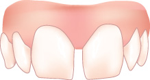
The maxillary central incisor erupts with three bumps on the incisal edge, called mamelons (Figure 9-35). They derive from the three developing lobes coming together. The mamelons become flattened due to attrition (wear), and the incisal edge becomes a flattened surface as well. At the gingival area of the crown on the labial surface, small curved lines run parallel to the CEJ. These are called imbrication lines (Figure 9-35). Most central incisors have imbrication lines.
Figure 9-35
Labial surface of the maxillary right central incisor with the mamelons, developmental depressions, and imbrication lines identified.

The crown of the maxillary central incisor is the longest of any of the maxillary teeth. The labial surface is convex, both mesial to distal and gingival to incisal. The lingual surface is concave, except the gingival one-third where the cingulum is present. The cingulum spreads toward the mesial and distal in an arch pattern, forming the mesial and distal marginal ridges. The mesial surface is slightly longer than the distal surface. The mesial-incisal angle is rather acute, at about a 90-degree angle, and the distal-incisal angle is more rounded. The root is about one-and-a-half to two times the length of the crown. The root appears constricted at the CEJ and then swells in the body, tapering suddenly at the apical portion. Therefore, it ends in a rather blunt apex. The root tends to incline distally
Maxillary Lateral Incisor
The maxillary lateral incisor is the second tooth from the midline and the smallest in the maxillary arch (Figure 9-36). It initially contacts the central incisor on the mesial and the primary canine (cuspid) on the distal. It resembles the maxillary central in most ways. The difference is primarily its size, the crown being about three-tenths smaller in all directions. The root also is smaller in all directions; however, the length has been known to be similar to that of the maxillary central. The crown of the lateral incisor appears narrower than the central, especially in females. The distal-incisal angle is more rounded than that of the central incisor, making the distal length much shorter than the mesial length.
Figure 9-36
Permanent dentition with maxillary lateral incisors identified and maxillary lateral incisor viewed from the five surfaces.

Except for the third molars, the maxillary lateral is the tooth with the most anomalies (extreme variations from the norm). The most frequent is the peg lateral. This is a diminutive, peg-shaped crown with a smooth surface lacking contact on the mesial and distal surfaces. Maxillary laterals are sometimes congenitally missing. When the tooth buds do not form agenesis occurs. Roots that are curved in unusual ways and distorted crowns may appear. Many of these deviations appear generation after generation.
Maxillary Canine (Cuspid)
The canine is often called the “cornerstone of the mouth” due to its placement, which is between the incisors and the bicuspids (Figure 9-37). It is the one tooth that turns the corner for the arch. The canine’s purpose is to tear the food, which is much different from the incisors, which bite or cut the food, and the premolars and molars, which chew or grind it.
Figure 9-37
Permanent dentition with maxillary canines (cuspids) identified and maxillary canine viewed from the five surfaces.

Canines
The canine (cuspid) is one of the most important teeth for animals, because it tears the food. The term canine is derived from the Latin term for dog (canus). The canines (cuspids) look much like dog’s teeth and therefore are named as canines.
The canine (cuspid) is the third tooth from the midline. Because of its placement and size, it is extremely important in supporting the muscles of the face. This is due to a bony ridge covering the labial portion of the roots called a canine eminence, which gives the face a cosmetic manifestation and contributes to a person’s appearance.
The root is the longest in the maxillary arch and therefore the most stable. The crown of the canine is convex on the facial surface, with a ridge running vertically. The incisal edge is fairly pointed and is off center, slightly toward the mesial (the name cuspid is derived from the long cusp that ends in a point on the incisal edge). The mesial surface of the canine (cuspid) is longer than the distal surface, and as they both turn toward the incisal edge, the angle is more rounded than that of the incisors. The lingual surface has two concave fossas, one toward the mesial and the other toward the distal, with a lingual ridge dividing them in the middle. On the outer sides of the fossa is a distal marginal ridge and a mesial marginal ridge (Figure 9-38A). On the lingual side of the tooth, toward the gingiva, is a cingulum (Figure 9-38B). The canine appears darker than the incisors because of the bulk of dentin.
Figure 9-38
Permanent maxillary canine with (A) labial view and (B) lingual view, showing anatomical landmarks.

Maxillary First Premolar (Bicuspid)
The facial cusp of the maxillary first premolar is much larger than the lingual cusp (Figure 9-39). It is longer and wider and appears from the facial side much like the cuspid. The cusps come together on the occlusal surface in a central groove. This central groove extends to the mesial and distal grooves. The mesial groove is bordered by the mesial marginal ridges, and the distal groove is bordered by the distal marginal ridge. The maxillary first premolar has a bifurcated root (two roots, one buccal and one lingual) that is slightly separated. Some first premolars have roots that are fused together; thus, one root has two canals. The roots have a depression on the mesial and distal sides running from the CEJ to the root bifurcation. The roots are shorter and in this aspect resemble the roots of the molars more than they do the roots of the cuspid.
Figure 9-39
Permanent dentition with maxillary first premolars (bicuspids) identified and maxillary first premolar viewed from the five surfaces.

This premolar is often considered for removal if the patient’s teeth are overcrowded and orthodontic treatment is needed. The position allows for movement from both anterior and posterior teeth. The orthodontist closes the space and the patient’s facial appearance is not changed by the removal of this tooth. Also, the depression in the root structure makes it more susceptible to periodontal disease; therefore, it is a better choice for removal than the second premolar.
Maxillary Second Premolar (Bicuspid)
The maxillary second premolar (Figure 9-40) resembles the first in all but the following variations: The cusps, one on the buccal and one on the lingual, are more equal in length. The lingual cusp is slightly shorter, but not as short as the cusp on the maxillary first bicuspid. The mesial buccal cusp slope is shorter than the distal buccal cusp slope. There is only one root and therefore only one root canal. There is a slight depression on the mesial root, but it is very shallow. The crowns of both the first and second bicuspids are wider bucco-lingually than mesio-distally. The second bicuspid is slightly more narrow mesial-distally than the first premolar.
Figure 9-40
Permanent dentition with maxillary second premolars (bicuspids) identified and maxillary second premolar viewed from the five surfaces.

Premolar (Bicuspid)
There are eight premolars: four in each arch, two in each quadrant. They are named the first and second premolars because of their positions from the midline. The first premolars, closest to the midline, line up in the fourth position. The second premolars line up in the fifth position from the midline. They are transitional teeth, placed between the cuspids and the molars. They look like the canines (cuspids) from the facial side; in fact, the buccal cusp functions much like a cuspid in tearing food, but the transitional teeth have an additional cusp on the lingual side (hence “bicuspids,” meaning two). The additional cusp aids in further breaking down the food or pulverizing it for the molars to chew. These posterior teeth are not as critical in personal appearance because of their placement. They do not always show when smiling or talking.
Maxillary First Molar
The maxillary first molar is often referred to as the “6-year molar” because of its eruption time (Figure 9-41). Often, parents do not realize that this is a permanent tooth because of its early eruption. The crown of the maxillary first molar appears square in shape with four primary cusps present: mesio-buccal, disto-buccal, mesio-lingual, and disto-lingual. There is a fifth cusp, the cusp of Carabelli, located on the largest cusp, the meso-lingual. This cusp is located about one-third the way down from the occlusal surface and appears as a “mini” cusp. The prominence of this cusp varies from tooth to tooth.
Figure 9-41
Permanent dentition with maxillary first molars identified and maxillary first molars viewed from the five surfaces.

The mesio-buccal and the disto-buccal cusps are divided by a buccal groove that extends about half the length of the crown and ends in a depression often called the buccal pit. The lingual cusps are slightly longer than the buccal cusps. The mesio-lingual cusp and the disto-lingual cusp are also divided by a lingual groove that travels about halfway down the crown on the lingual side, ending in a shallow depression called the lingual pit.
The root of the maxillary first molar is trifurcated. The first two roots, a meso-buccal root and a distal-buccal root, are placed on the buccal side. These buccal roots curve slightly toward each other. The third root is the largest and longest and is located on the lingual side. These three roots are spread out from each other and normally have one canal each.
On the occlusal surface of the maxillary first molars, the four primary cusps come together in a central fossa. There is an oblique (diagonal) ridge running across the occlusal surface that unites the distal cusp ridge of the mesio-lingual cusp and the lingual cusp ridge of the disto-buccal cusp. Another ridge, a transverse ridge, runs from the buccal cusp of the mesio-lingual cusp to the lingual cusp ridge of the mesio-buccal cusp. The occlusal surface also has a mesial ridge on the mesial of the occlusal surface and a distal ridge on the distal occlusal surface. This creates a surface with additional grooves and ridges to properly grind food.
Maxillary Second Molar
The second molar is called the “12-year molar” because of the time of eruption (Figure 9-42). It is similar to the first molar in many ways; however, it is smaller both in size of the crown and size of the root. The crown of the maxillary second molar has four cusps (no cusp of Carabelli). The cusps are located mesio-buccal, disto-buccal, mesio-lingual, and disto-lingual. The surface of the mesial of the tooth is greater across than the distal surface. The occlusal surface of the molars tapers down in size from the first molar toward the third molar.
Figure 9-42
Permanent dentition with maxillary second molars identified and maxillary second molar viewed from the five surfaces.

The occlusal surface, although smaller, is much like that of the first molar. It has more supplementary grooves than the first molar, making it more wrinkled in appearance. The roots are the same in number but smaller in size and not as spread apart as the roots of the first molar. Each root has one canal, as in the maxillary first molar.
Maxillary Third Molar
Developmental variations make it impossible to describe exactly what the third molar looks like. The maxillary third molar is called the “wisdom tooth” because it was thought that by the time these teeth erupted into the oral cavity a person would have obtained maturity or wisdom (Figure 9-43). Many people do not develop third molars. If third molars do develop, they may not erupt into the oral cavity because of lack of space in the posterior of the arches. This is the one tooth that, after careful diagnosis, the dentist may recommend be removed.
Figure 9-43
Permanent dentition with maxillary third molars identified and maxillary third molars viewed from the five surfaces.

When the tooth erupts normally, it resembles the second molar, only slightly smaller. It exhibits a more wrinkled appearance on the occlusal surface because many more supplemental grooves are usually present. The roots are normally fused together and vary in number.
Maxillary Molar
The word molar is derived from the Latin word molaris, referring to a millstone. This seems like an appropriate term for the teeth that chew, grind, or break down the food into tiny particles for swallowing. When normal eruption occurs, the molars are the first and last permanent teeth in the mouth. There are 12 molars in the oral cavity: 6 in each arch and 3 in each quadrant. They are called the first, second, and third molars because of their placement from the midline. The first molar is the closest to the midline. The molars are the strongest teeth in the arch due to the size of their crowns and the shape and size of their roots. The first molar is the largest and the strongest; this decreases toward the posterior, leaving the third molar the weakest and smallest of the molars. Just as the cuspids are considered the cornerstones of the anterior teeth, the first molars are considered the cornerstones of the developing occlusion in the posterior teeth. The molars do not replace any primary teeth and therefore are not succedaneous teeth. They erupt posterior to the deciduous dentition.
Mandibular Central Incisor
The mandibular central incisor is the least variable tooth in the mouth (Figure 9-44). It is also the smallest tooth in the dentition. It is smaller than the mandibular lateral incisor, which is not the case in the maxillary arch. The maxillary central incisor is larger than the maxillary lateral incisor.
Figure 9-44
Permanent dentition with mandibular central incisors identified and mandibular central incisor viewed from the five surfaces.

When the mandibular central incisor erupts, it has three mamelons on the incisal edge. These wear off, leaving a fairly straight incisal edge for cutting. The crown of the mandibular incisor has a labial surface that is convex but does not appear to have the developmental depressions and imbrication lines of the maxillary central. The crown is narrow and the incisal angle makes a sharp 90-degree angle as it extends down the mesial and distal surfaces. The lingual is concave and has a cingulum near the gingiva. It is relatively smooth, and the structures are not as prominent as in maxillary centrals. The root is straight and ends abruptly at the apex.
This tooth is the first from the midline; therefore, the mesial surface of each central incisor contacts its counterpart. The distal surface contacts the lateral in its prospective quadrant.
Mandibular Lateral Incisor
The anatomy of the mandibular lateral incisor so closely resembles that of the central incisor that a detailed description is unnecessary (Figure 9-45). The mandibular lateral incisor is slightly larger. The root is also larger and slightly longer. Concavities may be present on the mesial and distal of the root. If these occur, the mesial concavity is shallower.
Figure 9-45
Permanent dentition with mandibular lateral incisors identified and mandibular lateral incisor viewed from the five surfaces.

The crown of the lateral incisor is shaped the same as the central incisor except that the distal surface is not as long. The incisal distal angle is more rounded to accommodate this change in length. This tooth does not have the developmental abnormalities of the maxillary lateral.
Mandibular Canine (Cuspid)
The mandibular canine is the third tooth from the midline (Figure 9-46). It resembles the maxillary canine but is not as well developed. The crown of the tooth is approximately the same length as the maxillary canine, but the root is generally shorter (the root is still longer than the mandibular central and lateral roots). The cusp of the mandibular canine is not as well developed as the maxillary and not as sharp on the tip. The function, however, is the same: Both are designed to tear food.
Figure 9-46
Permanent dentition with mandibular canines (cuspids) identified and mandibular canine viewed from the five surfaces.

The distal cusp slope is longer than the mesial cusp slope. The cingulum and marginal ridges are not as pronounced as on the maxillary canine, and the pronounced buccal ridge helps give shape to the face.
The canine is the longest tooth in the mandibular arch. The single root provides for stability; it is the cornerstone of the mandibular arch. The root, with one canal, has deep depressions on both the mesial and distal surfaces.
Mandibular First Premolar (Bicuspid)
The mandibular first premolar is much more of a transitional tooth than the maxillary first premolar (Figure 9-47). It does not resemble the mandibular second premolar as much as the maxillary first premolar resembles the maxillary second premolar. It looks much more like the mandibular canine: It has two cusps, one buccal and one lingual. The lingual cusp is often nonfunctioning; therefore, the shape is much like the canine. The buccal cusp is larger in all directions and its convex surface is more pronounced. The mesial cusp slope is shorter than the distal cusp slope.
Figure 9-47
Permanent dentition with mandibular first premolars (bicuspids) identified and mandibular first premolar viewed from the five surfaces.

The occlusal surface has both the mesial and distal ridges and, as the buccal and lingual cusps incline toward the occlusal groove, a transverse ridge crosses the tooth.
The single straight root of the mandibular first premolar is slightly shorter than the mandibular second premolar and a great deal shorter than the root of the mandibular canine. It sometimes bifurcates slightly at the apex.
Mandibular Second Premolar (Bicuspid)
The buccal surface of the mandibular second premolar resembles the mandibular first premolar except it is not as long and it is wider (Figure 9-48). The lingual cusps are much more developed. Instead of one lingual cusp, it has two or possibly three functioning cusps. This tooth helps with the transition from cutting and tearing to chewing. The occlusal surface of the mandibular second premolar resembles the molars, while the first mandibular premolar resembles the canine.
Figure 9-48
Permanent dentition with mandibular second premolars (bicuspids) identified and mandibular second premolar viewed from the five surfaces.

The cusps of the lingual surface are shorter than the buccal cusps and are divided by a lingual groove. The mesio-lingual cusp is slightly larger than the disto-lingual cusp but more equal in size than the cusps of the mandibular first bicuspid.
The mandibular second premolar can be the two-cusp type, or bicanineate form. This is seen less often and it consists of a larger single buccal cusp and a lingual cusp. The occulsal surface groove pattern may be in the shape of an “H” or a “U” (sometimes called a “C” pattern) depending on whether the groove pattern is straight or mesio-distally curved (Figures 9-49A and B). The three-cusp type or tricanineate form is seen more often and consists of one buccal cusp and two lingual cusps, and the groove pattern on the occulsal surface resembles a “Y” (Figure 9-49C).
Figure 9-49
Different shapes of the occlusal surface of the permanent mandibular second premolar. (A) is the “H”-type shape, (B) is the “U”- or “C”-type shape, and (C) is the “Y”-type shape.

Three grooves divide the occlusal surface. The disto-buccal groove and the mesio-buccal groove come together with the lingual groove to form a “Y” shape on the occlusal surface. All the cusps slope into these grooves in the middle of the tooth. The mesial and distal ridges outline the sides of the occlusal surface.
The root is shorter than the maxillary cuspid root but longer than the mandibular first bicuspid root. The root has a single canal and inclines slightly toward the distal.
Mandibular Molars
The mandibular molars are the largest and strongest of the mandibular teeth.
Mandibular First Molar
The mandibular first molar or “6-year-molar” (Figure 9-50) normally erupts slightly before the maxillary first molar and is thought to be the keystone of the dental arch. It has the widest crown of any tooth in the dentition and is the largest mandibular tooth.
Figure 9-50
Permanent dentition with mandibular first molars identified and mandibular first molar viewed from the five surfaces.

There are normally five functioning cusps on the occlusal surface of this tooth (Figure 9-51). The mesio-buccal cusp is the bulkiest and the longest of the three buccal cusps; however, it is shorter than the lingual cusps. The disto-buccal cusp is a rounded cusp, found between the larger mesio-buccal cusp and the smaller distal cusp. The mesio-lingual cusp and the disto-lingual cusp are the longest and the sharpest of the five cusps. The mesio-lingual cusp may be slightly smaller than the disto-lingual cusp. All these cusps come together on the occlusal surface in the central fossa and are divided by a groove extending between one cusp and the next. For instance, the buccal groove coming from the central fossa extends between the mesio-buccal cusp and the disto-buccal cusp and ends halfway down the buccal surface of the crown of the tooth in a buccal pit (see Figure 9-15). These divisions make the occlusal surface appear as though five lobes come together. The mesial surface of the tooth is slightly concave, and the distal side is fairly straight.
Figure 9-51
The permanent mandibular molar shown with the occlusal anatomy identified.

The mandibular first molar has two roots: mesial and distal. The mesial root is the wider and the stronger of the two. It normally has two pulp canals, which is unusual because most of the teeth have one canal per root. The root is fairly flat in shape from the buccal to the lingual and tends to incline first toward the mesial of the tooth and then curve back toward the distal. The distal root is the smaller and weaker of the two. It is usually straight but occasionally will curve toward the mesial or the distal. It usually has a wider canal, and the outer surface of the root is more convex on the distal portion.
Mandibular Second Molar
The mandibular second molar is similar to the first molar but smaller (Figure 9-52). It has four cusps: mesio-buccal, disto-buccal, mesio-lingual, and disto-lingual. They are nearly the same size, but the mesio-buccal cusp is normally the largest and the disto-lingual cusp is normally the smallest. They are divided by the buccal groove on the buccal surface and the lingual groove on the lingual surface. Both these grooves travel down the outside portion of the crown, about one-half of the surface, and end in pits or shallow depressions. The occlusal surface exhibits more supplemental grooves than the first molar.
Figure 9-52
Permanent dentition with mandibular second molars identified and mandibular second molar viewed from the five surfaces.

The roots of the second molar are normally shorter than the first molar, but they do have more variations. The two bifurcated roots generally are closer together and may even be fused. They normally angle more toward the distal than the roots of the first mandibular molar. The mesial root is wider than the distal root and may have one or two canals (the distal root has one canal). They are shaped similar to the first molar, but the mesial root is flatter and the distal root is rounder.
Mandibular Third Molar
The mandibular third molar has many variations in shape and size (Figure 9-53). If it does develop properly and erupt, this molar resembles the second molar but is smaller. The mandibular third molar has a wrinkled surface and the roots are often fused together. The roots tend to angle toward the distal in almost a horizontal position and may be four or more in number and fused together. These teeth, like the maxillary third molars, are referred to as “wisdom teeth” and may not develop or erupt. The dentist must determine if it is to the patient’s advantage to keep these teeth. If they do erupt, they normally are difficult to keep plaque free because of their location and additional grooves.
Figure 9-53
Permanent dentition with mandibular third molars identified and mandibular third molar viewed from the five surfaces.
