W3: Colour Vision I
Overview
Role of colour vision (CV) in daily functioning
Review basic CV principles from a clinical perspective
Understand processes underlying abnormal CV and theoretical constructs in the design of clinical CV tests
Become familiar with the various CV tests
Recognise and differentiate specific CV defect patterns
Understand occupational & safety implications of abnormal CV
Clinical recording and reporting of results
Colour in everyday activities
Colour allows interpretation of information and signals
Connotative colour codes – colours convey specific meanings
Signals: lighting, signage, electronics
Examples: Red (stop/danger), Yellow (warning/caution), Green (go/safety)
Redundancy: information may also be conveyed by text, shape, or relative position
Denotative colour codes – colours have no inherent meaning; used to signify or organize
Use of colour in daily functioning
Colour is used in education
Colour used across many industries and occupations
Medicine and medical imaging
Mapping
Meteorology, etc.
CV - Basic principles
Colour is a subjective visual sensation
Psychological concepts: color described by three variables – Hue, Saturation, and Luminosity (brightness)
Psychophysical concepts: spectral sensitivity curves, wavelength discrimination, saturation discrimination, colour mixture
Physiological aspects of colour (refer to Visual Science 4 CV lecture notes)
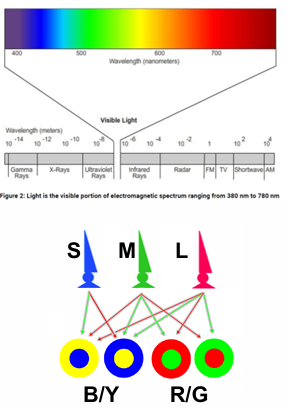
Visual system and wavelength sensitivity
Visual system is sensitive to visible light:
Normal CV:different hues; with all luminance and saturation variations, potential CV sensations number in the tens of thousands
Retina contains 3 cone photoreceptors:
Post-receptoral colour pathways: Red/Green and Blue/Yellow channels; Luminance channel
Channels: Red/Green, Blue/Yellow, and Luminance (often denoted as )
Colour processing
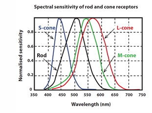
Colour processing is explained by the Young–Helmholtz theory (trichromacy)
Retina cone types and peak sensitivities:
Short-wavelength cones (S) – Cyanolabe; peak sensitivity
Medium-wavelength cones (M) – Chlorolabe; peak sensitivity
Long-wavelength cones (L) – Erythrolabe; peak sensitivity
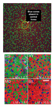
Retinal cone mosaic
Individual variations akin to a fingerprint
Typical ratio:
Approximately more L than M cones
S-cones are rare (≈ of cones)
Variation with eccentricity
No S-cones in central
Central vision can simulate a tritanopic CV defect if CV is measured in that region
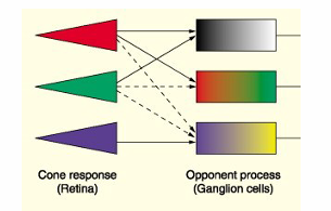
Post-receptoral colour pathways (Hering’s theory)
Inner retina, LGN, and primary visual cortex contribute to colour pathways
Red/Green channel (Parvocellular)
Blue/Yellow channel (Koniocellular)
Luminance channel (Magnocellular)
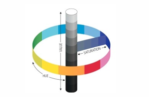
Colour space representation
Colour attributes model:
1) Hue – the distinctive name of the colour
2) Value (brightness) – represented by the perpendicular axis of the colour circle, from black to white
3) Saturation/Chroma – the percentage of “pure” hue in a colour
Perceptual colour attributes
Hue – associated with the dominant wavelength
e.g., red, yellow, blue
Saturation – amount of grey content; spectral colours are fully saturated
White/black/grey – fully desaturated
Value/Chroma (brightness) – related to perceived radiant energy; analogous to a dimmer switch
Colour properties
With three primary spectral wavelengths a match can be found for any reference wavelength
Primary colours commonly cited:
Red (650 nm), green (540 nm) and blue (460 nm)
CIE 1931 - standardised international system
Red (700 nm), green (546.1 nm) and blue (435.8 nm)
Metameric match: different spectral distributions that are perceived as identical colours.
Different spectral power distributions can be perceived as identical colours (metamers)
Colour obeys algebraic laws, including additive, subtractive, and scalar properties; Grassmann’s laws of metamers describe these relations
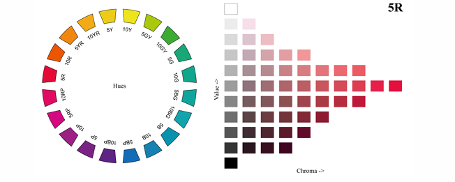
Colour space representation: Munsell system
Munsell colour system uses Hue/Value/Chroma, e.g. 5R 4/8
Hues: 10 subdivisions within 10 colour names → 100 hues in total
Values: 0 to 10 (dark to bright)
Chroma: 0 to variable limits depending on Hue/Value
Complex colour space
Conceptually, a 3D space with coordinates (x, y, z) satisfying
1931 CIE (x,y) chromaticity diagram
Spectral colours lie along the spectral locus curve where colours are fully saturated
Colour temperature described via the black body locus
Candle flame around appears more yellow
Incandescent around
LEDs around
Direct sunlight around
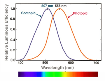
Psychophysics of colour
Relative luminous efficiency, , describes visual sensitivity across wavelengths
Under photopic conditions, a monochromatic stimulus at is perceived as brighter than other monochromatic stimuli of equal energy
Scotopic conditions V’(lambda)
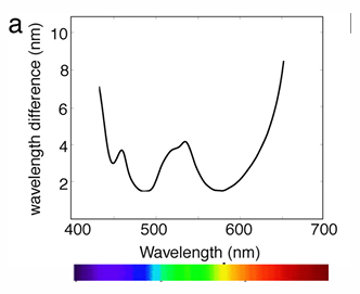
Wavelength discrimination
Wavelength difference that induces a noticeable hue difference
We can discriminate different hues with small wavelength changes in the range of approximately
More wavelength difference is required at the ends of the visible spectrum
Saturation discrimination
Just noticeable difference from white can be produced by a proportional mixture of a monochromatic light and white light
Yellow, approximately , is the most desaturated
The ratio of yellow to white must be high for the colour to appear different from white
Blue and red are more saturated; a small amount of these colours can make the mixture appear different from white
Other factors affecting colour perception
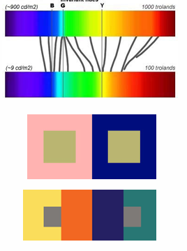
Bezold–Brücke (Bezold-Brucke) effect: hue changes with luminance
At high luminance, hues below appear bluer; hues above appear yellower
Invariant hues: examples include
Contrast effects on colour perception:
Simultaneous contrast (background effects)
Successive contrast (over time effects)
Stiles-Crawford effects: perceived colour and brightness depend on the part of the pupil through which light enters
Other factors affecting colour perception (physiology & anatomy)
Normal physiological variations:
Ocular media
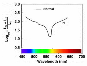
Age-related lens changes (cataract) increase absorption of short wavelengths, reducing blue-end sensitivity
Macular pigment (MP) – dietary pigments lutein/zeaxanthin accumulate in the RNFL
MPs absorb short-wavelength light before photoreceptor stimulation, altering blue sensitivity
Retinal eccentricity
Colour vision deteriorates toward the periphery
CV is relatively poor outside the central 2° of the visual field
Therefore, most CV tests are designed for foveal viewing
Next topic
Types, genetic and characteristics of abnormal colour vision