Lecture on Synaptic Integration
Overview of Lecture on Synaptic Integration
The primary focus of today's lecture is synaptic integration and its role in neuronal function.
Relevant reading is from SilverForm, Chapter 8.5.
Graded Potentials
Definition: Graded potentials are changes in membrane potential that vary in size, as opposed to being all-or-nothing.
Properties of Graded Potentials:
Graded: The magnitude of the potential depends on the amount of neurotransmitter released.
Small Release of Neurotransmitter: Opens a small number of ion channels, resulting in a small postsynaptic potential (PSP).
Large Release of Neurotransmitter: Opens a larger number of channels, leading to a larger PSP.
Decremental: Graded potentials decay in strength as they travel across the cell membrane.
Types of Potentials:
Excitatory Postsynaptic Potential (EPSP):
A depolarizing graded potential that brings the neuron closer to the threshold for firing an action potential.
Inhibitory Postsynaptic Potential (IPSP):
A hyperpolarizing graded potential that moves the neuron away from the threshold, making it less likely to fire an action potential.
Summation:
Graded potentials can combine; two smaller graded potentials can combine to form a larger graded potential.
Synaptic Integration
Purpose of Synaptic Integration:
The process of integrating multiple synaptic inputs to determine whether the neuron's initial segment will reach the threshold necessary to fire an action potential.
Diagram Discussion:
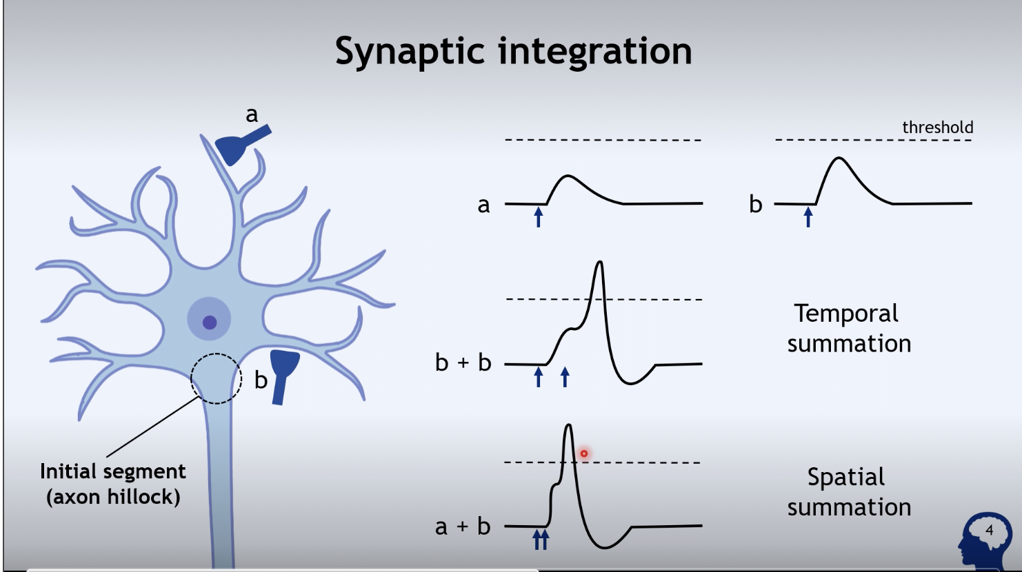
Two inputs, A and B, both are excitatory inputs causing EPSPs.
Mechanism of A and B:
At input A:
Stimulation results in the release of an excitatory neurotransmitter.
The neurotransmitter diffuses across the synaptic cleft and binds to ionotropic receptors, specifically non-specific monovalent cation channels, leading to rapid sodium influx and depolarization, resulting in a fast EPSP.
At input B:
The same process occurs, resulting again in an EPSP.
Comparison of A and B:
A's EPSP is smaller due to the decremental property of graded potentials; it travels further, thus degrading more compared to the closer input B.
Threshold and Action Potentials
Initial Segment and Axon Hillock:
The axon hillock decides whether an action potential will be fired based on membrane potential reaching threshold.
Threshold:
If the membrane potential reaches the critical threshold of -55 millivolts, an action potential is initiated.
Reaching Threshold:
Input B alone does not reach threshold due to its small EPSP.
Two methods to reach threshold:
Temporal Summation:
Firing input B twice in rapid succession results in the addition of two EPSPs, potentially elevating the potential to threshold.
Spatial Summation:
Firing inputs A and B simultaneously adds their EPSPs together to be enough to reach threshold.
Important Note:
Temporal and spatial summation are crucial, but the timing interval is critical for effective summation; if separated by too much time, they do not sum effectively.
Inhibitory Synapses
Inhibition via Input C:
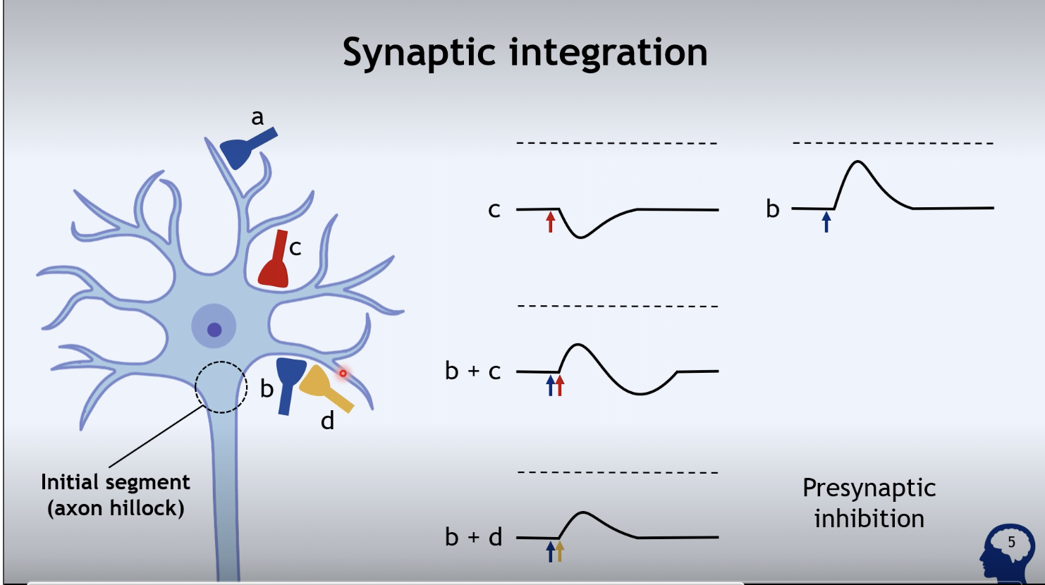
Stimulation of input C releases an inhibitory neurotransmitter.
This neurotransmitter binds to different ionotropic receptors with integral chloride channels, leading to rapid hyperpolarization, which is a fast IPSP.
Adding EPSPs and IPSPs:
When input B (EPSP) and input C (IPSP) are stimulated:
The EPSP from B rises the potential toward threshold, while the IPSP from C counters it, moving the potential away from threshold, thus inhibiting action potential firing.
Presynaptic Inhibition via Input D:
Input D does not synapse directly on the neuron but on the presynaptic terminal of input B.
Effect of Stimulation:
When D is stimulated alongside B, it reduces the amount of neurotransmitter released from B, resulting in a smaller EPSP due to reduced depolarization. This is termed presynaptic inhibition.
Types of Synapses
Axoaxonic Synapse (D):
Terminology can be misleading; it synapses onto the presynaptic terminal not the axon directly.
Typically inhibitory, limits neurotransmitter release.
Axosomatic Synapse (C):
Synapses directly onto the cell body (soma); typically serves inhibitory functions.
Axodendritic Synapse (A & B):
Synapses onto the dendritic spines, generally excitatory, allowing the neuron to receive inputs effectively.
Implications of Synaptic Integration
Physiological Relevance:
Neurons possess hundreds of excitatory and inhibitory synapses each evoking small EPSPs and IPSPs.
Individual postsynaptic potentials (PSPs) are small (usually a few millivolts) and not enough alone to reach action potential threshold.
Summation of these potentials is necessary:
Depolarization from EPSPs moves the cell towards threshold, increasing action potential likelihood.
Hyperpolarization from IPSPs moves the cell away from threshold, reducing action potential likelihood.
Summary of Synaptic Integration Role:
Determines if the initial segment reaches the threshold of -55 millivolts to trigger an action potential.
Conclusion and Activities
Learning Outcome Review:
The objective is to explain the role of synaptic integration in neuronal function thoroughly.
Preparation for Next Lecture: Discussions will continue on synaptic functions and their broader implications.