Sensory Pathway
Sensory Pathways
Definition: consist of a chain of neurons that transmit sensory information from receptor organs to the cerebral cortex, leading to perception of sensation.
Neurons Required: At least three serial neurons are necessary:
Primary Neurons: From the receptor to the dorsal root ganglion (DRG).
Secondary Neurons: From the DRG to the dorsal horn of the spinal cord.
Tertiary Neurons: From the dorsal horn to the thalamus.
General Sensory Pathway Structure
Key Components:
Dorsal Root Ganglion (DRG): Contains the cell bodies of primary sensory neurons.
Dorsal Horn: Site of synapse for primary and secondary neurons.
Ascending Tracts: Pathways that carry sensory information from the spinal cord to the brain.
Important Facts about Sensory Pathways
All sensory stimuli are processed in the brain, with the exception of olfaction (smell).
Sensory pathways relay at the thalamus, primarily the ventral posterolateral nucleus (VPL), except for olfactory (smell) signals.
Ascending Tracts
Spinothalamic Tract:
Transmits pain, temperature, and crude touch sensations (however, it's not the main function of this track).
Medial Lemniscus:
Functions: conveys critical sensory information, including:
Fine Touch: The ability to perceive and identify fine details on the skin.
Two-Point Discrimination: The capacity to distinguish between two closely spaced points on the skin.
Vibration Sense: The ability to detect vibrations applied to the skin.
Position Sense: Awareness of the position of body parts in space, also known as proprioception.
Weight Perception: The ability to perceive the weight of an object being held, contributing to conscious awareness of body position and posture.
Spinocerebellar Tracts:
Transmit unconscious proprioceptive information to the cerebellum to aid in movement coordination.
Spinothalamic Tract Detail
Structure:
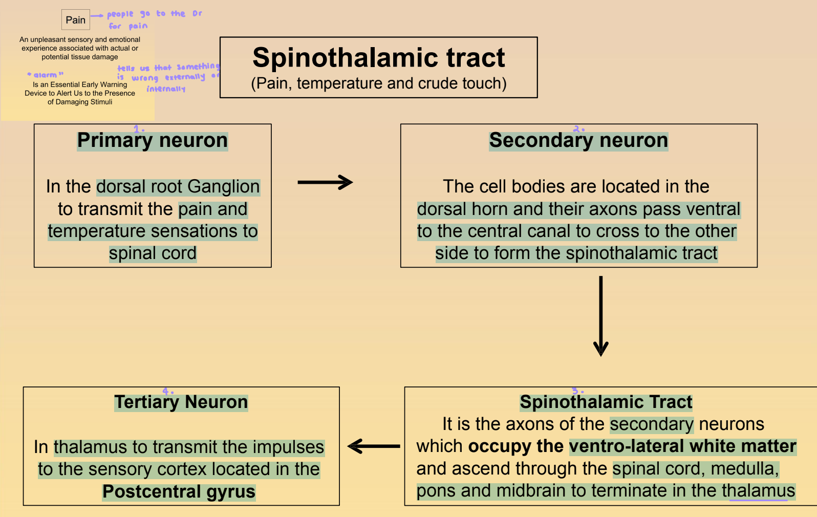
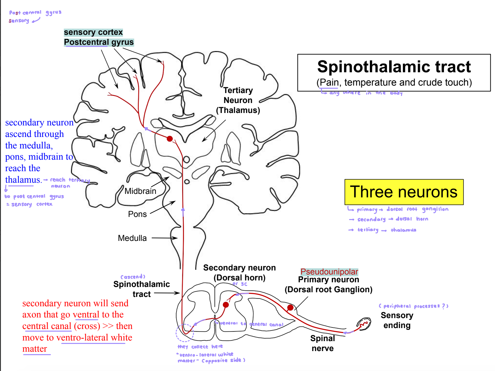
Primary Neuron: Cell bodies in the DRG; transmits pain and temperature information to the spinal cord.
Secondary Neuron: Located in the dorsal horn; crosses to the contralateral side near the central canal and forms the spinothalamic tract in the ventrolateral white matter.
Tertiary Neuron: Located in the thalamus; relays information to the sensory cortex in the postcentral gyrus.
Clinical Implications:
Chordotomy: Surgical procedure that can result in loss of pain and temperature sensation in the area contralateral to the lesion.
After a chordotomy, pain may initially disappear but can return due to the function of alternative pathways like the spinoreticulothalamic tract.
Medial Lemniscus System
Functions: Conveys information about:
The type of stimulus (what).
The precise location of the stimulus (where).
The temporal aspects of stimulus onset and cessation (when).
Clinical Application: Sensory testing can include tasks such as identifying fine touch or vibration, showing the functioning of this pathway.
Differences and Similarities between Spinothalamic and Medial Lemniscus Tracts
Similarities:
Primary neurons terminate in the dorsal horn of the spinal cord.
Secondary neurons cross to the opposite side at similar spinal levels.
Differences:
Spinothalamic tract is located in the ventrolateral white matter and conveys pain/temperature, while medial lemniscus is in the dorsal white matter and conveys fine touch/proprioceptive sensations.
Spinothalamic tract has a direct synapse in the thalamus (VPL); medial lemniscus involves synapses in the gracile and cuneate nuclei before reaching the thalamus.
Neuropathology
Lesions in Sensory Pathways:
Hemisection of the spinal cord results in:
Loss of pain/temperature sensation on the opposite side.
Loss of fine touch/proprioception on the same side.
Paralysis of muscles at the segmental level.
Conclusion
Understanding sensory pathways is crucial for diagnosing and treating sensory deficits and managing pain effectively. Neurological assessments often utilize knowledge of these pathways to identify lesions and plan interventions.