Endocytosis and Intracellular Vesicle Trafficking Course Notes
Core Concepts of Endocytosis and Membrane Permeability
Cell Membrane Overview: The lipid bilayer (cell membrane) is approximately thick and acts as a barrier that is poorly permeable to ions and macromolecules.
Uptake Mechanisms by Size:
- Ions and Small Molecules: Particles up to () can enter the cell through protein pumps, carriers, or channels.
- Macromolecules: Larger molecules, such as proteins ranging from , require membrane-bound carriers known as endosomes, which are typically in size. This process is defined as endocytosis.Definition of Endocytosis: The process of taking up extracellular material (fluids and macromolecules) through the invagination of the plasma membrane, followed by pinching off to form an intracellular vesicle.
Types of cellular uptake mechanisms
micropinocytosis: enables cells to take up fluid and nutrients
Clathrin-coated vesicle: allows uptake of receptors
Noncoated vesicle:
Caveolae: enables cells to redistribute their plasma membrane and takes up very specific ions
phagocytosis: enables cells to engulf large particles such as bacteria
Phagocytosis: Mechanics and Pathogen Interaction
Primary Function: The ingestion or removal of bacteria, typically sized between .
Professional Phagocytic Cells: These cells possess specific receptors capable of binding to antibodies that tag bacteria (opsonization).
Molecular Mechanism of Phagocytosis:
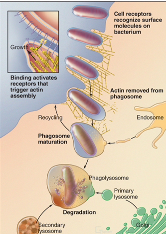
1. Receptor Activation: Receptors bind to the bacteria, triggering the activation of -actin polymerization.
2. Zippering: The plasma membrane protrudes and "zippers" tightly around the particle.
3. Sealing: The phagosome is sealed off from the external environment.
4. Maturation: -actin disassembles, and the phagosome is transported into the cell.
5. Degradation: The phagosome fuses with lysosomes to form a phagolysosome, where content is degraded.
Pathogenic Exploitation: Some bacteria induce their own phagocytosis to enter non-phagocytic mammalian cells:
- Yersinia: Causes Bubonic Plague (characterized by swollen lymph nodes).
- Listeria: Causes Listeriosis (fever, muscle aches, diarrhea).
- Shigella: Causes Shigellosis (diarrhea, fever, stomach cramps).
Macropinocytosis
Functions:
- Feeding: General uptake of extracellular fluid for feeding and removal of large numbers of growth factor receptors from plasma membrane.
- Cancer Metastasis: Cancer cells frequently utilize macropinocytosis for nutrient uptake to support rapid growth.Mechanism: Growth factors stimulate actin-driven protrusions of the plasma membrane called "ruffles” that are taken up as large vesicles.
Bacterial Induction: Salmonella can trigger macropinocytosis to enter the cell, causing Salmonellosis (diarrhea, vomiting, abdominal cramps).
Clathrin-Mediated Endocytosis (CME)
Function: receptor-mediated endocytosis allows the cell to take up specific macromolecules which are not abundant in the extracellular fluid
more than 25 different types of receptors
requires clathrin and adapter molecules
Major Types:
- Constitutive Receptor-Mediated: present all the time regardless of presence of macromolecule (like an escalator). Iron uptake - transferrin receptor, cholesterol uptake - LDL receptor
- Ligand-Induced Receptor-Mediated: can be stopped/turned on by growth signall]ing factors, e.g. EGF receptorStructure of Clathrin: Clathrin forms a three-legged structure called a triskelion, consisting of heavy chains and light chains.
CME Molecular Mechanism:
1. Selection (coat assembly and cargo selection): Adaptin molecules recruit specific receptors inside the cytosol (coat formation).
2. Bud Formation: Adaptin recruits clathrin (membrane starts to bend).
3. Vesicle formation: Recruitment of Dynamin, N-WASP and Arp2/3 leads to actin polymerization → pinches membrane → vesicle. plasma membrane cant produce its own dyamin as charged surfaces will repel eachother
4. Uncoating: Once inside, the vesicle is uncoated by HSC70 and Auxillin.
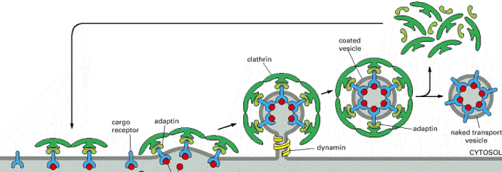
Dynamin Specifics: A large () protein that oligomerizes into spirals at the neck of the budding vesicle. It contains a PH (Pleckstrin Homology) domain, a GED (GTPase Effector Domain), and a PRD (Proline-Rich Domain). It is essential for vesicle fission.
Role of -Actin:
- Supports vesicle scission.
- Transports the vesicle away from the plasma membrane.
- Models of Invagination: The "Edge pushing model" and "Apical pulling model" describe actin's role in CCS (Clathrin-Coated Structure) invagination. Two alternative models for actin-dependent invagination of CCS
Caveolin-Mediated Endocytosis
Structure: Features small () invaginations known as Caveolae, which are highly enriched in cholesterol and the protein caveolin.
Formation: Caveolin self-associates to form a coat; this, combined with membrane cholesterol, induces membrane invagination.
Requirements: Dynamin and -actin are required for the internalization of caveolae.
Biological Function: Currently understood to function as a plasma membrane reservoir to balance membrane tension.
The Endocytic Pathway and Intracellular Trafficking
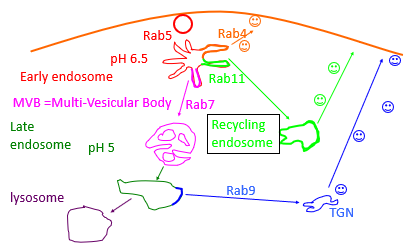
Organelle Progression:
- Early Endosome: Initial sorting station; maintained at approximately .
- MVB (Multi-Vesicular Body): Transition stage between early and late pathways.
- Late Endosome: Maturing compartment; maintained at approximately .
- Lysosome: Final degradation site.
- Recycling Endosome: Returns membranes/receptors back to the surface.
- TGN: Trans-Golgi Network.Rab Proteins:
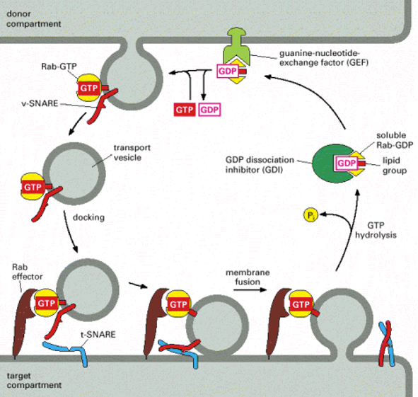
regulate compartmental specificity
recruit motor proteins that transport carriers on actin filaments or microtubules
recruit effectors: targeting and docking components (e.g. SNARES)
RAB’s and effector proteins are primary determinants of compartmental specificity
Rab protein classificiation:
Rab4 - receptor will end up going to the plasma membrane acting transmembrane protein
rab7 - sent to late endosome then degraded by lysosomes
rab 11- sent to the recycling endosome then return back to the plasma membrane
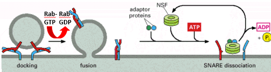
SNARE Proteins (SNAP Receptors):
protein complexes
transmembrane proteins
recruited by rabs and are specific for compartments
v-SNAREs and t-SNAREs on opposing membrane form trans-SNARE complex
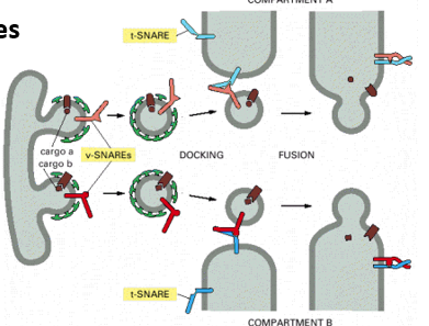
conversion of trans-SNARE to cis-SNARE complex provides energy for fusion of vesicle with target membrane
NSF (N-ethyl maleimide sensitive factor), an ATPase dissociate cis-SNARE complex and recycle SNAREs