Exs 323: Chap09 Circulatory
Overview of the Circulatory System
Respiratory system adds oxygen and removes carbon dioxide from the blood, while the circulatory system is responsible for the delivery of oxygenated blood and nutrients to tissues by their needs
Works alongside the pulmonary system (Cardiopulmonary system), emphasizing the role of the heart and lungs in oxygenating blood and supporting systemic circulation.
Primary Purposes:
Transport oxygen and nutrients to tissues, vital for cellular respiration and energy production.
Remove carbon dioxide and wastes from tissues, preventing toxic buildup.
Transport signaling molecules, including hormones, vital for body regulation.
Regulate body temperature, aiding in homeostasis during varying environmental conditions.
Key CV Adjustments during Exercise:
Increased cardiac output, essential for meeting elevated metabolic demands during physical activity.
Redistribution of blood flow from inactive organs to working muscles, optimizing oxygen delivery and nutrient supply to active tissues.
Structural Components of the Cardiovascular System
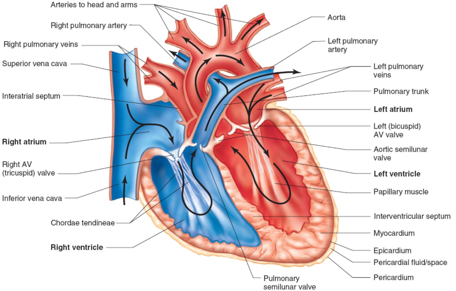
Heart: Pumps blood to create pressure, consisting of chambers (atria and ventricles) that ensure unidirectional blood flow.
Arteries and Arterioles: Carry oxygen-rich blood away from the heart, featuring muscular walls that control blood flow.
Capillaries: Sites of gas and nutrient exchange allow diffusion between blood and tissues.
Veins and Venules: Carry deoxygenated blood towards the heart, equipped with valves to prevent backflow.
Circulatory Response to Exercise
The heart consists of two main circuits: -
Pulmonary Circuit: The right side pumps deoxygenated blood to the lungs for reoxygenation.
Systemic Circuit: The left side pumps oxygenated blood to the body, delivering vital nutrients.
Heart Structure and Cardiac Cycle
Layers of Heart Wall:
Epicardium: Outer layer providing protection.
Myocardium: Thick, muscular middle layer responsible for contraction.
Endocardium: Inner layer lining the chambers.
Coronary arteries supply oxygen-rich blood to the myocardium to sustain its activity.
Myocardial infarction, or heart attack, arises from blockage in coronary blood flow, causing tissue damage.
Cardiac Cycle Phases:
Systole: Contraction phase; approximately 60-70 mL of blood ejected from ventricles per beat.
Diastole: Relaxation phase; heart fills with blood, crucial for effective pumping.
Heart sounds correspond to valve operations during systole and diastole, serving as indicators of heart health.
why only 1/3 of blood pressure?
Note that a rising heart rate results in a greater time reduction in diastole, whereas systole is less affected.
Blood Pressure Regulation
Blood pressure can be increased by the following factors:
a. ↑ in blood volume
b. ↑ in heart rate
c. ↑ blood viscosity
d. ↑ in stroke volume
e. ↑ peripheral resistance
Mean Arterial Pressure (MAP):
Indicates overall blood pressure; influenced by cardiac output and vascular resistance, providing insight into perfusion status.
Normal values are approximately 120/80 mmHg, representing healthy cardiovascular function.
Short-term regulation:
Sympathetic nervous system and baroreceptors regulate blood pressure, allowing rapid response to physical demands or stressors.
Long-term Regulation:
Governed by kidneys managing blood volume, crucial for sustaining blood pressure over time.
Cardiac Output Regulation
Cardiac Output Formula:
Q = Heart Rate (HR) x Stroke Volume (SV), illustrating the heart’s efficiency in delivering blood.
Key influencers of HR and SV include autonomic nervous system modulations, with sympathetic stimulation increasing both parameters during exercise.
Stroke Volume Factors:
End-Diastolic Volume (EDV)(pre-load): Volume of blood in ventricles at end of diastole; higher EDV leads to increased stroke volume.
Afterload: The pressure the ventricle must overcome to eject blood; significant in determining stroke volume.
Contractility: Strength of ventricular contraction, influenced by the sympathetic nervous system, enhancing cardiac output during physical exertion.
End-Diastolic Volume (EDV)(pre-load)
Dependent on venous return
Venous return increased by:
–Venoconstriction
Via SNS
–Skeletal muscle pump
Rhythmic skeletal muscle contractions force blood in the extremities toward the heart
One-way valves in veins prevent backflow of blood
–Respiratory pump
Changes in thoracic pressure pull blood toward heart
Result: Frank-Starling mechanism →Greater EDV results in a more forceful contraction
Blood Flow Dynamics
Blood flow is governed by pressure differences and resistance; resistance is influenced by vessel diameter, blood viscosity, and vessel length, impacting overall circulation efficiency.
So our pressure gradient generated by systole is driving blood from the LV (Left ventricle) towards the RA (Right Atrium).
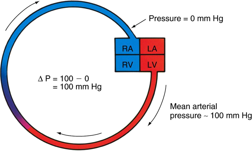
Blood flow dynamics change during exercise due to various physiological adaptations :

Changes in Blood Flow During Exercise
During exercise, cardiac output increases significantly, with substantial redistribution towards active muscles: - Up to 80–85% of cardiac output during maximal exercise is redirected to muscle tissues, leading to decreased flow to non-active organs.
Regulation of blood flow is facilitated through nitric oxide as a potent vasodilator, enhancing blood supply to working muscles.
Oxygen Delivery and Recovery
Oxygen delivery increases due to heightened cardiac output and blood flow redistribution towards active tissues, crucial for sustaining energy production during exercise.
Transitioning from rest to exercise causes a rapid increase in heart rate, stroke volume, and cardiac output, with gradual recovery in HR and SV post-exercise as the body returns to homeostasis.
Final Thoughts
Understanding how the cardiovascular system adjusts to exercise helps explain performance in sports and activity-related physiology, providing insights into training adaptations and recovery strategies.
Focus on adjustments during different intensities and durations of exercise and their implications for optimal training and performance enhancement strategies.
Rest to exercise / exercise to recovery
At the onset of exercise:
–Rapid increase in HR, SV, cardiac output
–Plateau in submaximal (below lactate threshold) exercise
During recovery
–Decrease in HR, SV, and cardiac output toward resting levels
–Time to return depends on:
Duration and intensity of exercise
Training state of subject
extra -
why only 1/3 of blood pressure?
Note that a rising heart rate results in a greater time reduction in diastole, whereas systole is less affected.
ST segment Depression - & myocardial escimeia
what are 2 major alterations of CVS during exercise?
frank staling mechanism → adjusts stroke volume
Autonomic nervous system is what’s regulating Heart rate and stroke volume
Red Blood Cells - deliver oxygen
Arteriovenous difference (AV difference) - how much oxygen is extracted from RBC