Life Science Lab + The Microscope and Microscopy Techniques
The Scientific Method is the process of objectively establishing facts through observation, experimentation, and repetition.
Step 1: Make an Observation
From your experiences, thoughts, or reading
Step 2: Ask a question
Why does this phenomenon occur?
Step 3: Formulate one or more hypotheses
What are the possible causes of this phenomen
Step 4: Develop testable prediction(s)
If (hypothesis 1,2,3…) is the cause of this phenomenon, then I expect….
IF, THEN statement
Step 5: Design an Experiment
Only test one hypothesis per experiment
Independent variable: The cause- its value is independent of other variables in your study.
Dependent variable: The effect – its value is dependent on the changes within the experiment
Control variable: Any variable(s) that are held constant in a research study.
Experiments must be repeatable, with clear, transparent procedures
Step 6: Collect Data
Record your results, challenges, and any changes to the procedure
Step 7: Analyze the data
Statisitical Tests: T-Tests, ANOVA, Regression, etc
T-test: compares the two means in a group
ANOVA: compaes the mean across 3+ groups
Regression: Anaylsis the relationship between two variables
Graphs: A visual representation of the data.
Independent variable on x-axis, dependent variable on y-axis
Step 8: Draw conclusion(s)
Does your data support your hypothesis?
All data and conclusions even if our predictions are not supported
What is a microscope?
An instrument used to see and enhance the resolution (clarity, sharpness) of an object.
Used to magnify an object that we cannot see with the human eye
Primary kinds of microscopes: Dissecting (stereo), Compound, SEM (Scanning Electron Microscopes), and TEM (Transmission Electron Microscope)
Types of Microscopes
There are many different types of microscopes, including Dissection (Stereoscopic), Compound, Fluorescence, Digital, Automatic Imaging, Confocal, Phase Contrast, SEMs or (Scanning Electron Microscopes), and lastly TEMs (Transmission Electron microscope).
In LifeScience, however, we will learn about and use just two
The Dissection (Stereoscopic) Microscope
Light BOUNCES OFF the specimen
Used to view larger, thick, solid or opaque specimens
Used in the dissection of small animals
Defining characteristics
Often have a simple or no built-in light source
Two focus knobs
1 or 2 ocular lens
Large specimen stage
The Compound Light Microscope
Light shines THROUGH the specimen
Used to view very small, thin, or transparent objects/specimens
Used to study the life histories and identity of microorganisms
Defining characteristics
Have a built-in adjustable light source
Coarse and Fine focus knobs
1 or 2 ocular lens, Multiple objective lenses
Adjustable stage for specimens
Parts of the Stereo-microscope
Ocular Lens
Objective Lens
Zoom Knob
Focus Knob
Arm
Lights (Not always)
Stage
Base
Light is bounced off the specimen and reflected into the lens for viewing
The Compound Microscope
Used for viewing at high magnification (4-1000x)
Magnifies the image twice
Composed of two or more lenses: a primary magnification lens (ocular) and a secondary lens system (objective)
Light is passed through an object and is then focused by the primary and secondary lens.
A compound Microscope has four different objective lenses:
4x = Low Power
10x = Medium Power
40x = High Power
100x = Oil Immersion
Ocular lens itself is approximately 10x
Parts of the Compound Microscope
Ocular Lens
Objective Lens (4x-100x)
Coarse Adjustment Knob
Fine Adjustment Knob
Stage Adjustment knobs
Condenser Lens
Specimen Clamps
Light Source
Arm
Stage (Mechanical)
Base
Compound Microscope – How Does it Work?
Light passes through primary and secondary lenses resulting in the viewed image becoming inverted and backward.
When adjusting, move the mechanical stage to the opposite direction from the desired field of view.
Left Right
Right Left
Up Down
Down Up
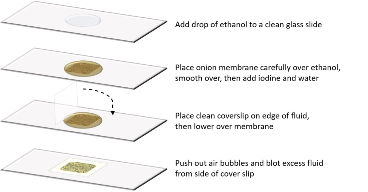
How to Prepare a Wet Mount