Fundamentals of Physiology: Epithelial Cell Function Study Guide
General Characteristics of Epithelial Cells
Definition and Location:
Epithelial cells cover body surfaces and line body cavities.
They form the interface between the internal environment of the body and the external environment.
Primary Functions of Epithelial Cells:
Protection: They form a protective barrier against physical damage and pathogens.
Selective Barrier: They act as a gateway, controlling what enters and exits the body or specific organs.
Gland Formation: Epithelial cells are specialized to form glands and secrete products.
Regulation: They regulate the exchange of molecules between different compartments.
Absorption: They are specialized for taking up nutrients or fluids, such as in the epithelial lining of the small intestine.
Structural Components of Epithelial Tissue:
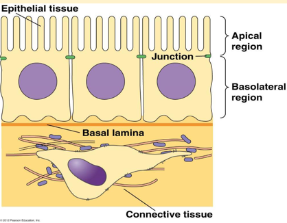
Apical Region: The surface facing the lumen or the exterior.
Basolateral Region: The surface attached to the basement membrane and adjacent cells.
Basal Lamina: A specialized layer of extracellular matrix that supports the epithelial cell layer.
Connective Tissue: Situated beneath the basal lamina, containing cells like fibroblasts.
Junctions: Specialized protein complexes that hold the cells together and facilitate communication.
Distinction Between Epithelial and Endothelial Cells:
Epithelial Cells:
Line both internal and external surfaces of the body.
Can be classified by shape: squamous, cuboidal, and columnar.
Can be organized into layers: simple (single layer) or stratified (multiple layers).
Endothelial Cells:
These are a specialized type of epithelial cells.
They specifically line the inner surfaces of blood vessels and lymph vessels.
simple squamous epithelium
Cell Polarity and the Domains of Epithelial Cells
Concept of Polarity:
Epithelial cells exhibit distinct polarity, meaning each surface (domain) has a specialized morphology and unique functions.
Apical Domain (Surface):
This domain faces the lumen or external environment and contains specialized structures:
Glycocalyx:
A carbohydrate coating made of glycoproteins and glycolipids.
In epithelial cells, it functions in cell-cell recognition, intercellular adhesion, and communication.
In endothelial cells, it regulates vascular permeability and interactions between blood and the endothelial cells.
Cilia:
Motile, fine hair-like cytoplasmic structures.
Length typically ranges between .
Function: Moving particles and fluid along the epithelial surface.
Locations: Trachea, large bronchi, and uterine tubes.
Microvilli:
Irregular projections of the cell membrane, approximately in length.
They are closely packed in cells with an absorptive function to increase surface area.
Locations: Small intestine and kidneys.
Lateral Domain:
This domain is responsible for cell-to-cell contact and adherence.
Tight Junctions (Zonula Occludens): Prevents the leakage of material between cells.
Adherens Junctions (Anchoring): Uses cadherins to bind cells together.
Desmosomes (Macula Adherens): Strong anchoring junctions using intermediate filaments.
Gap Junctions: Channels for communication and transport of small ions/molecules between cells.
Basal Domain:
This domain faces the underlying connective tissue.
Functions:
Acts as an adhesion interface between epithelial cells and the extracellular matrix (ECM) using integrins and hemidesmosomes.
Serves as a sieve or permeability barrier controlling entry and exit from cells.
Controls cell organization and specialization.
Membrane Proteins and Disease Examples
Cystic Fibrosis (CF):
Nature of Disease: A life-shortening, autosomal recessive genetic disorder that affects multiple systems, primarily the lungs. multisystem disease, often difficult to treat
Symptoms: Difficulty breathing, coughing up thick sticky mucus blocking airways, frequent lung infections, and elevated sweat chloride levels.
Molecular Mechanism:
Caused by a mutation in the CFTR (Cystic Fibrosis Transmembrane Conductance Regulator) protein.
CFTR is a transmembrane protein expressed on the apical region of epithelial cells lining the lungs, gut, and exocrine glands.
Normal CFTR Function:
Transports ions across the epithelial surface to create an osmotic gradient maintaining salt-water balance on many body surfaces.
This allows water to flow into the luminal space, keeping mucus hydrated.
goblet cells and submucosal glands produce mucous
mucous is effectively transported, facilitates optimal mucociliary clearance and airway defence.
Effect of CFTR Mutation:
Reduced channel number, function or both.
Water flow is inhibited, leading to dehydration of the airway surface liquid.
Result: Build-up of static, thick mucus on the outside of cells, static mucous.
less surface liquid
Decreased bicarbonate transport leads to an acidic pH.
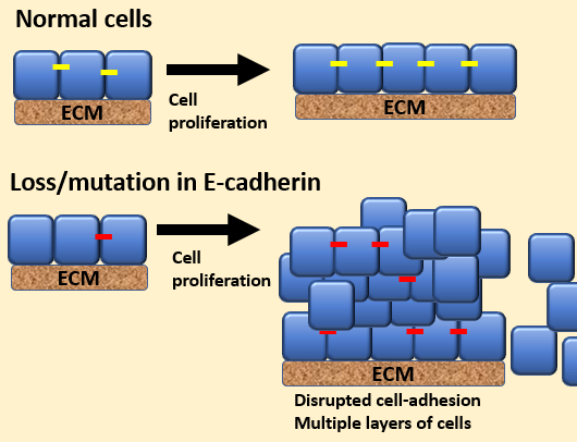
E-Cadherin and Cancer:
TM proteins, major constitutions of adherens junction, important for cell-cell binding
cadherins mediate intercellular adhesion
in normal cells proliferation/growth ceases when cells come in contact with one another - contact inhibition
Malignant Transformation:
If is lost or mutated, cells lose contact inhibition.
cells undergo malignant transformation, uncontrolled cell proliferation, tumor formation
Specialized Functions in Various Systems
1. Barrier Function (Skin):
Maintains a physical barrier against environmental insults, microbial invasion, chemicals, toxins, and allergens.
Facilitates communication between internal and external environments.
Example (Eczema): Damaged skin barrier leads to loss of moisture, inflammation, and penetration of allergens/bacteria.
2. Gas Exchange (Lungs):
Occurs across the alveolar epithelium.
Type I Pneumocytes: Extremely thin cells lining of the alveoli; united by tight junctions to facilitate gas exchange.
Type II Pneumocytes: Produce a thin layer of surfactant that covers the alveolar surface to prevent collapse.
3. Absorption (Gastrointestinal System):
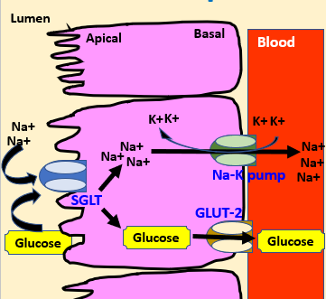
Tall cells forming a single later (simple) with apical microvilli increase surface area for absorption.
Tight junctions ensure selective absorption by preventing material from passing between cells.
allows only selection of material to be absorbed
abnormal expression levels/activity of NA-K pump in diabetes, alzheimer’s disease, hypertension and tumours
Transport Mechanisms:
Apical membrane: Uses SGLT (Sodium-Glucose Linked Transporter) to bring glucose and into the cell.
Basolateral membrane: Uses GLUT-2 for glucose exit into the blood and the pump to maintain ion gradients.
4. Reabsorption (Kidneys):
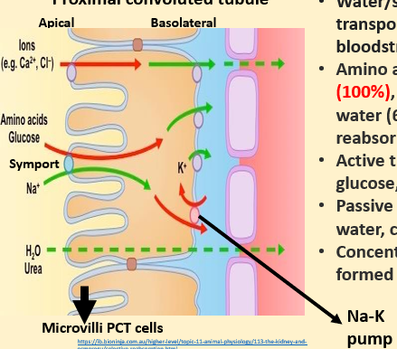
water/solutes transported into bloodstream
Occurs in the Proximal Convoluted Tubule (PCT) where cells have extensive microvilli.
Substances Reabsorbed:
Average reabsorption: of glucose and amino acids; of salts and water.
Active transport is used for glucose and amino acids; passive transport is used for water and chloride ions.
Goal: To form concentrated urine while returning essential solutes to the bloodstream.
5. Secretion (Pancreas):
Endocrine Function:
Islets of Langerhans contain Alpha cells and Beta cells.
Diabetes Mellitus Type 1: Occurs when Beta cells are destroyed, stopping insulin secretion.
Exocrine Function:
Pancreatic acinar cells secrete digestive enzymes, water, and a high concentration of into the duodenum.
Acinar Cell Injury: Caused by alcohol, smoking, or high-fat diets increase stress on acinar cells. loss of exocrine pancreatic function - reduces absorption of nutrients, malnutrition
Result: Malnutrition due to reduced nutrient absorption and chronic damage known as pancreatitis.