C2.1 Chemical signalling (HL) Notes
C2.1.1 receptors
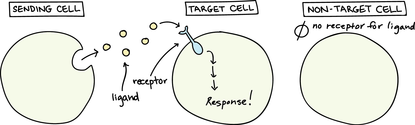
Receptors as Proteins with Binding Sites for Specific Signalling Chemicals
Receptors are highly specific, meaning they only bind to particular molecules, called ligands.
This specificity ensures that cells respond accurately to the right signals.
Definition
ReceptorA receptor is a protein that detects and responds to specific signals. They bind to signalling molecules(ligands) to initiate a cellular response.
Definition
LigandA ligand is a molecule that binds to a specific site on a receptor protein.
Analogy
Imagine you're in a crowded room, and someone calls your name.
You immediately turn to see who it is.
This is similar to how cells communicate: they "listen" for specific signals using receptors.
Receptors
Receptors are proteins found either:
On the cell membrane (for ligands that cannot pass through the membrane, like hydrophilic molecules).
Inside the cell (for ligands that are hydrophobic and can diffuse through the membrane, like steroid hormones).
Receptors have specific binding sites that fit the structure of their ligand like a lock and key.
This specificity ensures that only the correct ligand binds to its receptor, triggering the appropriate cellular response.
Steps of Interaction
Binding: The ligand binds to the receptor's ligand-binding site.
Conformational Change: The receptor changes shape, activating it.
Signal Transmission: The activated receptor triggers a response inside the cell.
Tip
Receptors and enzymes both have specific binding sites, but receptors do not alter their ligands.
They simply transmit the signal.
Why Receptor-Ligand Interactions Matter
Receptor-ligand interactions are essential for many biological processes, including:
Hormonal Regulation: Hormones like insulin bind to receptors to control blood sugar levels.
Nerve Signalling: Neurotransmitters bind to receptors to transmit signals between neurons.
Immune Responses: Cytokines bind to receptors to coordinate immune activity.
Tok
How does the specificity of receptor-ligand interactions reflect the broader principle of structure determining function in biology?
Can you think of other examples where this principle applies?
Self Review
What is a ligand? Give two examples.
Explain how receptors are specific to their ligands.
Describe the role of neurotransmitters as ligands in the nervous system.
C2.1.2 cell signalling by bacteria in quorum sensing
Cell Signalling by Bacteria in Quorum Sensing
Imagine a crowded room where everyone whispers.
At first, the whispers are too faint to hear.
But as more people join in, the collective sound grows louder until it triggers a response.
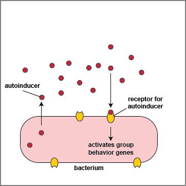
This is how quorum sensing works in bacteria, a process that enables them to coordinate activities based on population density.
Definition
Quorum SensingA communication system used by bacteria to coordinate group behaviors based on population density through the release and detection of signaling molecules.
How Quorum Sensing Works
Production of Signaling Molecules: Each bacterium releases small chemical signals called autoinducers.
Diffusion and Accumulation: These molecules diffuse freely in the environment. As the bacterial population grows, the concentration of autoinducers increases.
Detection: When the concentration of autoinducers reaches a critical threshold, they bind to specific receptors in the bacteria.
Response Activation: This binding triggers changes in gene expression, leading to coordinated behaviors like biofilm formation or bioluminescence.
Analogy
Think of autoinducers as text messages sent by each bacterium.
When enough messages are sent, the group decides to act together.
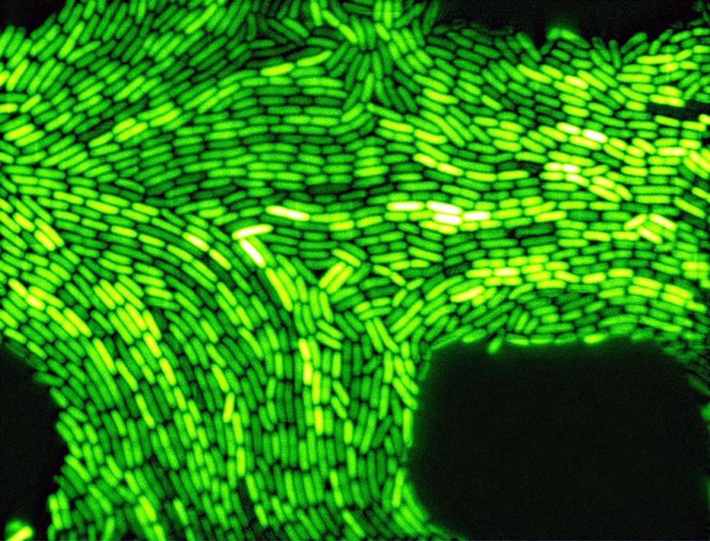
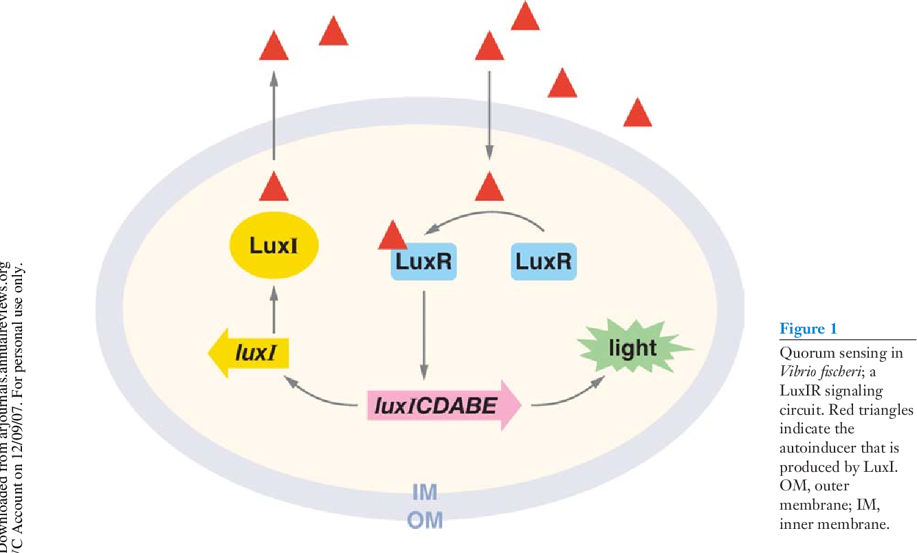
Bioluminescence in Vibrio Fischeri Is A Classic Example
Vibrio fischeri, a marine bacterium, uses quorum sensing to produce light through bioluminescence.
This process is most famously observed in its symbiotic relationship with the bobtail squid.
The Role of Autoinducers
Low Population Density: When V. fischeri bacteria are sparse, the concentration of autoinducers is too low to trigger a response.
High Population Density: In dense populations, such as those in the squid's light organ, autoinducers accumulate and bind to a cytoplasmic receptor protein called LuxR.
Gene Activation: The LuxR-autoinducer complex binds to DNA, activating genes responsible for producing the enzyme luciferase.
Light Production: Luciferase catalyzes a chemical reaction that emits light, with over 80% of the energy released as visible light.
Note
Free-living V. fischerido not emit light because it would waste energy without a functional purpose.
However, in the squid's light organ, bioluminescence helps the squid camouflage itself by mimicking moonlight on the ocean surface, reducing the risk of predation.
Mutualistic Relationship with the Bobtail Squid
Bacterial Benefit: The squid provides V. fischeri with a nutrient-rich environment, supplying sugars and amino acids.
Squid Benefit: The light emitted by the bacteria helps the squid avoid predators.
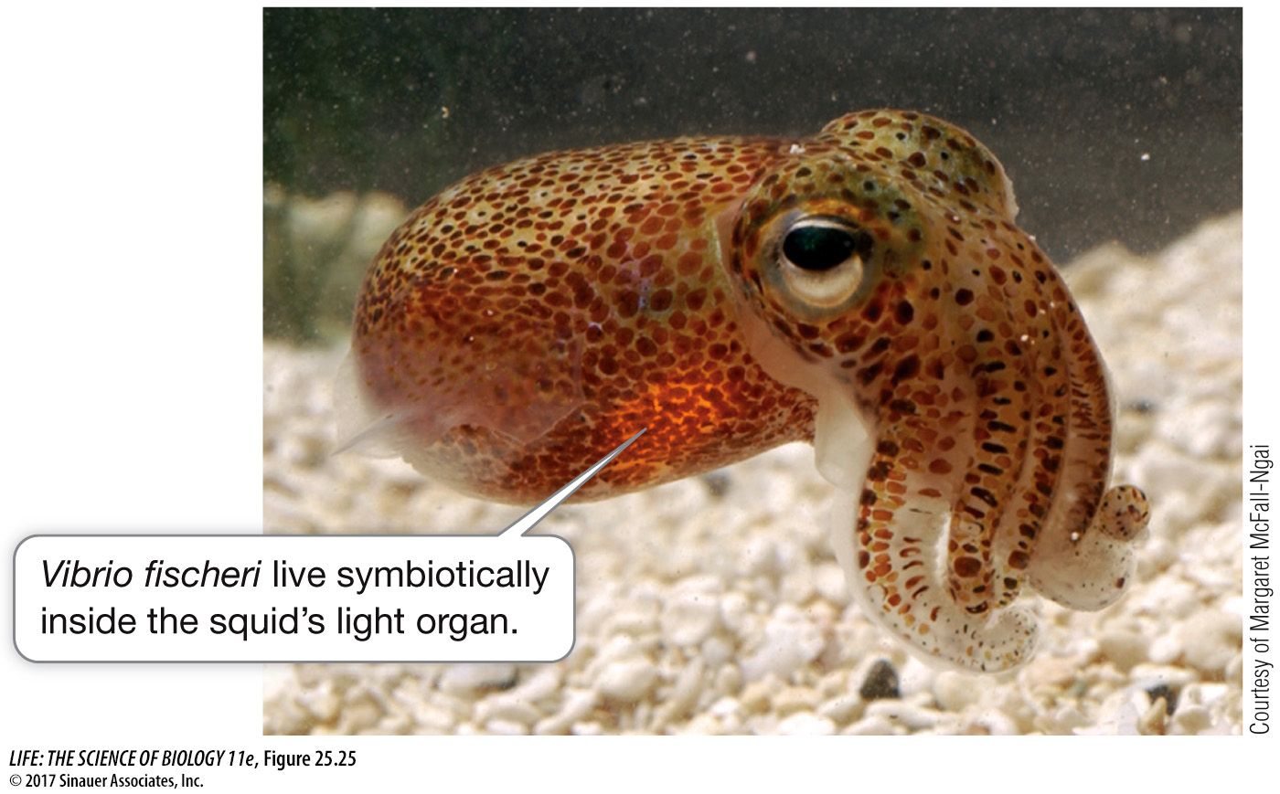
Example
The bobtail squid and Vibrio fischeri demonstrate a classic example of mutualism, where both organisms benefit from the relationship.
Why Quorum Sensing Matters
Quorum sensing enables bacteria to perform tasks that are ineffective at low densities but highly efficient when coordinated across a large population.
Key Activities Regulated by Quorum Sensing
Biofilm Formation: Bacteria produce glue-like substances to form protective layers, such as dental plaque.
Virulence Factor Production: Pathogenic bacteria coordinate the release of toxins to overwhelm a host's immune system.
Bioluminescence: As seen in Vibrio fischeri, light production is triggered only at high densities.
Warning
Don't confuse quorum sensing with simple chemical signaling.
Quorum sensing specifically involves population-density-dependent behaviors.
Mechanism of Quorum Sensing in Vibrio Fischeri
Autoinducer Production: V. fischeri synthesizes an autoinducer molecule (Autoinducer-3 or AI-3).
LuxR Activation: At high concentrations, the autoinducer binds to LuxR receptor,forming a complex. The binding of AHL to LuxR activates the lux operon, a set of genes responsible for producing the bioluminescent proteins.
Gene Expression: The LuxR-autoinducer complex binds to DNA, activating genes for luciferase production.
Bioluminescence: The enzyme luciferase catalyzes the light-emitting reaction. The light emission occurs when luciferase catalyzes the oxidation of a substrate, producing light as a by-product.
Note
The lux operon is a prime example of gene regulation through quorum sensing.
As the population grows and AHL binds to LuxR, the lux operon is activated, allowing the bacteria to produce bioluminescence proteins in large quantities.
Tip
Remember that quorum sensing is a two-step process: detecting autoinducers and responding through gene expression changes.
Broader Implications of Quorum Sensing
Quorum sensing is not limited to Vibrio fischeri.
It is a widespread mechanism in bacteria, with significant ecological and medical implications.
Applications and Challenges
Medical Research: Understanding quorum sensing can lead to novel treatments that disrupt bacterial communication, preventing biofilm formation or reducing virulence.
Biotechnology: Harnessing quorum sensing could improve synthetic biologyapplications, such as engineered bacteria for environmental cleanup.
Antibiotic Resistance: Targeting quorum sensing offers an alternative to traditional antibiotics, potentially reducing the evolution of resistant strains.
Tok
How might disrupting quorum sensing in bacteria raise ethical questions?
Consider the balance between preventing infections and preserving natural microbial ecosystems.
Self Review
How does quorum sensing enable Vibrio fischeri to produce light?
Explain the relationship between Vibrio fischeri and the Hawaiian bobtail squid.
C2.1.3 hormones, neurotransmitters, cytokines and calcium ions
Cells Communicate Using Signalling Chemicals That Regulate Physiological Processes
In multicellular organisms, these signalling chemicals are diverse and can be grouped into several functional categories.
These include:
Hormones
Neurotransmitters
Cytokines
Calcium ions.
Analogy
In a bustling city, traffic lights control the flow of cars, walkie-talkies keep police officers connected, and emergency sirens alert everyone to urgent situations.
Each of these signals serves a unique purpose, ensuring the city runs smoothly.
Similarly, your body relies on a variety of signalling chemicals to coordinate its activities.
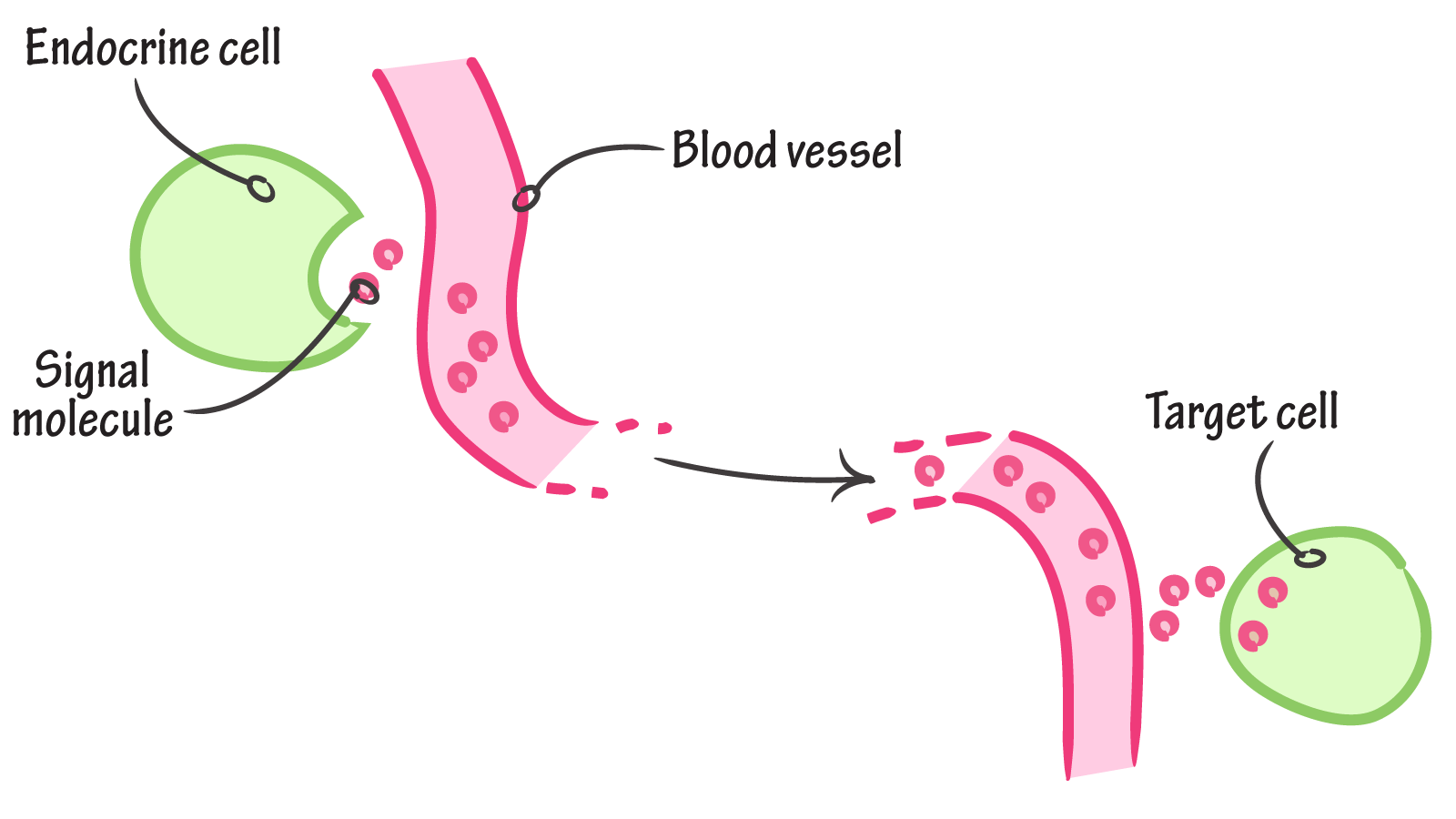
Hormones Are The Long-Distance Messengers
Definition
HormoneHormones are chemical messengers secreted by endocrine glands into the bloodstream.
Hormones are like the postal service of your body, delivering messages over long distances.
They are produced by endocrine glands and travel through the bloodstream to reach target cells.
Example
Insulin, produced by the pancreas, regulates blood sugar levels by signaling cells to absorb glucose.
Key Features of Hormones
Wide Reach: Hormones can affect cells far from their origin.
Specificity: Only target cells with the right receptors respond to a hormone.
Long-Lasting Effects: Hormones can remain active for hours or even days.
Slow Action: Hormonal responses are slower compared to other signals, as they rely on blood circulation.
Note
Hormones are classified based on their chemical structure, such as peptides (e.g., insulin), steroids (e.g., testosterone), or amines (e.g., thyroxin).
Tip
Remember: Endocrine glands release hormones into the bloodstream, while exocrine glands use ducts to secrete substances like sweat or saliva.
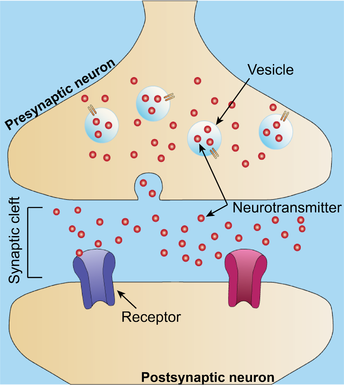
Neurotransmitters Are The Rapid Communicators
Neurotransmitters are the body's instant messaging system, transmitting signals across synapses,the tiny gaps between neurons.
They enable rapid communication within the nervous system.
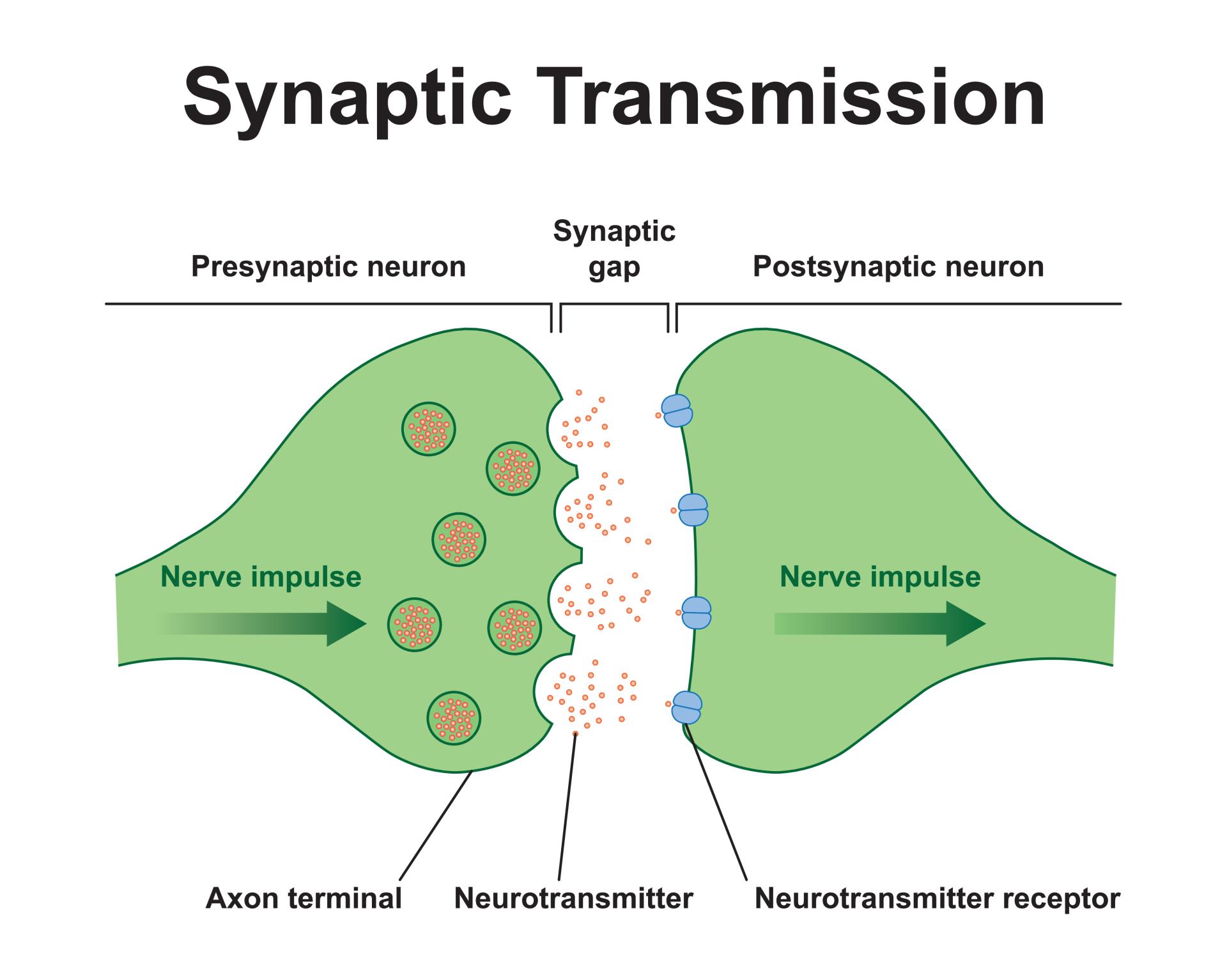
Definition
NeurotransmitterNeurotransmitters are chemical signals used by neurons to communicate across synapses, the tiny gaps between nerve cells or between nerve cells and other target cells.
Example
Acetylcholine is a neurotransmitter that triggers muscle contractions by transmitting signals from motor neurons to muscle fibers.
Key Features of Neurotransmitters
Short Distance: Neurotransmitters act across synapses, typically just 20-40 nanometers wide.
Fast Action: Signals are transmitted in milliseconds, enabling quick responses.
Short-Lived Effects: Neurotransmitters are quickly broken down or reabsorbed,ensuring precise control.
Localized Impact: They affect only the postsynaptic neuron or muscle fiber they bind to.
Warning
Don't confuse neurotransmitters with hormones!
While both are signalling molecules, neurotransmitters act over short distances and have rapid, short-lived effects.
Analogy
Think of neurotransmitters as text messages - quick, direct, and meant for a specific recipient.
In contrast, hormones are like public announcements broadcasted to a wide audience.
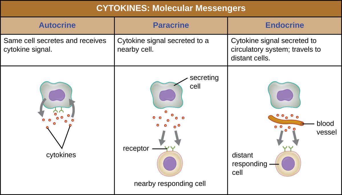
Cytokines Are The Immune System Coordinators
Cytokines are small proteins that act as the immune system's communication network.
They coordinate responses to infections, inflammation, and other immune processes.
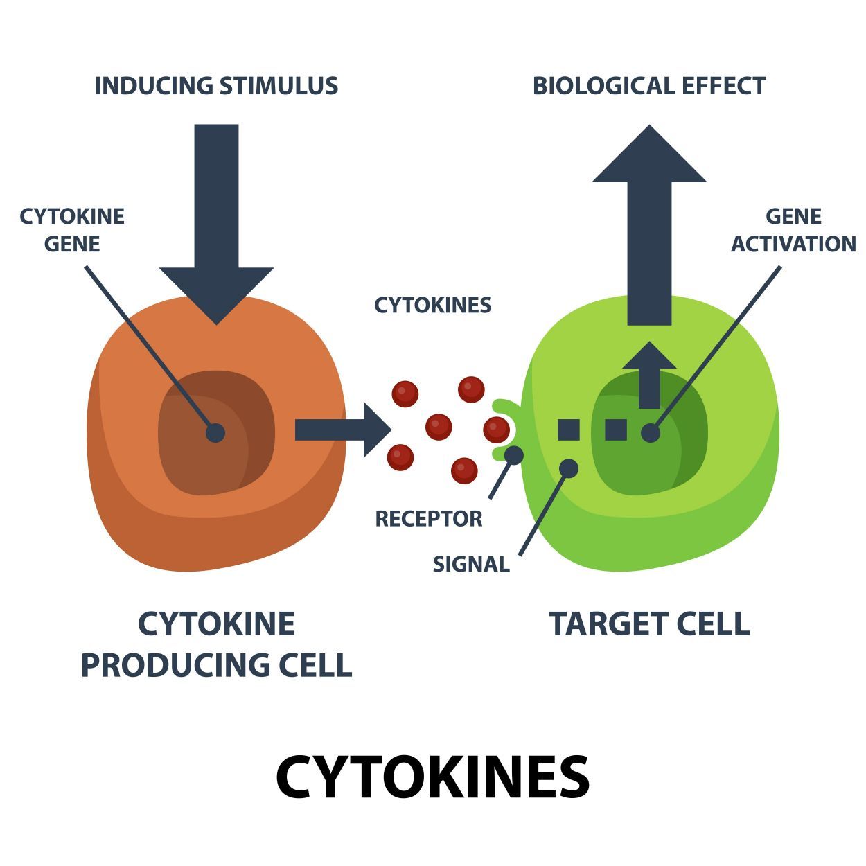
Definition
CytokineCytokines are small proteins that mediate and regulate immune responses, inflammation, and cell signaling
Example
Interleukin-1 (IL-1) is a cytokine that promotes inflammation by signaling immune cells to the site of an infection.
Key Features of Cytokines
Local or Short-Range Action: Cytokines often act on nearby cells or the cell that produced them.
Diverse Effects: They regulate immune responses, cell growth, and even embryonic development.
Multiple Sources: Cytokines can be produced by various cell types, including immune cells and epithelial cells.
Complex Interactions: A single cytokine can have different effects depending on the target cell and receptor.
Note
Cytokines usually act locally (paracrine signaling) but can have systemic effects.
Note
Cytokines play a critical role in inflammation.
However, excessive cytokine activity can lead to conditions like cytokine storms, which are associated with severe diseases such as sepsis.
Tok
How does the specificity of cytokine-receptor interactions compare to the specificity of neurotransmitter-receptor interactions?
What does this reveal about the evolution of communication systems in the body?
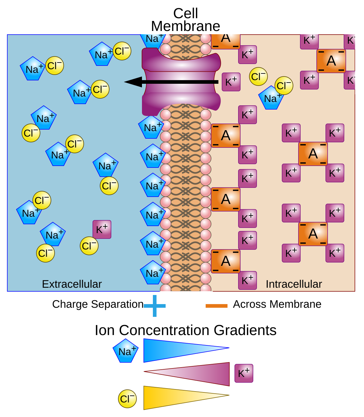
Calcium Ions Are The Universal Signal
Calcium ions are not traditional molecules like hormones or neurotransmitters but play a crucial role as secondary messengers in cellular signaling.
Calcium ions () are versatile signalling molecules involved in a wide range of cellular processes, from muscle contraction to neurotransmitter release.
Example
In muscle fibers, ions are released from the sarcoplasmic reticulum in response to a nerve impulse, triggering the contraction of muscle fibers by interacting with proteins like troponin.
Key Features of Calcium Ions
Intracellular and Extracellular Roles: ions function both inside and outside cells.
Rapid and Reversible: levels can change quickly, allowing for fast responses.
Wide Range of Functions: ions are involved in muscle contraction, neurotransmitter release, and even gene expression.
Highly Regulated: concentrations are tightly controlled by channels, pumps, and storage organelles like the sarcoplasmic reticulum.
Warning
A common misconception is that calcium ions only play a role in bone health.
While they are essential for bone structure, their signalling functions are equally critical.
Self Review
Can you explain how calcium ions contribute to both muscle contraction and neurotransmitter release?
What similarities and differences can you identify?
Comparing the Four Categories
Category | Distance | Speed | Duration | Examples |
|---|---|---|---|---|
Hormones | Long (via bloodstream) | Slow | Long-lasting | Insulin, thyroxin |
Neurotransmitters | Short (across synapses) | Fast | Short-lived | Acetylcholine, dopamine |
Cytokines | Local or short-range | Variable | Variable | Interleukin, interferon |
Calcium Ions | Intracellular or extracellular | Fast | Short-lived | Muscle contraction, neurotransmitter release |
Note
While these categories have distinct roles, they often interact.
For instance, calcium ions can mediate the effects of hormones and neurotransmitters within target cells.
Similarly, cytokines can influence the release of hormones during stress or inflammation.
Tok
In what ways do the different categories of signalling chemicals reflect the interconnectedness of biological systems?
How might this influence our understanding of health and disease?
Self Review
What are the key differences between hormones and neurotransmitters in terms of distance and duration of action?
How do cytokines differ from hormones in terms of their range of action and sources?
Why are calcium ions considered a universal signalling molecule?
C2.1.4 chemical diversity of hormones and neurotransmitters
Chemical Diversity Enables Hormones and Neurotransmitters to Perform Specialized Roles
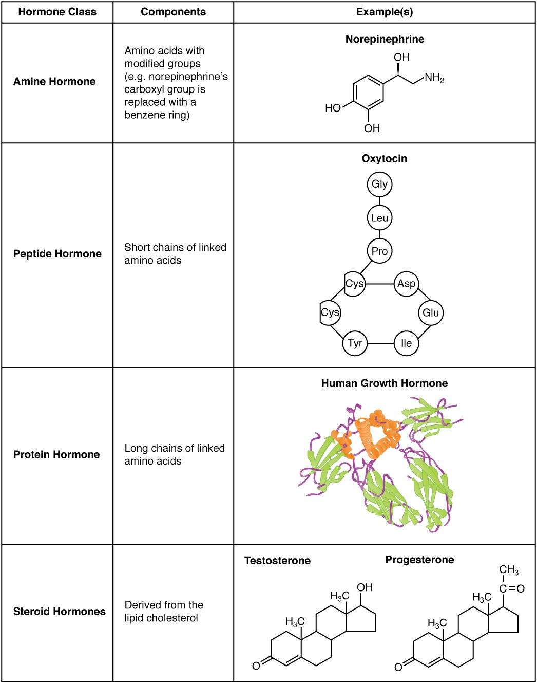
Hormones and neurotransmitters are classified into various chemical groups based on their structure, including amines, proteins, steroids, amino acids, peptides, and nitrous oxide.
Hormones Are The Chemical Messengers of the Endocrine System
Hormones are signalling chemicals produced by specialized cells and transported through the bloodstream to target cells.
They regulate a wide range of processes, from growth and metabolism to reproduction.
They can be classified into three main chemical groups: amines, peptides, and steroids.
Amines
Amines are hormones derived from amino acids, particularly those involving modifications to amino groups.
These hormones are generally water-soluble and often have rapid, short-term effects.
Example
Adrenaline (Epinephrine): A hormone produced by the adrenal glands that prepares the body for the fight-or-flight response by increasing heart rate, dilating airways, and increasing blood flow to muscles.
Thyroxine (T4): A hormone produced by the thyroid gland, crucial for regulating metabolism and growth.
Proteins and Peptides
Proteins and peptides are chains of amino acids that act as signaling molecules.
These are typically water-soluble and bind to receptors on the cell surface to initiate a cellular response.
Example
Insulin: A peptide hormone produced by the pancreas that helps regulate glucose levelsin the blood by promoting glucose uptake in cells.
Growth Hormone (GH): Stimulates growth, cell reproduction, and regeneration in the body.
Steroids
Steroid hormones are derived from cholesterol and are lipid-soluble, meaning they can pass through cell membranes and bind to intracellular receptors.
Steroid hormones typically have longer-lasting effects.
Example
Cortisol: Produced by the adrenal glands, cortisol helps regulate metabolism, immune response, and stress reactions.
Testosterone: A male sex hormone that regulates male reproductive functions, muscle mass, and bone density.
Estrogen: Female sex hormones that regulate the female reproductive system and secondary sexual characteristics.
Chemical Diversity of Neurotransmitters Shapes Neuronal Communication
Neurotransmitters also show a vast chemical diversity.
These chemicals transmit signals between neurons at synapses, and they can be categorized into several groups based on their structure.
Each group of neurotransmitters acts differently, influencing various aspects of neuronal communication.
Definition
NeurotransmitterNeurotransmitters are chemical signals used by neurons to communicate across synapses, the tiny gaps between nerve cells or between nerve cells and other target cells.
Amino Acids
Amino acids are the building blocks of proteins and are among the most common neurotransmitters.
These are typically fast-acting and can directly influence the postsynaptic membrane.
Example
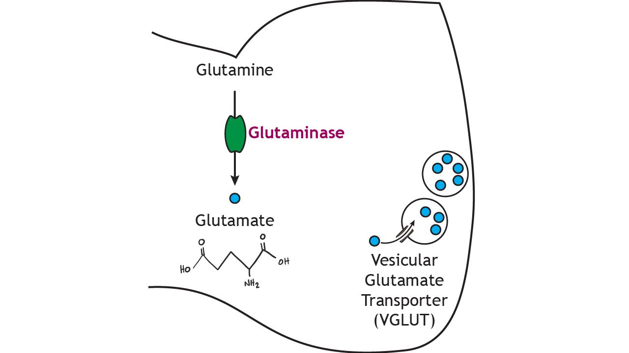
Glutamate: The primary excitatory neurotransmitter in the brain, involved in learning and memory.
GABA (Gamma-aminobutyric acid): The primary inhibitory neurotransmitter in the brain, responsible for calming neural activity and preventing over-excitation.
Peptides
Peptides, similar to their role as hormones, can also act as neurotransmitters.
They typically have longer-lasting effects compared to amino acid neurotransmitters.
Example
Substance P: Involved in the transmission of pain signals.
Endorphins: Neurotransmitters that act as natural painkillers and mood elevators.
Warning
Peptides are short chains of amino acids (e.g., insulin), while proteins are longer chains (e.g., GH).
Both fall under the same category in this context.
Amines
Amines are also important neurotransmitters that derive from amino acids.
They include some of the best-known neurotransmitters and have a wide range of effects on mood, alertness, and attention.
Example
Dopamine: Involved in reward, motivation, and motor control.
Serotonin: Regulates mood, appetite, and sleep.
Nitric Oxide (NO)
Nitric oxide is a gaseous neurotransmitter, which is highly unique compared to other neurotransmitters.
It diffuses directly across membranes, rather than binding to specific receptors.
Example
Nitric Oxide (NO): Regulates blood vessel dilation (widening of blood vessels) and plays a role in memory and immune function.
Tip
Remember that neurotransmitters can be broken down or reabsorbed quickly, unlike hormones, which often have longer-lasting effects.
Why Such Diversity?
The wide range of chemical structures among hormones and neurotransmitters is essential for their specialized roles.
1. Specificity of Action
Each signalling molecule must have a unique shape and chemical properties to bind selectively to its receptor.
This ensures that signals are precise and do not interfere with one another.
Example
Insulin binds specifically to insulin receptors, while epinephrine targets adrenergic receptors.
2. Solubility and Transport
The chemical nature of a signalling molecule determines how it is transported and where it acts.
Water-soluble hormones (e.g., peptides) cannot cross cell membranes and must bind to surface receptors.
Lipid-soluble hormones (e.g., steroids) can diffuse through membranes and act on intracellular receptors.
Warning
Don't confuse the solubility of hormones with their mechanism of action.
Remember, water-soluble hormones (amines, peptides) bind surface receptors, while lipid-soluble hormones (steroids) enter the cell and bind intracellular receptors.
3. Speed and Duration of Action
The diversity in chemical structure also influences how quickly and how long a signalling molecule acts.
Neurotransmitters work rapidly and have short-lived effects.
Hormones often have slower, longer-lasting effects.
Analogy
Think of neurotransmitters as text messages, fast and short-lived, while hormones are like letters, taking longer to deliver but having a more sustained impact.
Tok
How does our understanding of chemical signalling influence ethical decisions in medicine, such as the use of performance-enhancing drugs or hormone replacement therapy?
Self Review
What are the three main chemical groups of hormones? How do they differ in structure and function?
Why do hormones and neurotransmitters need to be chemically diverse?
How does the structure of a signalling molecule influence its function and transport?
Can you identify examples of hormones and neurotransmitters in your own body and describe their roles?
C2.1.5 localized and distant effects of signalling molecules
Localized and Distant Effects of Signalling Molecules
Imagine you're in a crowded room.
To communicate with someone nearby, you whisper.
But to reach someone across the room, you shout or use a microphone.
Cells face similar challenges when sending signals.
They rely on signalling molecules to communicate, whether the target is right next door or far away.
Definition
Signalling moleculesSignalling molecules are chemicals that transmit information between cells, triggering specific responses in target cells.
Localized Signalling: Neurotransmitters
Definition
NeurotransmitterNeurotransmitters are chemical signals used by neurons to communicate across synapses, the tiny gaps between nerve cells or between nerve cells and other target cells.
How Neurotransmitters Work
Release: When a nerve impulse reaches the end of a presynaptic neuron, neurotransmitters are released into the synaptic gap.
Diffusion: They travel a short distance (20-40 nm) across the gap.
Binding: Neurotransmitters bind to receptors on the postsynaptic neuron, triggering a response.
Example
Acetylcholine is a neurotransmitter that binds to receptors on muscle cells, causing them to contract.
Key Characteristics of Neurotransmitters
Short Distance: They act over extremely short distances, ensuring precise communication between neurons.
Rapid Action: Signals are transmitted in milliseconds, allowing for quick responses.
Short-Lived Effects: Neurotransmitters are quickly broken down or reabsorbed, preventing prolonged effects.
Note
Don't confuse neurotransmitters with hormones.
Neurotransmitters act locally and quickly, while hormones have broader and longer-lasting effects.
Definition
HormonesHormones are chemical messengers that travel through the bloodstream to regulate various physiological processes.
Distant Signalling: Hormones
How Hormones Work
Secretion: Hormones are released into the bloodstream by endocrine glands.
Transport: They travel throughout the body, reaching distant target cells.
Binding: Hormones bind to specific receptors on target cells, triggering a response.
Example
Insulin is a hormone produced by the pancreas that regulates blood glucose levels by acting on liver, muscle, and fat cells.
Key Characteristics of Hormones
Long Distance: Hormones can affect cells far from their origin.
Slower Action: Hormonal effects take longer to manifest compared to neurotransmitters.
Long-Lasting Effects: Hormones can remain active in the bloodstream for minutes to hours, influencing processes like growth, metabolism, and reproduction.
Tip
Remember that hormones only affect target cells with specific receptors, ensuring that their actions are precise despite widespread distribution.
Comparing Neurotransmitters and Hormones
Feature | Neurotransmitters | Hormones |
|---|---|---|
Distance | Short (synaptic gap) | Long (through bloodstream) |
Speed | Rapid (milliseconds) | Slower (seconds to hours) |
Duration | Short-lived | Long-lasting |
Specificity | Localized to one or a few target cells | Widespread, but only affects cells with specific receptors |
Analogy
Think of neurotransmitters as text messages sent to a specific friend, while hormonesare like a public announcement broadcasted to everyone in the area.
Other Examples of Signalling Molecules
Cytokines
Role: Small proteins involved in immune responses, inflammation, and cell growth.
Action: Act locally or on nearby cells, rarely traveling long distances.
Example
Interleukin-1 is a cytokine that promotes inflammation in response to infection.
Calcium Ions (Ca)
Role: Act as signalling molecules in muscle contraction and neurotransmitter release.
Action: Operate within cells or between closely connected cells.
Example
In muscle cells, calcium ions trigger contraction by binding to specific proteins.
Why Does This Matter?
Coordination: Signalling molecules ensure that cells work together, whether it's coordinating a muscle contraction or regulating blood sugar levels.
Adaptation: By using different types of signals, organisms can respond to both immediate and long-term challenges.
Tok
How do the localized effects of neurotransmitters and the widespread effects of hormones reflect the balance between precision and efficiency in biological systems?
C2.1.6 transmembrane receptors vs intracellular receptors
Differences Between Transmembrane and Intracellular Receptors
Cell signaling involves receptors that recognize signaling molecules and initiate a cellular response.
These receptors can be classified into transmembrane receptors (in the plasma membrane) and intracellular receptors (in the cytoplasm or nucleus).
The location and structure of these receptors determine how they interact with ligands and trigger specific pathways.
Definition
ReceptorA receptor is a protein that detects and responds to specific signals. They bind to signalling molecules(ligands) to initiate a cellular response.
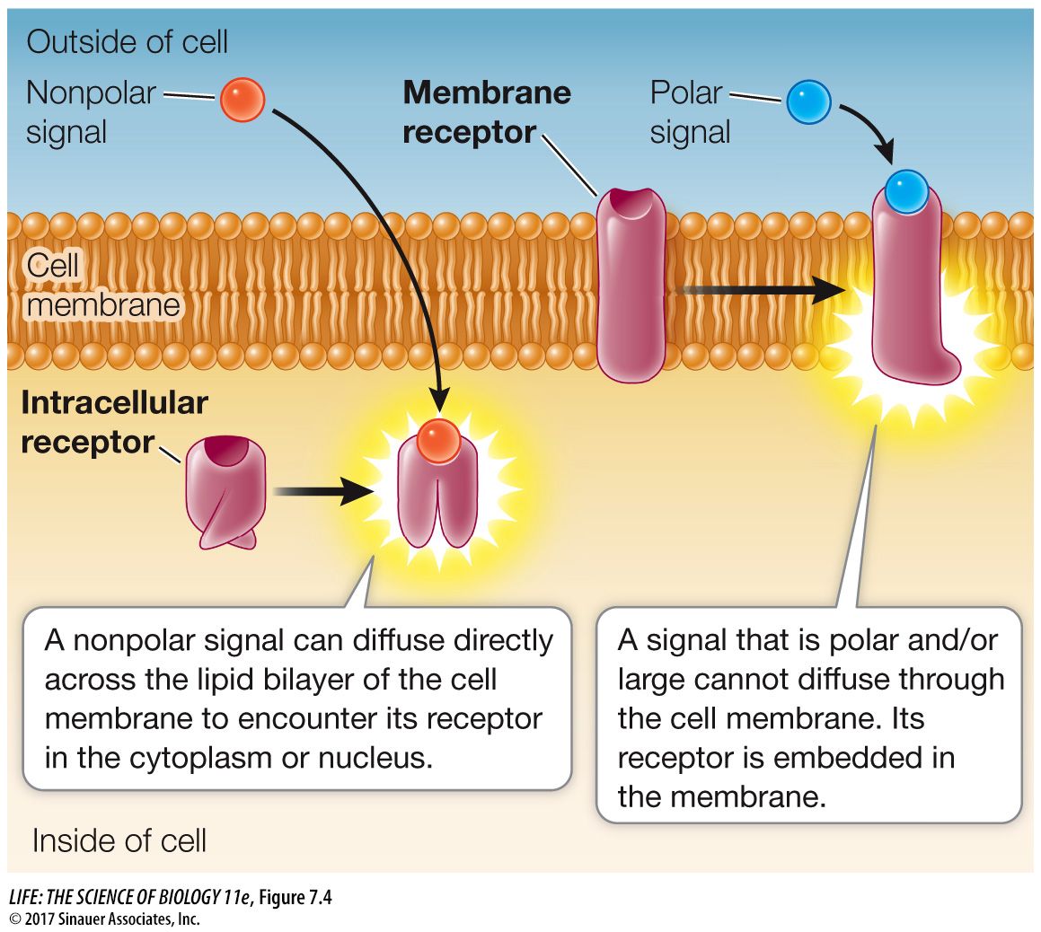
Transmembrane Receptors Are Gatekeepers of the Plasma Membrane
Definition
Transmembrane receptorsTransmembrane receptors are embedded in the plasma membrane and interact with signalling molecules that cannot cross the membrane.
Structure and Distribution of Amino Acids
Hydrophobic Core: The part of the receptor that spans the membrane contains hydrophobic amino acids, allowing it to interact with the lipid bilayer.
Hydrophilic Regions: The regions exposed to the extracellular and intracellular environments are composed of hydrophilic amino acids, enabling interaction with aqueous solutions.
Example
Adenosine Receptor:
This transmembrane receptor has seven alpha helices that traverse the membrane.
Its ligand-binding site is exposed to the outside, allowing adenosine to bind and trigger a response inside the cell.
Interaction with Signalling Molecules
Hydrophilic Ligands: Transmembrane receptors bind to ligands that are hydrophilic and cannot cross the lipid bilayer, such as peptides or neurotransmitters.
Signal Transduction: Binding of the ligand induces a conformational changein the receptor, activating intracellular signalling pathways.
Example
G-Protein-Coupled Receptors (GPCRs)
When a ligand binds to a GPCR, it activates a G protein, which then triggers a cascade of intracellular events, such as the production of cyclic AMP (cAMP).
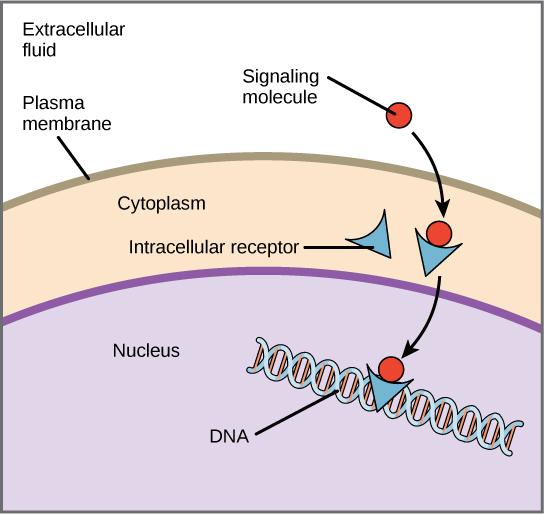
Intracellular Receptors Are The Cell's Internal Responders
Definition
Intracellular receptorsIntracellular receptors are located within the cytoplasm or nucleus and interact with signalling molecules that can cross the plasma membrane.
Structure and Distribution of Amino Acids
Hydrophilic Surface: Intracellular receptors have hydrophilic amino acids on their surface, allowing them to remain soluble in the aqueous environment of the cytoplasm or nucleus.
Example
Oestradiol Receptor
This receptor binds to the steroid hormone oestradiol in the cytoplasm.
The hormone-receptor complex then moves to the nucleus, where it regulates gene expression.
Interaction with Signalling Molecules
Hydrophobic Ligands: Intracellular receptors bind to ligands that are hydrophobic and can diffuse through the lipid bilayer, such as steroid hormones.
Direct Gene Regulation: The ligand-receptor complex often acts as a transcription factor, binding to DNA and influencing gene expression.
Example
Testosterone
This hormone diffuses into target cells, binds to its receptor in the cytoplasm, and the complex then enters the nucleus to activate genes involved in muscle growth and development.
Key Differences Between Transmembrane and Intracellular Receptors
Feature | Transmembrane Receptors | Intracellular Receptors |
|---|---|---|
Location | Embedded in the plasma membrane | Located in the cytoplasm or nucleus |
Ligand Type | Hydrophilic (cannot cross membrane) | Hydrophobic (can cross membrane) |
Amino Acid Distribution | Hydrophobic core; hydrophilic ends | Hydrophilic surface |
Signal Transduction | Indirect, via secondary messengers | Direct, often regulates gene expression |
Example | G-protein-coupled receptors (GPCRs) | Steroid hormone receptors |
Why Do These Differences Matter?
Specificity of Response: The location and structure of receptors ensure that only specific signalling molecules can trigger a response.
Adaptation to Ligand Properties: Transmembrane receptors are adapted to hydrophilic ligands that cannot cross the membrane, while intracellular receptors are suited forhydrophobic ligands that can diffuse through the lipid bilayer.
Diversity of Cellular Responses: Transmembrane receptors often initiate rapid, short-term responses (e.g., ion channel opening), whereas intracellular receptors typically regulate long-term processes like gene expression.
Tip
Remember that the distribution of hydrophilic and hydrophobic amino acids in a receptor is critical to its function and location.
This principle also applies to other membrane proteins, such as transporters and enzymes.
Tok
The relationship between structure and function is a recurring theme in biology. How does this principle apply to other biological molecules, such as enzymes or DNA?
Can you think of examples where a small change in structure leads to a significant change in function?
Self Review
Why do transmembrane receptors have hydrophobic regions, while intracellular receptors do not?
How does the ability of a signalling molecule to cross the plasma membrane determine the location of its receptor?
Can you provide an example of a signalling molecule that interacts with each type of receptor?
Contrast the speed and effect of signals mediated by transmembrane receptors versus intracellular receptors.
C2.1.7 initiation of signal transduction pathways by receptors
Initiation of Signal Transduction Pathways by Receptors
Definition
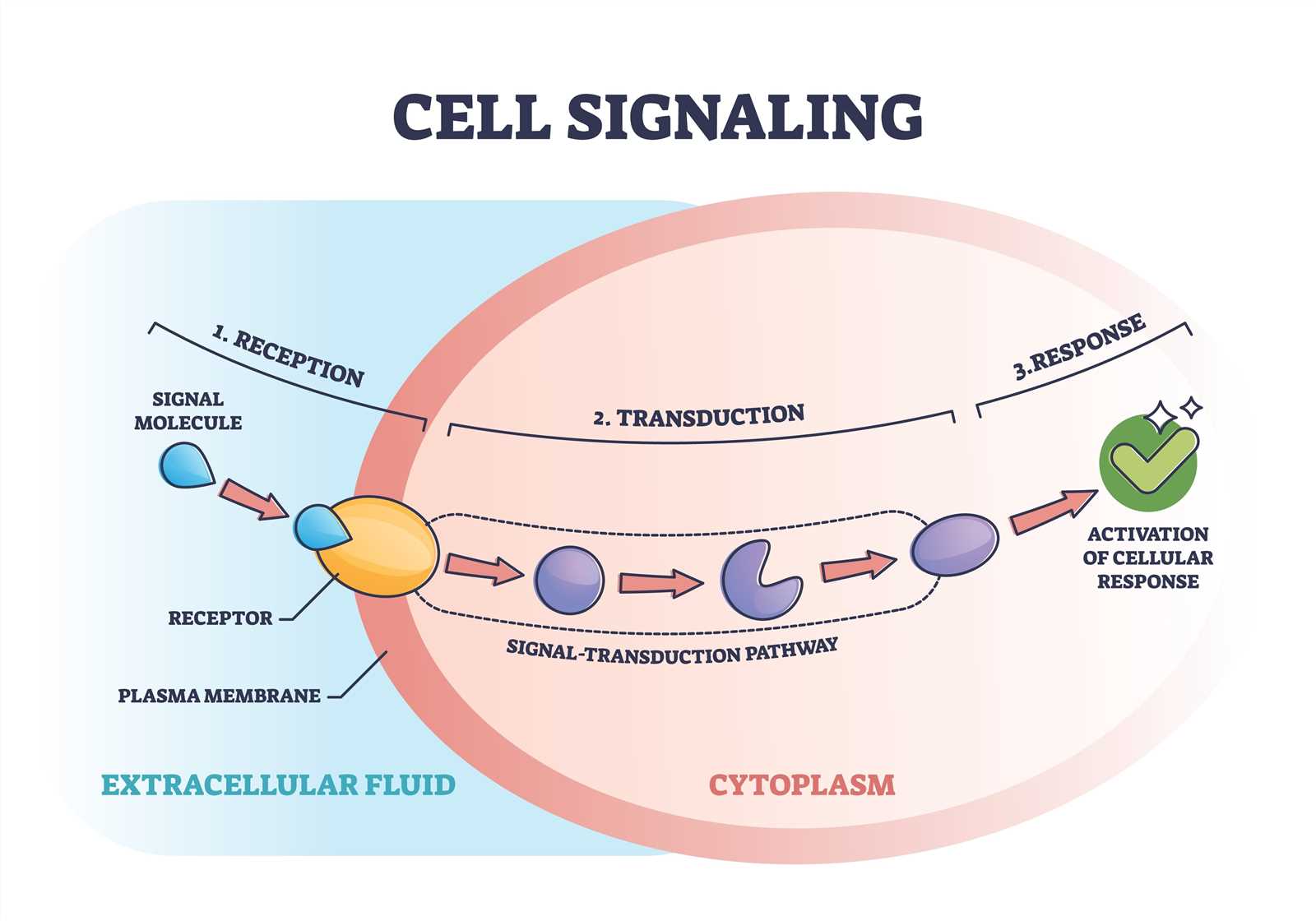
Signal transductionSignal transduction is the process by which cells respond to external signals, converting them into a cascade of chemical responses inside the cell.
Signal transduction is the process by which a cell converts an external signal into a specific response.
It begins when a signaling molecule (ligand) binds to a receptor on the cell surface or inside the cell.
This binding triggers a series of molecular events, often involving multiple steps, to produce a cellular response.
Signal transduction pathways are essential for coordinating cellular activities and ensuring that cells respond appropriately to their environment.
Analogy
Imagine you're in a crowded room, and someone whispers your name.
Instantly, your attention shifts.
This is similar to how cells respond to signals in their environment.
But how does a cell "hear" these signals and respond appropriately?
The answer lies in signal transduction pathways.
Note
Signal transduction pathways are highly specific and efficient, ensuring that cells respond accurately to their environment.
The Three Stages of Signal Transduction
Reception: The signaling molecule binds to a receptor.
Transduction: The signal is relayed through a series of molecules inside the cell.
Response: The cell performs a specific action, such as activating a gene or releasing a substance.
Tip
Think of signal transduction as a relay race.
The ligand starts the race by binding to the receptor, which then passes the "baton" (the signal) through various molecules until the final runner (the effector) completes the task.
Types of Receptors
Receptors are proteins that recognize and bind specific signaling molecules. They can be classified into two main types:
Transmembrane Receptors: Located in the plasma membrane, these receptors bind signaling molecules that cannot cross the membrane, such as proteins and peptides.
Intracellular Receptors: Found inside the cell, these receptors bind signaling molecules that can diffuse through the membrane, such as steroid hormones.
Transmembrane Receptors: Relaying Signals Across the Membrane
Transmembrane receptors span the plasma membrane, with one part exposed to the extracellular environment and another part inside the cell.
When a signaling molecule binds to the extracellular domain, it causes a structural change in the receptor, activating its intracellular domain.
Analogy
Think of the transmembrane receptor as a doorbell.
When you press it (ligand binding), it sends a signal to the house (cell), triggering a response (e.g., someone answering the door).
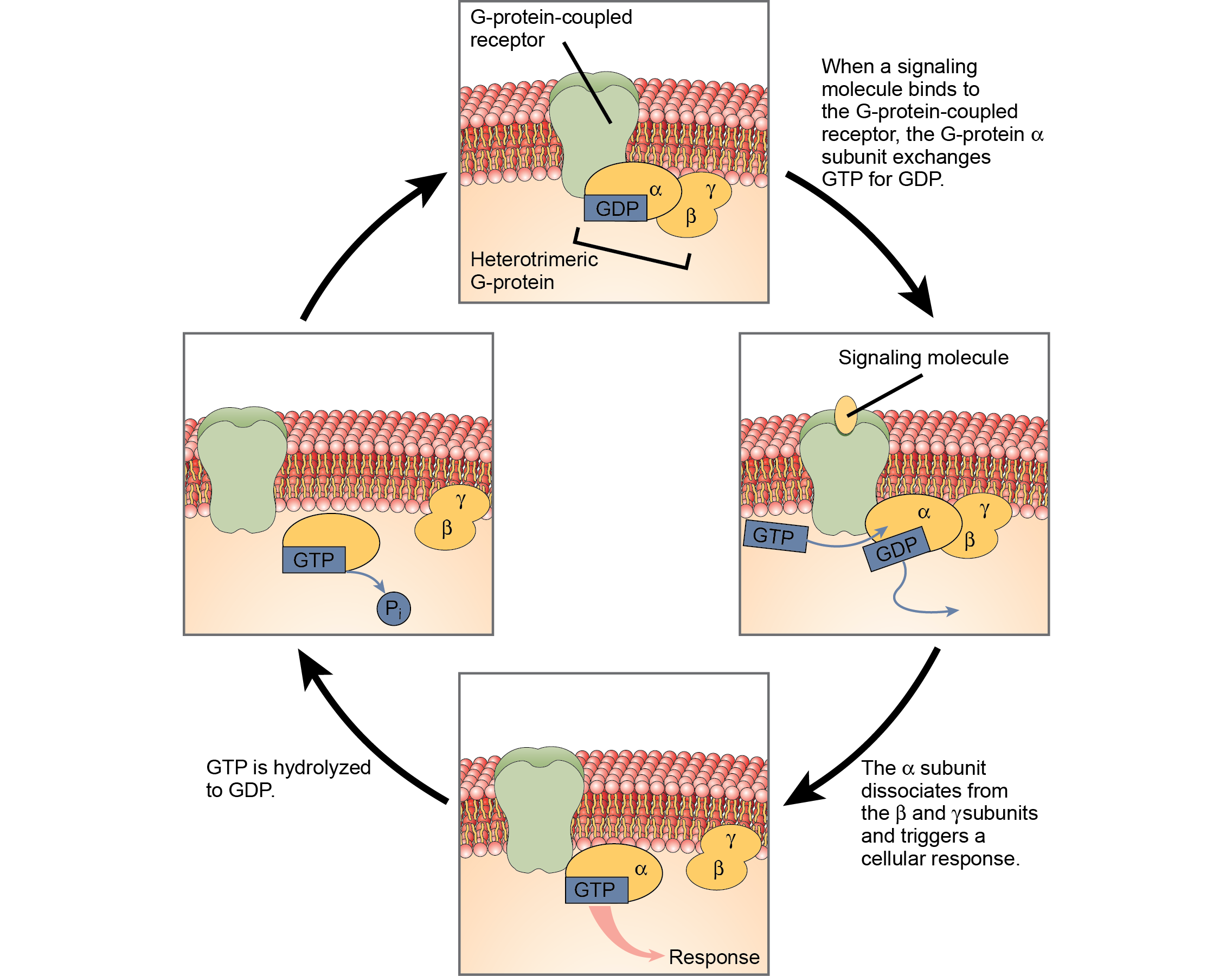
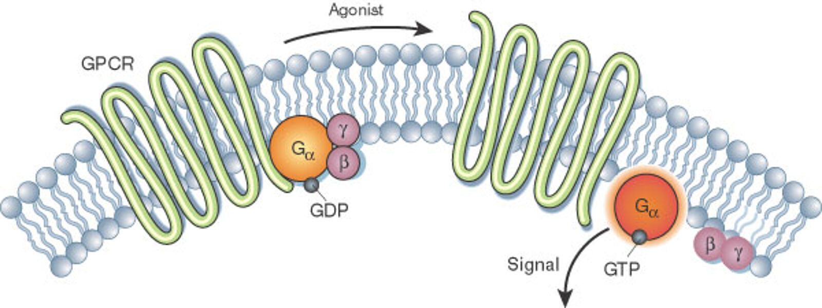
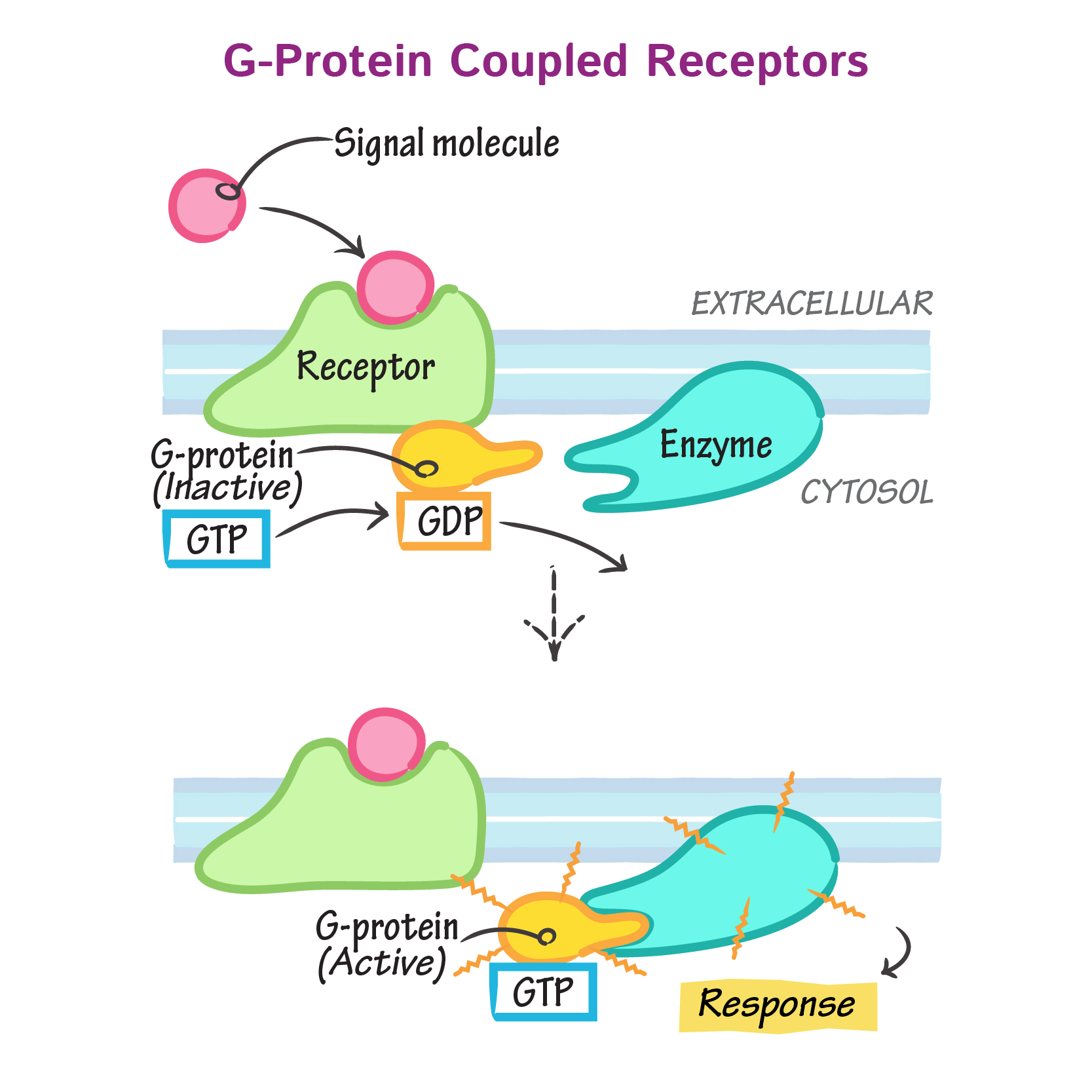
G-Protein-Coupled Receptors (GPCRs)
GPCRs are one of the most common types of transmembrane receptors. They work in partnership with a G protein, which is attached to the inner side of the plasma membrane.
Ligand Binding: A signaling molecule binds to the GPCR, causing a conformational change in the receptor.
G Protein Activation: The activated receptor causes the G protein to exchange GDP for GTP, activating the G protein.
Signal Relay: The activated G protein dissociates into subunits, which interact with other proteins in the cell, such as enzymes or ion channels, to propagate the signal.
Example
When epinephrine (adrenaline) binds to its GPCR on liver cells, it activates a G protein.
This, in turn, activates the enzyme adenylyl cyclase, which converts ATP into cyclic AMP (cAMP), a secondary messenger.
cAMP amplifies the signal, leading to the breakdown of glycogen into glucose.
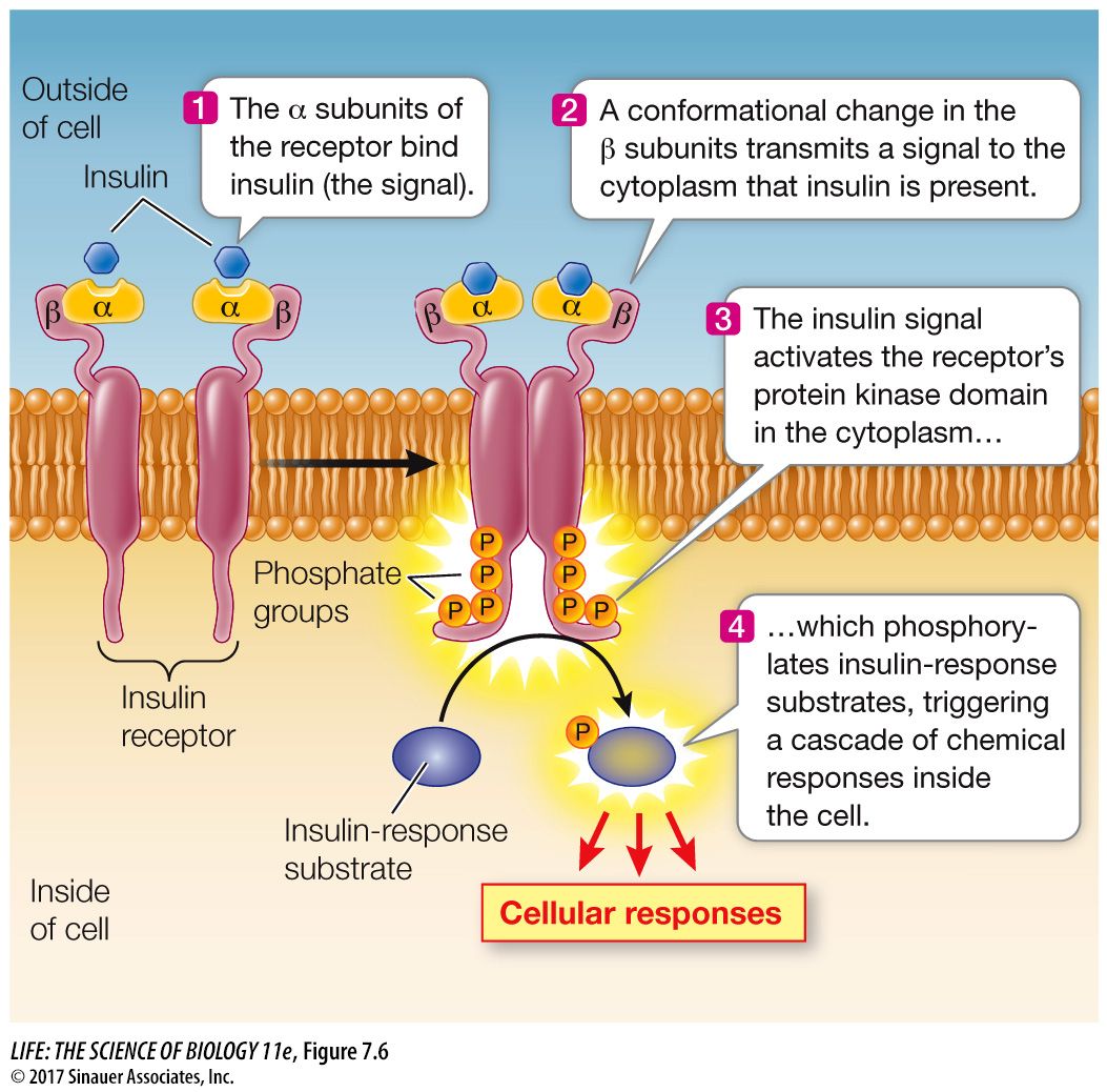
Receptor Tyrosine Kinases (RTKs)
RTKs are another type of transmembrane receptor. They have an enzymatic domain that adds phosphate groups to specific proteins, a process called phosphorylation.
Ligand Binding: A signaling molecule binds to the RTK, causing two receptor molecules to dimerize (pair up).
Activation: The dimerized receptors phosphorylate each other, activating their kinase domains.
Signal Relay: The phosphorylated receptor recruits and activates other proteins in the cell, triggering a cascade of events.
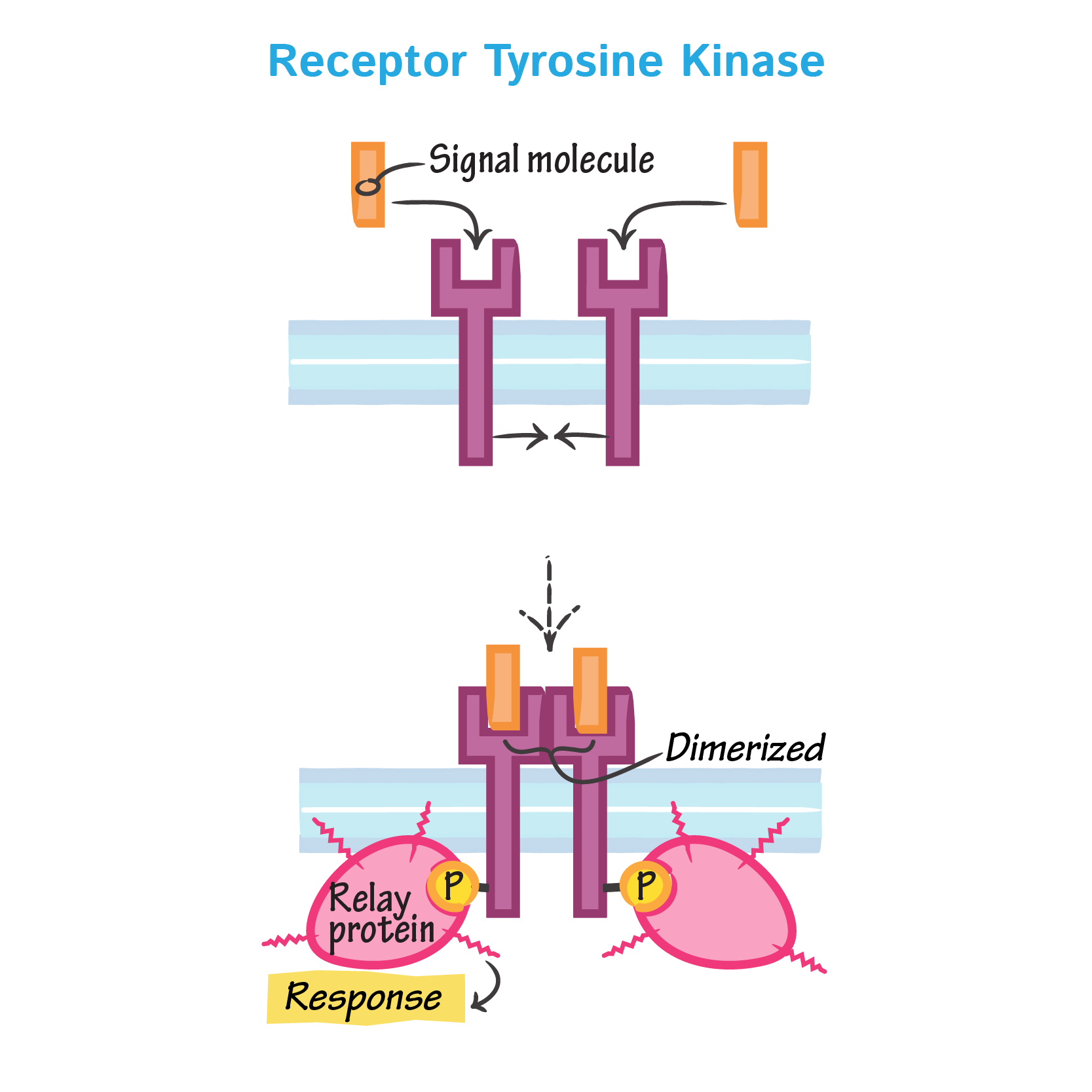
Example
The insulin receptor is an RTK.
When insulin binds, it activates the receptor, leading to the insertion of glucose transporters into the plasma membrane, allowing glucose to enter the cell.
Intracellular Receptors: Directly Influencing Gene Expression
Some signaling molecules, like steroid hormones, can cross the plasma membrane and bind to receptors inside the cell.
These receptors often act as transcription factors, directly influencing gene expression.
Ligand Binding: The signaling molecule enters the cell and binds to its receptor in the cytoplasm or nucleus.
Complex Formation: The ligand-receptor complex binds to specific DNA sequences, promoting or inhibiting the transcription of target genes.
Response: The cell produces specific proteins in response to the signal.
Example
Testosterone binds to its receptor in the cytoplasm.
The hormone-receptor complex then enters the nucleus and activates genes involved in muscle growth and development.
Tok
How do signal transduction pathways illustrate the balance between specificity and amplification in biological systems?
Can you think of other systems where a small input leads to a large output?
Self Review
What happens when a signaling molecule binds to a receptor on a cell?
C2.1.8 transmembrane receptors for neurotransmitters and changes to membrane potential
Transmembrane Receptors for Neurotransmitters and Changes to Membrane Potential
Transmembrane receptors for neurotransmitters play a critical role in cellular communication by converting chemical signals into electrical responses.
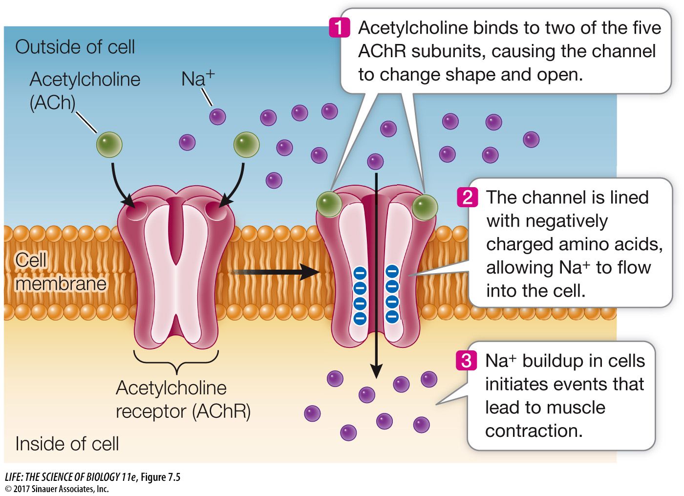
One classic example is the acetylcholine receptor (AChR), a type of ligand-gated ion channel.
The binding of acetylcholine (a neurotransmitter) to its receptor triggers a series of events that alter the membrane potential, leading to downstream cellular responses.
Analogy
Imagine flipping a light switch.
The action is small, but it triggers a cascade of events that illuminate a room.
In biology, neurotransmitters act like this switch, initiating changes in a cell by binding to transmembrane receptors.
These receptors are essential for communication between neurons and other cells, such as muscle fibers.
Definition
Membrane potentialMembrane potential is the difference in electrical charge between the inside and outside of a cell.
Note
Transmembrane receptors are proteins that span the plasma membrane, with one part exposed to the extracellular environment and another facing the cytoplasm.
They act as gatekeepers, receiving signals from the outside and transmitting them into the cell.
These receptors are highly specific, binding only to particular molecules, called ligands.
A ligand is a molecule that binds to a specific site on a receptor, much like a key that fits into a lock.
The Acetylcholine Receptor: A Classic Example
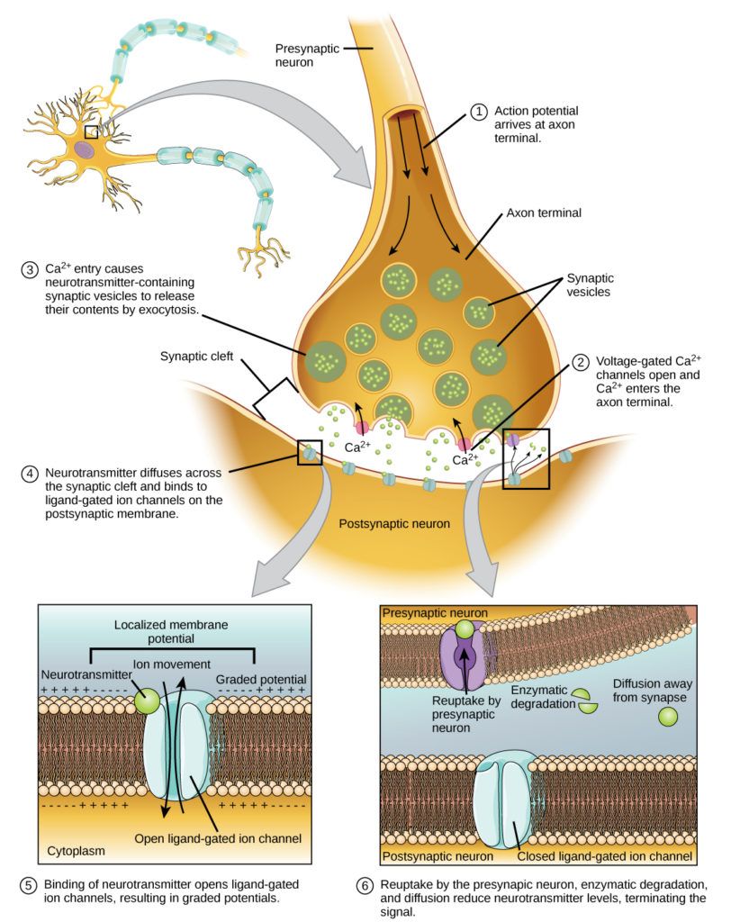
Acetylcholine is a neurotransmitter that plays a crucial role in transmitting signals across synapses, particularly at the junctions between neurons and muscle fibers.
The acetylcholine receptor is a transmembrane protein that acts as both a receptor and an ion channel.
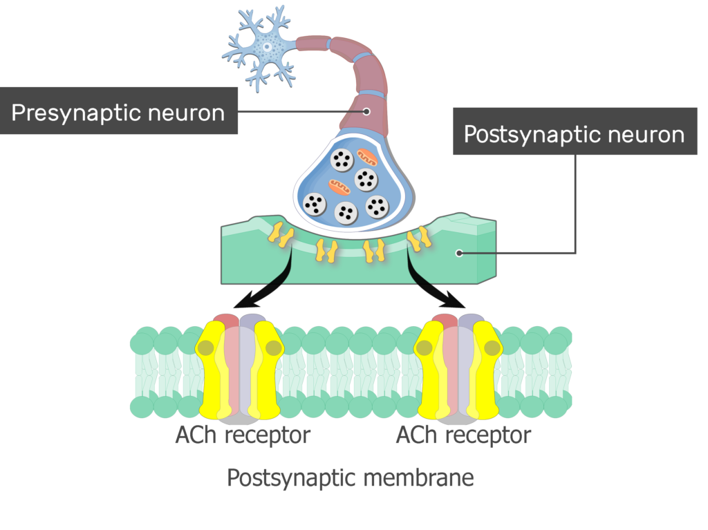
How It Works
Binding of Acetylcholine: When acetylcholine is released into the synaptic gap, it diffuses across and binds to the receptor on the postsynaptic membrane.
Conformational Change: This binding causes the receptor to change shape, opening an ion channel within the protein.
Ion Flow: Positively charged ions, such as sodium (), flow into the cell through the open channel.
Depolarization: The influx of ions reduces the negative charge inside the cell,causing a change in membrane potential. This local depolarization can trigger an action potential or muscle contraction.
Definition
DepolarizationA change in the membrane potential of the presynaptic neuron, making it more positive.
Note
The acetylcholine receptor is a ligand-gated ion channel, meaning it opens in response to the binding of a specific ligand (acetylcholine).
Exam_technique
Quick review of the steps to remember the action of acetylcholine receptor
Step 1: Acetylcholine (ACh) is released from the presynaptic neuron.
Step 2: Acetylcholine binds to the acetylcholine receptor on the postsynaptic membrane.
Step 3: The binding of ACh causes the receptor ion channel to open.
Step 4: Positively charged ions (Na⁺ or Ca²⁺) flow into the postsynaptic cell.
Step 5: This influx of positive ions leads to depolarization of the membrane.
Step 6: The signal can be propagated if the depolarization reaches a threshold, causing an action potential.
Changes in Membrane Potential
At rest, the inside of a cell is more negative compared to the outside.
When ions flow through the receptor, this balance shifts, creating a depolarization.
If the depolarization is strong enough, it can trigger an action potential, a rapid electrical signal that travels along the neuron or muscle fiber.
Tip
Depolarization occurs when the membrane potential becomes less negative (or more positive) due to the influx of positively charged ions.
Why Is This Important?
The ability of neurotransmitters to change membrane potential is critical for:
Neural Communication: Transmitting signals between neurons.
Muscle Contraction: Initiating the contraction of muscle fibers.
Reflexes and Responses: Enabling rapid responses to stimuli.
Example
When you touch a hot surface, sensory neurons release neurotransmitters that bind to receptors in motor neurons.
This triggers a rapid response, causing your muscles to contract and pull your hand away.
Broader Implications
Transmembrane receptors are not limited to neurotransmitters. They play roles in:
Hormonal Signaling: Receptors for hormones like insulin regulate glucose uptake.
Immune Responses: Cytokine receptors help coordinate immune activity.
Cell Growth and Division: Growth factor receptors control cell proliferation.
Tok
How do the structural differences between transmembrane and intracellular receptors reflect their roles in cell signaling? Consider how these differences might influence the speed and specificity of the signals they transmit.
Self Review
What are transmembrane receptors, and how do they work?
How does the acetylcholine receptor change membrane potential?
Why is the ability to change membrane potential important for cells?
C2.1.9 transmembrane receptors that activate g proteins
Transmembrane Receptors That Activate G Proteins
GPCRs are a large family of transmembrane receptors found in the plasma membrane of cells.
They are called "G protein-coupled" because they work in partnership with G proteins, molecular switches that relay signals inside the cell.
Definition
G protein-coupled receptorsA large family of transmembrane receptors that detect external signals and activate intracellular G proteins, initiating cellular responses.
Structure of GPCRs
Seven-Helix Structure: GPCRs span the membrane with seven alpha-helices.
Ligand-Binding Site: Located on the extracellular side, where signaling molecules (ligands) bind.
Intracellular Domain: Interacts with G proteins to transmit the signal into the cell.
Analogy
Think of a GPCR as a doorbell.
The ligand is the person pressing the button, the receptor is the bell, and the G protein is the wiring that carries the signal to the chime inside the house.
How Do GPCRs Work?
1. Ligand Binding
The process begins when aligand -such as ahormone, neurotransmitter, or sensory molecule - binds to the receptor's extracellular site.
Example
Epinephrine (adrenaline) binds to its receptor on liver cells to trigger the release of glucose into the bloodstream.
2. Activation of the G Protein
Inactive State: The G protein is bound to guanosine diphosphate (GDP) and is inactive.
Conformational Change: Ligand binding causes the GPCR to change shape, activating the G protein.
GDP to GTP Exchange: The alpha subunit of the G protein releases GDP and binds guanosine triphosphate (GTP), activating the G protein.
Warning
Don't confuse GDP and GTP with ATP.
While ATP is the cell's main energy currency, GTP specifically activates G proteins.
3. Signal Propagation
Subunit Separation: The activated G protein splits into two parts: the alpha subunit (with GTP) and the beta-gamma dimer.
Effector Activation: These subunits interact with target proteins in the membrane, such as enzymes or ion channels, to trigger the next steps in the signaling pathway.
Example
In the case of epinephrine, the activated G protein stimulates adenylyl cyclase, an enzyme that converts ATP into cyclic AMP (cAMP), a secondary messenger.
4. Termination of the Signal
Hydrolysis of GTP: The alpha subunit hydrolyzes GTP back to GDP, inactivating itself.
Reassembly: The G protein subunits reassemble, and the system returns to its resting state, ready for the next signal.
Tip
To remember the steps, think of the acronym L-A-S-T: Ligand binding, Activation, Signal propagation, Termination.
GPCRs Are Key to Sensory Perception, Hormonal Regulation, and Neurotransmission
GPCRs are incredibly versatile and are involved in a wide range of physiological processes, including:
Sensory Perception: Detecting light, odors, and tastes.
Hormonal Responses: Regulating metabolism, growth, and stress responses.
Neurotransmission: Mediating communication between neurons.
Examples of GPCRs and Their Signaling Molecules
GPCR Type | Signaling molecule | Mechanism of Action | Response |
|---|---|---|---|
β-Adrenergic Receptor | Epinephrine (Adrenaline) | Activates adenylyl cyclase via Gαs, increasing cAMP levels and promoting fight-or-flight responses. | Heart rate increase |
Acetylcholine Receptor | Acetylcholine | Activates phospholipase C via Gαq, leading to IP₃ and DAG production, causing smooth muscle contraction. | Parasympathetic responses |
Rhodopsin | Light | Activates G proteins in photoreceptor cells, triggering a visual signal cascade. | Vision in Retina |
Histamine Receptor | Histamine | Activates G proteins to regulate inflammation and allergic responses. | Inflammatory response in tissues |
A Closer Look: The Epinephrine Pathway
Ligand Binding: Epinephrine binds to its GPCR on the cell membrane.
G Protein Activation: The G protein exchanges GDP for GTP and activates adenylyl cyclase.
cAMP Production: Adenylyl cyclase converts ATP into cAMP, which acts as a secondary messenger.
Signal Amplification: cAMP activates protein kinase A (PKA), which phosphorylates enzymes to break down glycogen into glucose.
Rapid Response: Within seconds, glucose is released into the bloodstream, providing energy for the "fight or flight" response.
Tok
How does the specificity of GPCRs reflect the broader principle of structure-function relationships in biology?
Can you think of other examples where small structural changes have significant functional impacts?
Self Review
Can you outline the steps of GPCR activation and explain how the signal is terminated?
What is the role of the Gα subunit in the signaling pathway?
C2.1.10 mechanism of action of epinephrine (adrenaline) receptors
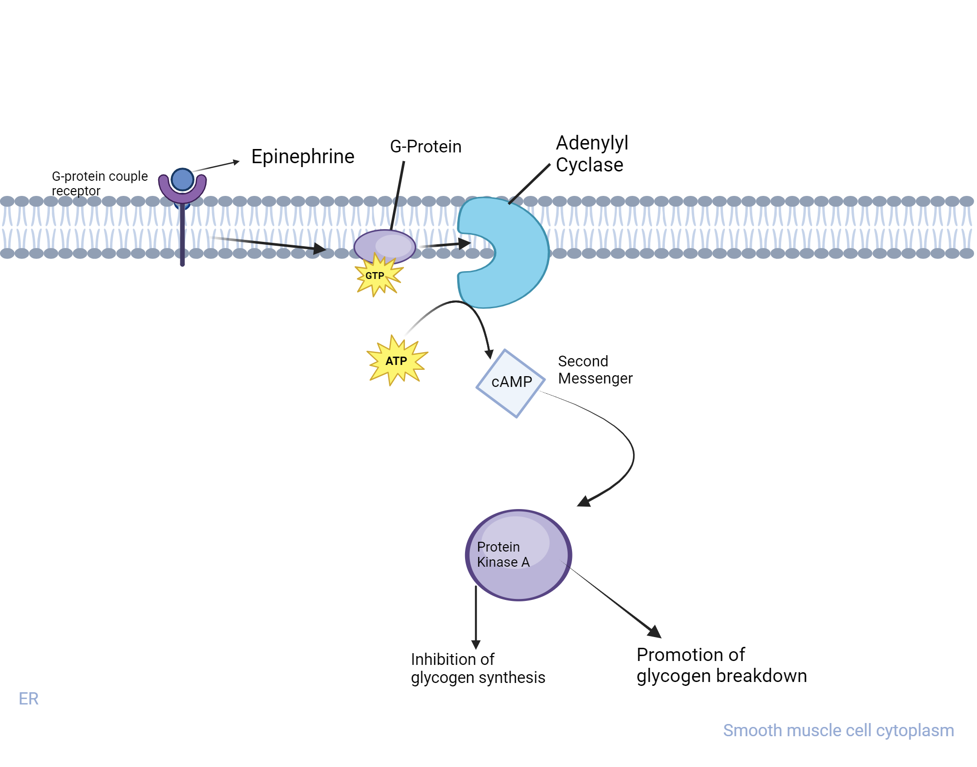
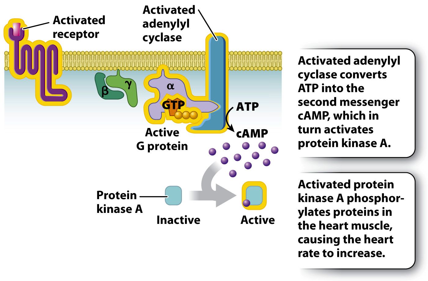
Epinephrine Triggers Rapid Body Responses Through cAMP Signaling
Imagine you're running a race. Your heart pounds, your breathing quickens, and energy surges through your muscles.
This rapid response is orchestrated by epinephrine (also known as adrenaline), a hormone that prepares your body for action.
But how does epinephrine trigger these changes so quickly?
The answer lies in a sophisticated communication system involving receptors, G proteins, and a second messenger called cyclic AMP (cAMP).
Hint
The terms "adrenaline" and "epinephrine" are used interchangeably.
"Adrenaline" is more common globally, while "epinephrine" is prevalent in North America.
"Adrenaline" comes from Latin ad = at and ren = kidney
"Epinephrine" comes from old Greek epi = above and nephros = kidney, respectively.
Epinephrine Activates the Fight-or-Flight Response via Adrenergic Receptors
Definition
EpinephrineEpinephrine, also known as adrenaline, is a hormone and neurotransmitter produced by the adrenal glands.
Epinephrine plays a critical role in the fight or flight response, activating various physiological changes in response to stress or danger.
The action of epinephrine is mediated through specific receptors, known as adrenergic receptors, which are part of the G protein-coupled receptor (GPCR) family.
Step 1: Epinephrine Binds to Its Receptor
Epinephrine is a hormone secreted by the adrenal glands during stress or excitement.
It travels through the bloodstream and targets specific cells, such as those in the heart, liver, and muscles.
These target cells have specialized proteins called G-protein-coupled receptors (GPCRs) embedded in their plasma membranes.
When epinephrine binds to its receptor, it triggers a conformational change (a change in shape) in the receptor.
This change is the first step in transmitting the signal into the cell.
Tip
GPCRs are the most common type of receptor in humans, involved in detecting hormones, neurotransmitters, and even light.
Step 2: Activation of the G Protein
Attached to the inner side of the plasma membrane is a G protein, which acts as a molecular switch.
It consists of three subunits: alpha (α), beta (β), and gamma (γ).
In its inactive state, the alpha subunit is bound to a molecule called GDP (guanosine diphosphate).
When the receptor changes shape, it interacts with the G protein, causing the GDP to be released and replaced by GTP (guanosine triphosphate).
This exchange activates the G protein, which then dissociates into two parts: the alpha subunit (now bound to GTP) and the beta-gamma dimer.
Note
Don't confuse GDP with GTP. GDP is the inactive form, while GTP is the active form that powers the G protein.
Step 3: Activation of Adenylyl Cyclase
The activated alpha subunit of the G protein moves along the plasma membrane and binds to an enzyme called adenylyl cyclase.
This enzyme is responsible for converting ATP (adenosine triphosphate) into cyclic AMP (cAMP), a critical second messenger.
Definition
Second MessengerA second messenger is a molecule that relays signals from the cell surface to the interior and amplifies the signal inside the cell.
Step 4: cAMP Amplifies the Signal
Once produced, cAMP diffuses through the cytoplasm and activates a protein called protein kinase A (PKA).
PKA is an enzyme that phosphorylates (adds phosphate groups to) other proteins, altering their activity.
This phosphorylation cascade leads to a variety of cellular responses, depending on the target cell:
In liver cells, it activates enzymes that break down glycogen into glucose, increasing blood sugar levels.
In heart cells, it enhances the contraction of heart muscles, increasing heart rate and blood flow.
In muscle cells, it promotes the breakdown of glycogen to provide energy for muscle contraction.
Analogy
Think of cAMP as a megaphone.
The original signal (epinephrine binding to the receptor) is like a whisper, but cAMP amplifies it so the entire cell can "hear" and respond.
Step 5: Termination of the Signal
The effects of epinephrine are temporary, and thesignal must be turned offto restore normal cellular function. This is achieved through several mechanisms:
Hydrolysis of GTP: The GTP bound to the alpha subunit is hydrolyzed back to GDP, inactivating the G protein.
Degradation of cAMP: An enzyme called phosphodiesterase breaks down cAMP into AMP, stopping the activation of PKA.
Dissociation of Epinephrine: The hormone eventually detaches from the receptor, returning it to its inactive state.
Warning
Students often mistake cAMP as the primary messenger.
Remember, epinephrine is the first messenger, and cAMP is the second messengerinside the cell.
cAMP Amplifies Epinephrine's Signal for a Rapid and Robust Response
The use of cAMP as a second messenger allows for signal amplification.
A single molecule of epinephrine can lead to the production of thousands of cAMP molecules, each of which can activate multiple PKA molecules.
This amplification ensures a rapid and robust response to the hormone.
Standardized Naming in Science Promotes Global Collaboration and Clarity
The dual naming of epinephrine and adrenaline highlights the importance of international cooperation in science.
While both terms are widely used, standardization ensures clarity and consistency in scientific communication.
Tok
How do naming conventions in science reflect cultural and historical influences?
Why is it important for scientists to agree on standardized terminology?
Tok
How might a drug that blocks adenylyl cyclase affect the body's response to epinephrine?
What potential therapeutic applications could this have?
Self Review
Can you explain why cAMP is called a "second messenger"? What role does it play in the signal transduction pathway?
What is the role of G proteins in epinephrine signaling?
C2.1.11 transmembrane receptors with tyrosine kinase activity
Tyrosine Kinase Receptors Mediate Growth, Metabolism, and Differentiation Signals
Transmembrane receptors are proteins that span the cell membrane, allowing them to transmit signals from the outside to the inside of a cell.
One important class of these receptors is the tyrosine kinase receptors.
These receptors play a critical role in cellular communication, especially in processes like growth, metabolism, and differentiation.
Definition
Tyrosine kinase receptorsA class of transmembrane receptors that have an enzymatic domain capable of phosphorylating tyrosine residues on themselves or other proteins, initiating a signaling cascade inside the cell.
Insulin Regulates Blood Glucose by Promoting Cellular Glucose Uptake
Insulin is a peptide hormone produced by the pancreas.
It regulates blood glucose levels by promoting the uptake of glucose into cells, especially in muscle and fat tissues.
Tip
Insulin is a peptide hormone, meaning it is made up of amino acids and is hydrophilic.
This prevents it from crossing the cell membrane, so it relies on a receptor to convey its signal.
Structure of the Insulin Receptor
The insulin receptor is atransmembrane proteincomposed of two main parts:
An extracellular domain that binds insulin.
An intracellular domain with tyrosine kinase activity.
Tip
The insulin receptor is a dimer, meaning it consists of two identical subunits.
This structure is crucial for its activation.
Binding of Insulin to Its Receptor
When insulin binds to the extracellular domain of its receptor, it triggers a series of events:
Dimerization: The two subunits of the receptor come together, forming a dimer.
Activation of Tyrosine Kinase: The intracellular domains of the receptor undergo a conformational change, activating their tyrosine kinase activity.
Autophosphorylation: The receptor phosphorylates specific tyrosine residues on its own intracellular domain.
Analogy
Think of the insulin receptor as a lock and insulin as the key.
When the key (insulin) fits into the lock (receptor), it "unlocks" the receptor's enzymatic activity, allowing it to phosphorylate tyrosine residues.
Movement of Glucose Transporters to the Plasma Membrane
One of the critical outcomes of AKT (Protein kinase B) activation is the movement of vesicles containingglucose transporters (GLUT4)to the plasma membrane.
How Does This Happen?
Vesicle Mobilization: AKT phosphorylates and inactivates proteins that inhibit vesicle movement, allowing vesicles containing GLUT4 to move towards the plasma membrane.
Vesicle Fusion: The vesicles fuse with the plasma membrane, inserting GLUT4 transporters into the membrane.
Glucose Uptake: GLUT4 transporters facilitate the entry of glucose into the cell by facilitated diffusion.
Example
In muscle cells, the insertion of GLUT4 transporters increases glucose uptake, providing energy for activities like exercise.
In fat cells, glucose is stored as glycogen or converted into lipids for long-term energy storage.
Why Is This Process Important?
The insulin signaling pathway is essential for maintaining normal blood glucose levels.
When this pathway is disrupted, it can lead to conditions like insulin resistance and type 2 diabetes.
Tok
How does the specificity of the insulin receptor illustrate the broader principle of structure-function relationships in biology?
Can you think of other examples where a small change in structure leads to a significant change in function?
Self Review
What is the role of tyrosine kinase activity in the insulin receptor?
How does the activation of AKT lead to increased glucose uptake in cells?
Why is the movement of GLUT4 transporters to the plasma membrane critical for glucose homeostasis?
c2.1.12 intracellular receptors that affect gene expression
Intracellular Receptors Regulate Gene Expression by Activating Target Genes
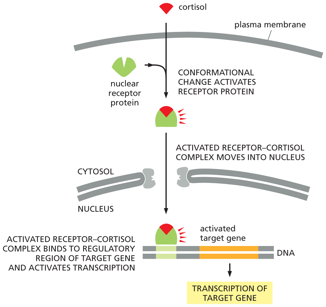
Some signaling molecules, such as steroid hormones, are hydrophobic and can pass through the plasma membrane of a cell.
These molecules bind to intracellular receptors, which are typically located in the cytoplasm or nucleus.
Upon binding, these receptors become activated and directly influence gene expressionby interacting with specific DNA sequences, leading to the transcription of certain genes.
Analogy
Consider a conductor leading an orchestra.
The conductor doesn't play an instrument but guides the musicians to create a symphony.
Similarly, intracellular receptors guide cells by activating specific genes, ensuring the right proteins are produced at the right time.
Analogy
Intracellular receptors are like conductors, directing cellular activities by activating specific genes.
How Do Intracellular Receptors Work?
Note
Intracellular Receptors Activate Gene Expression by Forming Hormone-Receptor Complexes
Hormone Entry
Steroid hormones, such as oestradiol, progesterone, and testosterone, are hydrophobic.
This allows them to diffuse through the plasma membrane and enter the cell.
Binding to Receptor
Once inside, the hormone binds to a specific intracellular receptor in the cytoplasm or nucleus.
This binding activates the receptor, changing its shape to expose a DNA-binding domain.
Formation of Hormone-Receptor Complex
The activated receptor and hormone form a hormone-receptor complex.
This complex acts as a transcription factor, a molecule that can bind to DNA and influence gene expression.
DNA Binding and Gene Activation
The hormone-receptor complex moves into the nucleus (if it isn't already there) and binds to specific DNA sequences called hormone response elements (HREs).
Binding to HREs initiates or enhances the transcription of target genes, leading to the production of specific proteins that carry out the hormone's effects.
Tip
Steroid hormones can directly influence gene expression because they can cross the cell membrane and interact with intracellular receptors.
Examples of Steroid Hormones and Their Effects
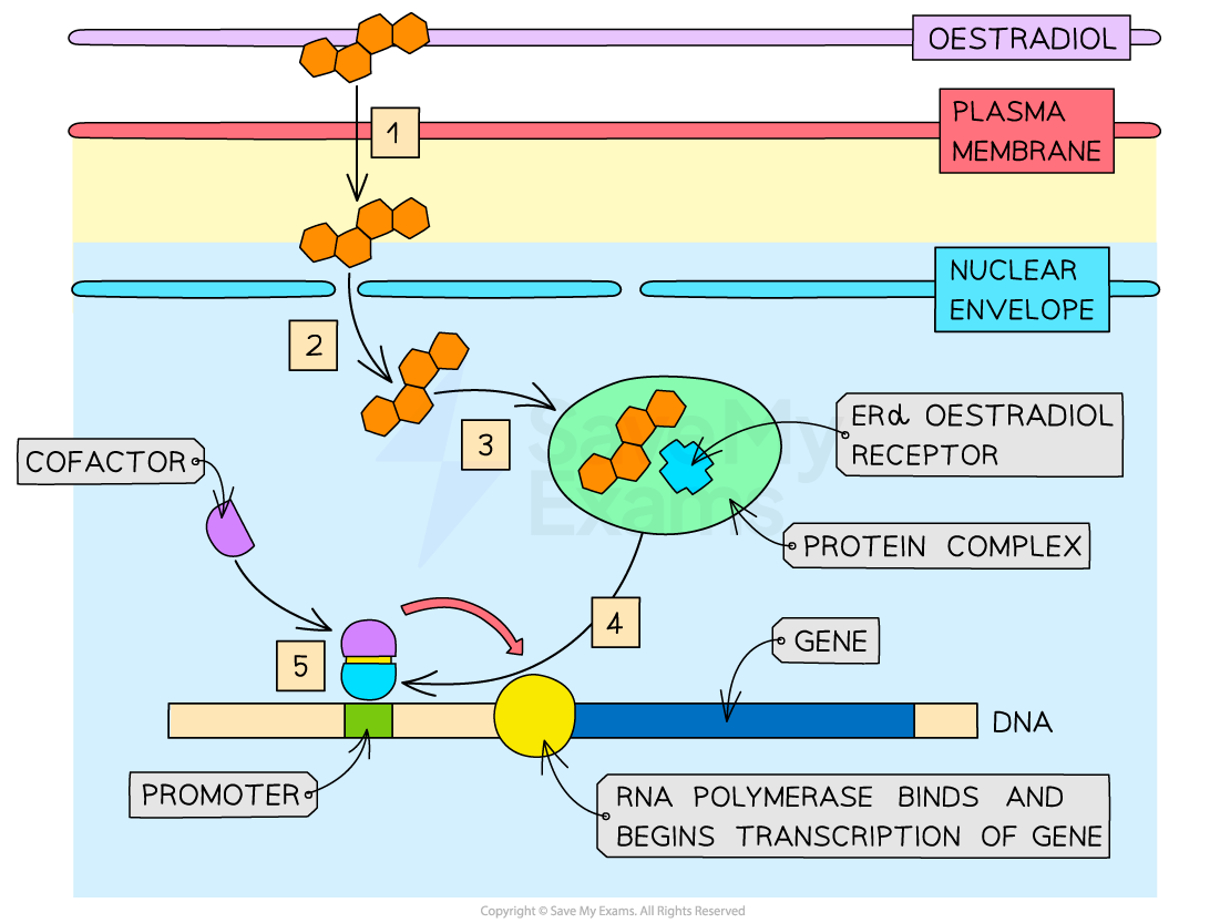
1. Oestradiol
Oestradiol is a key hormone in the regulation of the menstrual cycle and reproductive processes.
Target Cells: Oestradiol acts on cells in the ovary, uterus, and brain.
Mechanism: In the hypothalamus, oestradiol binds to its receptor, forming a complex that enters the nucleus and enhances the transcription of the gonadotropin-releasing hormone (GnRH) gene.
Outcome: This increases the production of GnRH, which stimulates the release of luteinizing hormone (LH) and follicle-stimulating hormone (FSH), critical for ovulation.
Example
During ovulation, high levels of oestradiol trigger a surge in LH by enhancing GnRH production.
This surge is essential for the release of an egg from the ovary.
Note
More on oestradiol next.
2. Progesterone
Progesterone is essential for maintaining the uterine lining during pregnancy.
Target Cells: Progesterone acts on cells in the uterus.
Mechanism: It binds to its receptor in the cytoplasm, forming a complex that moves into the nucleus and activates genes involved in maintaining the endometrium.
Outcome: This ensures the uterine lining remains thick and supportive for a developing embryo.
Warning
Don't confuse progesterone's role with oestradiol.
While oestradiol prepares the uterus for ovulation, progesterone maintains the uterine lining after ovulation.
3. Testosterone
Testosterone is involved in the development of male secondary sexual characteristics and other physiological processes.
Target Cells: Testosterone acts on a variety of cells, including those in muscle, bone, and the prostate.
Mechanism: It binds to the androgen receptor, forming a complex that enters the nucleus and activates genes responsible for protein synthesis and muscle growth.
Outcome: This leads to increased muscle mass, bone density, and the development of male characteristics.
Example
In prostate cells, testosterone activates genes involved in the production of enzymes and proteins essential for cell growth and function.
Why Are Intracellular Receptors Important?
Direct Gene Regulation: Unlike many other signalling pathways, steroid hormones can directly influence gene expression without relying on secondary messengers.
Long-Lasting Effects: The changes induced by steroid hormones are often sustained, affecting processes like growth, development, and reproduction.
Specificity: Each hormone-receptor complex targets specific genes, ensuring that only the necessary proteins are produced.
Applications and Implications
Understanding intracellular receptors has practical applications in medicine and biology.
Hormone Therapies: Treatments for conditions like hormone imbalances, infertility, and certain cancers rely on manipulating steroid hormone pathways.
Drug Development: Synthetic hormones or hormone blockers are designed to target specific receptors, offering treatments for conditions like breast cancer or prostate enlargement.
Evolutionary Insights: The conservation of steroid hormone pathways across species highlights their fundamental role in biology.
Tok
How does our understanding of steroid hormones influence ethical decisions in sports, such as the use of performance-enhancing drugs?
Self Review
Why are steroid hormones able to pass through the cell membrane, whereas peptide hormones cannot?
Can you explain how oestradiol and progesterone differ in their roles during the menstrual cycle?
C2.1.13 effects of the hormones oestradiol and progesterone on target cells
Oestradiol and Progesterone Regulate Gene Expression in Specific Target Cells
Hormones like oestradiol and progesterone play key roles in regulating processes in specific target cells by binding to intracellular receptors.
Their effects are highly localized to particular cell types, such as cells in the hypothalamus or the endometrium, and involve the regulation of gene expression.
Tip
Remember that steroid hormones like oestradiol can pass through cell membranes because they are lipid-soluble.
Oestradiol: Regulating Hormone Release in the Hypothalamus
Definition
OestradiolA form of estrogen that promotes the growth of the uterine lining (endometrium) and regulates FSH and LH via feedback loops.
Oestradiol is a steroid hormone produced mainly in the ovaries.
It plays a central role in the menstrual cycle, influencing reproductive organs and the brain.
The hypothalamus is a small but critical region in the brain that regulates hormone release.
Gonadotropin-Releasing Hormone (GnRH): The hypothalamus produces GnRH, which triggers the pituitary gland to release luteinizing hormone (LH) and follicle-stimulating hormone (FSH).
Example
During the follicular phase of the menstrual cycle, rising oestradiol levels eventually trigger a surge in LH, leading to ovulation.
Mechanism of Action
Entry into the Cell: Oestradiol diffuses through the plasma membrane of hypothalamic cells.
Binding to Receptor: It binds to an intracellular receptor in the cytoplasm.
Activation of Gene Expression: The hormone-receptor complex enters the nucleusand acts as a transcription factor, enhancing the production of GnRH mRNA.
Warning
Don't confuse positive and negative feedback.
Oestradiol's effect depends on its concentration and the timing within the menstrual cycle.
Progesterone Maintains the Endometrium
Progesterone is another steroid hormone, primarily produced by the corpus luteum in the ovaries after ovulation.
Its main role is to prepare and maintain the uterine lining (endometrium) for a potential pregnancy.
Note
If pregnancy does not occur, progesterone levels drop, leading to the shedding of the endometrium during menstruation.
How Does Progesterone Affect the Endometrium?
Thickening the Lining: Progesterone stimulates the endometrial cells to proliferate and thicken, creating a nutrient-rich environment for a fertilized egg.
Preventing Contractions: It inhibits uterine muscle contractions, ensuring the embryo can implant and grow undisturbed.
Analogy
Progesterone is like a caretaker, ensuring the "home" (endometrium) is ready to host a guest (embryo) or dismantling it if no guest arrives.
Mechanism of Action
Diffusion into Cells: Progesterone diffuses through the plasma membrane of endometrial cells.
Binding to Receptor: It binds to an intracellular receptor in the cytoplasm.
Gene Activation: The hormone-receptor complex enters the nucleus and activates genes involved in cell proliferation and secretion of nutrients.
Example
Progesterone activates the gene for insulin-like growth factor, which supports cell growth and maintenance of the endometrial lining.
Warning
It's a common misconception that progesterone only affects the uterus.
While its primary role is in the reproductive system, it also influences other tissues, such as the breasts and brain.
Tok
How do the feedback mechanisms of oestradiol and progesterone reflect broader biological principles, such as homeostasis and regulation?
Self Review
What is the role of oestradiol in GnRH regulation during the menstrual cycle?
How does progesterone affect the endometrial cells during the luteal phase?
C2.1.14 regulation of cell signalling pathways by positive and negative feedback
Feedback Mechanisms Fine-Tune Cell Signaling Pathways
Definition
Feedback InhibitionFeedback inhibition is a process where the end product of a metabolic pathway inhibitsan enzyme involved in its own synthesis.
Analogy
Imagine your body as a finely tuned orchestra.
Each instrument (or cell) plays its part, guided by signals that ensure harmony.
But what keeps this orchestra from playing too loudly or too softly?
The answer lies in feedback mechanisms--the conductors of cellular communication.
Positive Feedback Amplifies the Signal
Positive feedback occurs when the end product of a process enhances its own production, creating a self-reinforcing cycle.
This type of feedback leads to a greater change in the same direction, which can drive processes to completion.
Analogy
Think of it like a snowball rolling down a hill, growing larger as it gathers more snow.
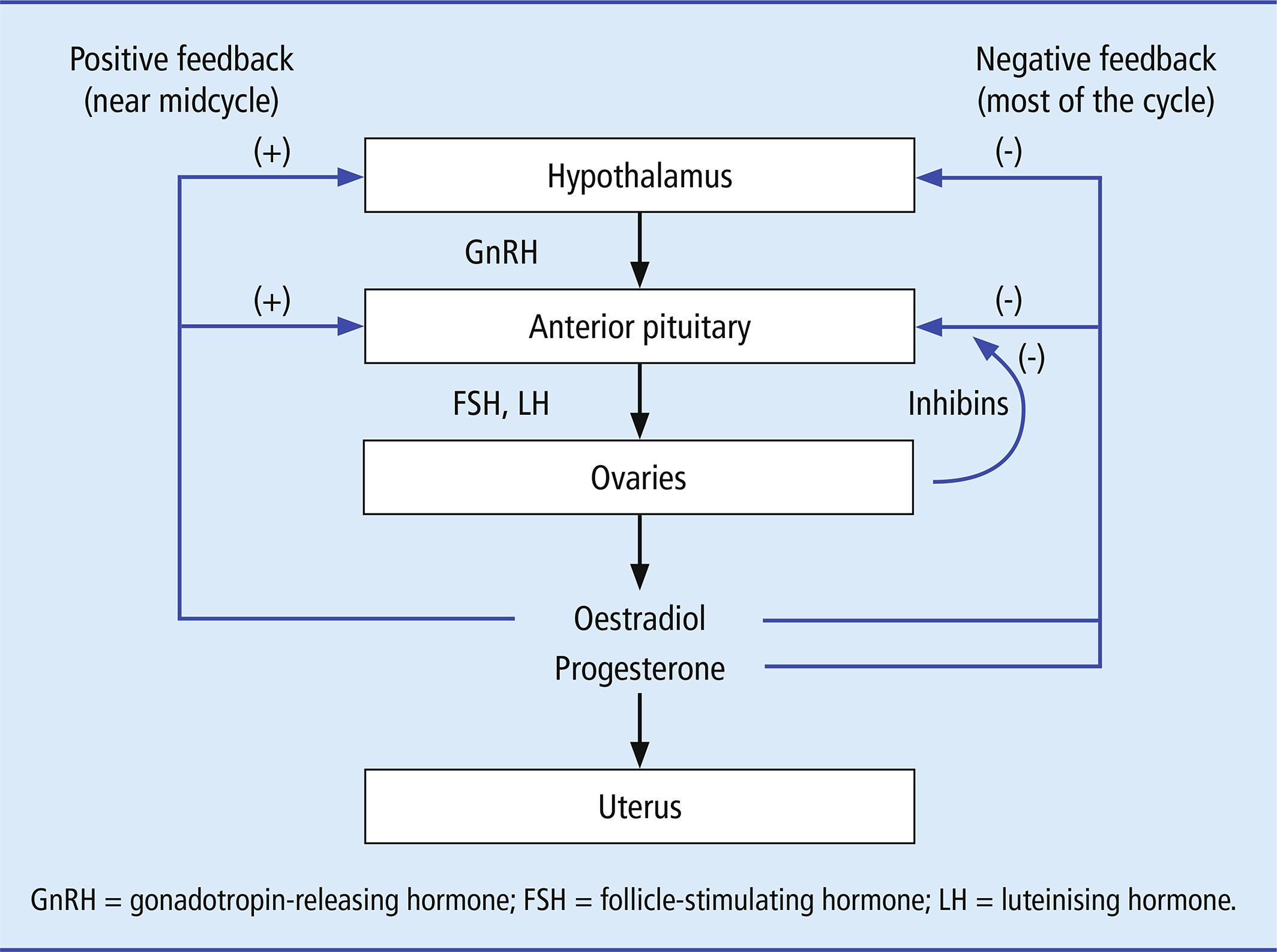
Example: Menstrual Cycle (Oestradiol and LH Surge)
In the menstrual cycle, rising levels of oestradiol stimulate the hypothalamus to release gonadotropin-releasing hormone (GnRH), which prompts the anterior pituitary to release luteinizing hormone (LH).
As LH levels rise, it triggers the LH surge, which causes ovulation, the release of an egg from the ovary.
The rising oestradiol levels initially stimulate more GnRH and LH release, creating a positive feedback loop that intensifies until ovulation occurs.
Note
Example: Calcium-Induced Calcium Release
Calcium ions (Ca²⁺) play a critical role in muscle contraction, neurotransmitter release, and other cellular processes.
In muscle cells, calcium is stored in the endoplasmic reticulum (ER).
When a signal triggers the release of calcium, it binds to inositol trisphosphate (IP₃) receptors on the ER, causing more calcium to be released.
This increase in calcium further activates nearby IP₃ receptors, amplifying the release.
Example
In heart muscle cells, calcium-induced calcium release ensures a strong contraction, vital for pumping blood efficiently.
Warning
Don't confuse positive feedback with homeostasis.
Positive feedback amplifies changes, while homeostasis typically stabilizes conditions.
Negative Feedback Restores Balance
Negative feedback occurs when the end product of a process inhibits its own production, maintaining stability by preventing overactivity.
This is the most common form of feedback and works to maintain stability.
Analogy
Imagine a thermostat in your home.
When the temperature rises above a set point, the thermostat turns off the heater to cool things down.
Example: Testosterone Regulation
Testosterone production is controlled by a negative feedback loop involving the hypothalamus, pituitary gland, and testes.
The hypothalamus releases gonadotropin-releasing hormone (GnRH), which stimulates the pituitary gland to produce luteinizing hormone (LH).
LH acts on the testes to produce testosterone.
As testosterone levels rise, they signal the hypothalamus and pituitary to reduce GnRH and LH production, decreasing testosterone synthesis.
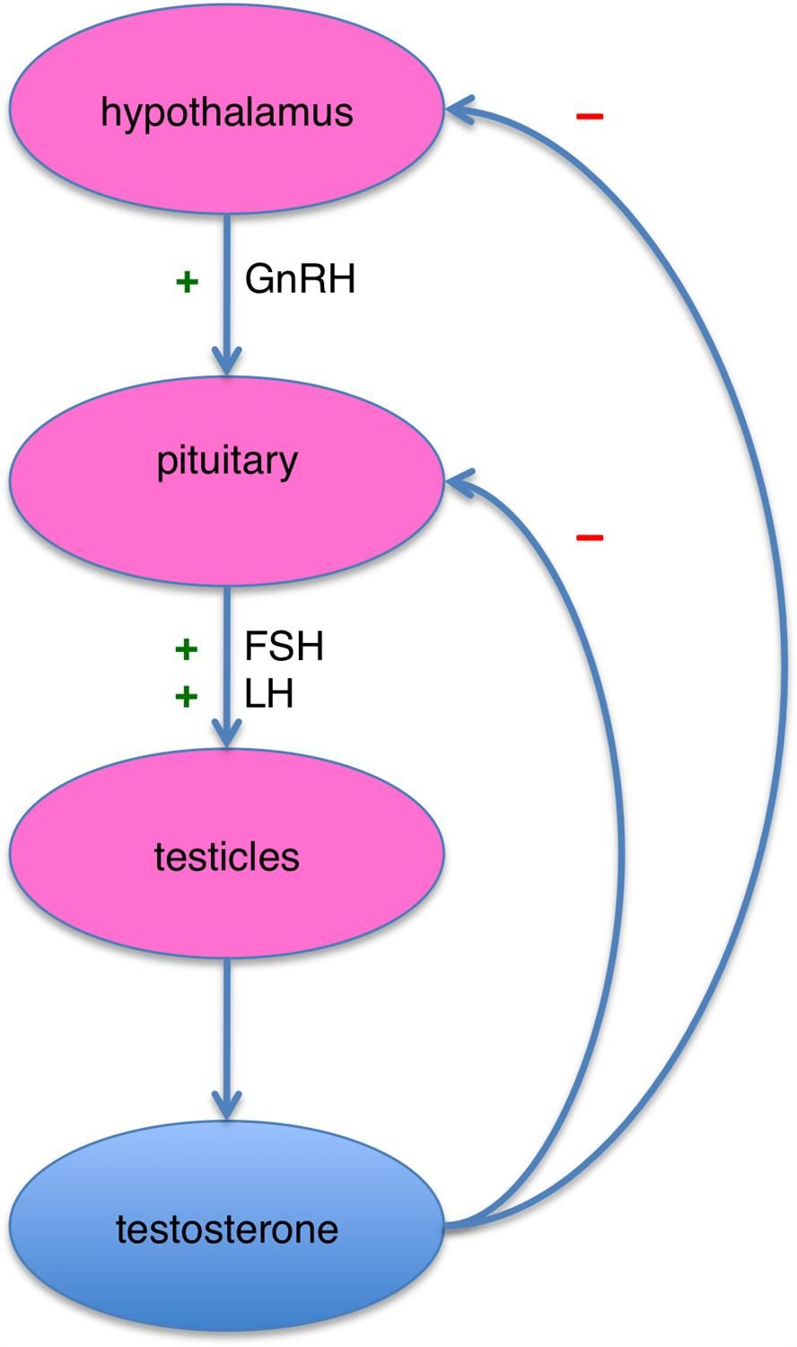
Example
This feedback loop ensures testosterone levels remain balanced, preventing excessive hormone production that could disrupt normal physiological functions.
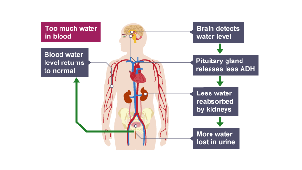
Example: Antidiuretic Hormone (ADH) Regulation
When the body becomes dehydrated, the hypothalamus detects the increased concentration of solutes in the blood and signals the pituitary to release antidiuretic hormone (ADH).
ADH promotes the reabsorption of water by the kidneys, reducing the amount of water lost in urine, which helps to increase blood volume and reduce blood solute concentration.
As blood volume increases and solute concentration decreases, the secretion of ADH is inhibited, thereby reducing water reabsorption and restoring homeostasis.
This is a negative feedback loop that ensures blood volume and osmolarity are kept within optimal ranges.
Tip
When studying feedback loops, always identify the stimulus, response, and outcome.
This will help you determine whether the loop is positive or negative.
Why Feedback Matters in Cell Signalling
Feedback mechanisms are essential for:
Precision: Ensuring signals are neither too weak nor too strong.
Adaptability: Allowing cells to respond to changing conditions.
Stability: Maintaining homeostasis in dynamic environments.
Tok
How do feedback mechanisms in biology compare to those in other systems, such as economics or climate regulation?
What can we learn from these parallels?