3.1.2 (8.5) The Human Heart (OCR A-level Biology)
The human heart is part of a double circulatory system
This means that blood passes through the heart twice
Deoxygenated blood from organs goes through heart, goes to lungs, blood becomes oxygenated, then passes through heart again and goes to other organs
Cycle repeats
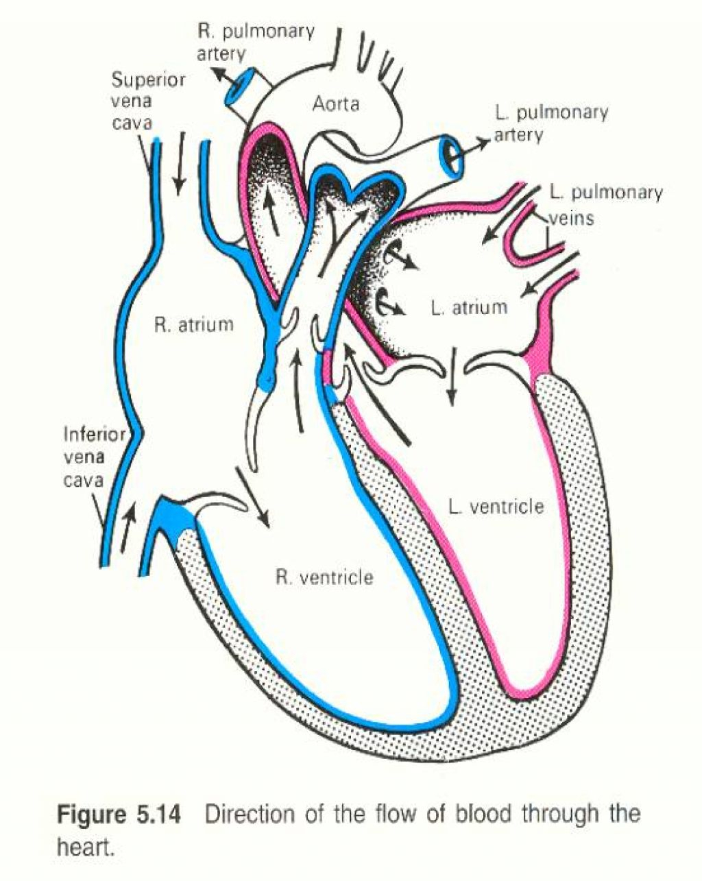
LABELLING THE HEART
Sides of the heart are flipped in diagrams, Left side of heart is displayed as the right and vice versa
RIGHT SIDE
Blood arrives through Vena Cava (vein) through either the superior (from brain) and inferior (from other organs) and enters Right Atrium, blood then passes through Tricuspid (or Atrioventricular) valve into Right Ventricle and then passes through Semi-lunar valve and exits through Pulmonary artery
LEFT SIDE
Blood arrives through Pulmonary vein and enters Left Atrium, blood then passes through Bicuspid (or Atrioventricular) valve into Left Ventricle and then passes through Semi-lunar valve and exits through Aorta
METHODS TO REMEMBER
To remember Atrium + Ventricle placement
A+V stacked makes a diamond, meaning the Atrium should be on top and the ventricle on the bottom
To remember blood flow of veins + Arteries
Vein contains ‘in’ meaning blood is flowing in to the heart
Artery starts with ‘A’ for blood flowing away from the heart
To remember which sides of the heart are which
If you were to hold a diagram of a heart against yourself with the heart facing away from you, the left side of the diagram is on your own left
You are looking at the diagram as if it is in your own chest
To remember valves
Tricuspid and Bicuspid can be replaced by the term atrioventricular valve if you cant remember which sides they belong on
Tricuspid = right
Bicuspid = left
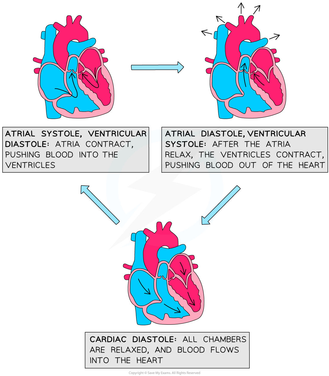
 THE CARDAIC CYCLE
ATRIAL SYSTOLE
Contraction of atria
Pressure increase in atrias, causes AV vales to open and blood flows into ventricles
VENTRICULAR SYSTOLE
Contraction of ventricle
Pressure increases in ventricles, causes SL valves to open and blood to be forced out through arteries
DIASTOLE
Relaxation of Atria + Ventricles
Blood flows passively into the heart
ELECTRICAL RHYTHM OF THE HEART
Wave of electrical excitation begins at the Sino-Atrial node (SAN)
This causes Atria to contract and a heartbeat to be initiated
Layer of non-conducting tissues prevents this wave from immediately reaching ventricles
Wave of electrical excitation picked by Atrio-Ventricular node (AVN)
Imposes a slightly delay before stimulating bundle of his (conducting tissues) which is made up of purkinje fibres
Bundle of His separates into two branches and conducts the wave of electrical excitation to the hearts apex (bottom of heart)
Triggers contraction of ventricles to efficiently empty ventricles
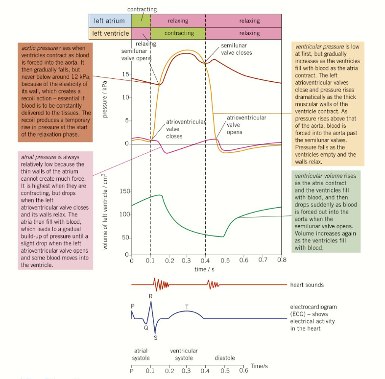
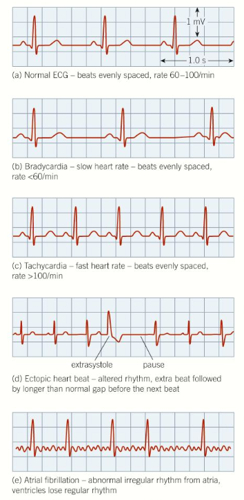
ELECTROCARDIAGRAMS
BRADYCARDIA
Heartbeats are too slow
Common in fit people
Abnormal treated by pacemaker to keep heartbeat steady
TACHYCARDIA
Heartbeats are too rapid
Normal during exercise
Abnormal treated by medication or in severe cases, surgery
ECTOPIC HEARTBEAT
Heartbeats outside of normal rhythm
ATRIAL FIBRILATION
Rapid, incomplete contraction of atria
Less effective pumping of blood
Example of Arrhythmia (abnormal heart rhythm)