Body Systems
Cell Theory
1) Every living thing is made up of cells
2) The cell is the basic unit of structure and function
3) All cells come from other cells
Cell Structures
Mitochondria: The mitochondria is powerhouse of the cell. It uses oxygen and glucose in cellular respiration.
Endoplasmic reticulum: Transport, also called “intracellular highway”. The rough endoplasmic reticulum contains ribosomes, while the smooth ER doesn’t.
Golgi apparatus: This packages and exports proteins. A vesicle is the package that can be sent out of the cell. Ribosomes build protein and send it through the ER. The proteins then go to the Golgi bodies where they are packaged for export.
Lysosomes: Contains digestive enzymes which break things down. It is also called the ‘suicide sac’.
Vacuole: Storage area for water and other substances. Plant cells usually have a large central vacuole.
Cell Organelles and Their Functions
1) Nucleus: Stores genetic information of the cell.
2) Nucleolus: Makes ribosomes.
3) Cytoplasm: Contains the contents of the cell.
4) Cytosol: Gel-like matrix that holds water and nutrients.
5) Cytoskeleton: Helps in structure, support and transport of the cell.
6) Ribosomes: Make proteins.
7) Rough endoplasmic reticulum: Assists in protein synthesis and modification for the endomembrane system.
8) Smooth endoplasmic reticulum: Detoxifies the cell and makes lipids.
9) Golgi apparatus: Sorts and ships proteins.
10) Lysosomes: Remove unwanted wastes and materials from the cell.
11) Peroxisome: Regulate biochemical pathways that involve oxidation.
12) Vacuole: Stores water and nutrients.
13) Vesicles: Transport materials.
14) Cell membrane: A thin, flexible barrier that protects the cell.
15) Cell wall: Rigid barrier that protects the cell. Only present in plant cells.
16) Chloroplasts: Makes food using the process of photosynthesis.
The Digestive System
Phases of digestion:
Ingestion
Movement
Mechanical and chemical digestion
Absorption
Elimination
Mouth:
Teeth mechanically break down food into small pieces.
Tongue mixes food with saliva (contains amylase, an enzyme which helps break down starch)
Epiglottis: a flap-like structure at the back of the throat. Closes over the trachea, preventing food from entering it. It is located in the pharynx.
Esophagus:
Approximately 20 cm long
Functions include secreting mucus and moving food from the throat to the stomach using muscle movements called peristalsis.
Stomach:
J-shaped muscular bag that stores the food and breaks it down into tiny pieces.
Mixes food with gastric juices that contain enzymes to break down proteins and lipids.
Hydrochloric acid in the stomach kills bacteria.
Food found in the stomach is called chyme.
Small intestine:
Roughly 7 meters long
Lining of the intestine walls have finger-like projections called villi to increase surface area for absorption of nutrients.
The villi are covered in microvilli which further increase surface area for absorption.
Nutrients from the food pass into the bloodstream through the walls of the small intestine.
Absorbs:
80% ingested water
Vitamins
Minerals
Carbs, proteins and lipids
Secretes digestive enzymes.
Large intestine:
About 1.5m long
Absorbs nutrients left behind by the small intestine
The end of the large intestine is the rectum (short term storage which holds feces before expelled)
Functions:
Bacterial digestion and fermentation of carbohydrates
Absorbs additional water
Concentrates wastes
Accessory Organs — The Glands
Not part of the path of ingested food, but play a critical role in digestion.
Includes the liver, gall bladder and pancreas.
Liver:
Directly affects digestion by producing bile.
Bile aids in the digestion of fat.
Filters out toxins and waste including drugs, alcohol, and poison.
Gall bladder:
Stores bile from the liver and releases it into the small intestine.
Fatty diets can cause the formation of gall stones
Pancreas:
Produces digestive enzymes to digest fats, carbs and proteins.
Regulates blood sugar by producing insulin.
Extra Notes
Digestion is the process by which food and drink are broken down into their basic chemical structures in the gastrointestinal tract.
Carbs are broken down into simple sugars and glucose.
Fats are broken down into fatty acids and glycerol.
Proteins are broken down into their amino acids.
The gastrointestinal tract is a series of hollow organs connecting from the mouth to the anus.
Stop 1: Mouth
Teeth claw the food and chop it into tiny pieces.
Saliva is produced (containing enzymes) to break down starch particles.
Saliva makes the food easier to swallow.
Stop 2: Stomach
Mixes digestive juices with the swallowed food.
Enzymes in the digestive juices start the break down of any protein particles.
Can hold up to 1L of food and liquid and can store it for about 3.5 hours.
Chyme is the mixture of gastric juices and hydrochloric acids.
Stop 3: Duodenum
First part of the small intestine.
Bile disperses fat globules into tiny fat droplets, then enzymes break them down into smaller pieces.
The break down of protein and starch particles is completed.
Stop 4: Ileum (small intestine)
In the second part of the small intestine, sugars, proteins, vitamins and minerals are in particles small enough to be absorbed.
All that’s left is a liquid of undigested waste material and water.
Villi cover the inside surface of the ileum, and increase surface area through which nutrients are able to be absorbed into the bloodstream.
Stop 5: Colon (large intestine)
Liquid of undigested food and water passes through here.
Most of the water is absorbed and passed onto the kidney to be excreted.
Stop 6: Anus
Food waste that was stored in the rectum is expelled through the anus.
The Respiratory System
The respiratory system consists of the nose, pharynx, larynx, trachea, bronchi and lungs. The primary function of this system is to furnish oxygen for individual tissue cells, and to take away the waste products and carbon dioxide produced by those same cells.
External and Internal Respiration
External respiration is the process of inhaling oxygen into the lungs, and exhaling carbon dioxide. That process includes the ventilation of the lungs and the exchange of air in the lungs and blood within the capillaries of the alveoli of the lungs.
Internal respiration is the metabolic process by which living cells use blood flowing through the capillaries, absorbing the oxygen they need and releasing the carbon dioxide they create.
The nose
The nose has 5 functions:
Serves as an air passageway.
Warms and moistens inhaled air.
It’s cilia and mucous membrane trap dust, pollen, bacteria and foreign matter.
Contains olfactory receptors, which smell odours.
Aids in phonation and voice quality.
The pharynx
The pharynx is the correct term for the throat. It is a muscular and membranous tube that is about 5 inches long, extending downwards from the base od the skull. It eventually becomes the esophagus. The nasopharynx is behind the nose; the oropharynx is behind the mouth; the laryngopharynx is behind the larynx.
The larynx
The larynx, commonly called the voice box, is located at the upper end of the trachea. The larynx contains vocal cords, which produce sound.
The trachea
The trachea or windpipe is a smooth muscular tube leading from the larynx to the bronchi. C-shaped cartilage rings prevent the trachea from crushing.
The trachea is the passageway for air to and from the lungs. It is lined with cilia (hairs), which sweep foreign matter out of the pathway. It is only about 1 inch in diameter and 4.5 inches long.
The bronchi
The bronchi are the 2 main branches at the bottom of the trachea providing a passageway for air to the lungs. The trachea divides into the right and left bronchus, and further divides into the bronchial tree.
As the branches of the bronchial tree get smaller, the 2 primary bronchi become bronchioles, and then very small alveolar ducts. The left bronchus is smaller than the right bronchus, because room is needed to accomodate the heart.
If a foreign body is inhaled or aspirated, it usually lodges in the larger right bronchus or enters the right lung. In the presence of infection, the bronchi sometimes become inflamed, resulting in a diagnosis of bronchitis.
The lungs
The lungs are 2 spongy organs located in the thorax (chest cavity). They consist of elastic tissue, filled with an interlacing network of blood vessels, tubes and sacs that carry air. Each lung is divided into lobes: the right lung is divided into 3 lobes while the left lung is divided into 2 lobes. The left lung has an indentation called the cardiac depression or notch, for placement of the heart.
At the end of each bronchiole are the alveoli. The lungs contain about 300 million alveoli sacs, which are the air cells where the exchange of oxygen and carbon dioxide takes place within the capillaries.
The base of the lungs rest on the diaphragm, a muscular wall separating the thorax from the abdominal cavity. It is involved in drawing downward in the chest during inhalation and pushing upward during exhalation.
Vital Signs
Vital signs, essential elements for determining an individual’s state of health, include temperature, pulse, respiration and blood pressure. A deviation from normal of any or all vital signs indicates a state of illness, and can be used by a physician in a diagnosis, prognosis (prospects of survival and recovery) and treatment.
The normal respiration rate for a 5 year old is 20-25 breaths per minute. For someone 15 years or older, it is 15-20 breaths per minute.
Tidal volume refers to the amount of air inhaled or exhaled during normal breathing — about 500 ml. Total lung capacity is 3.6 - 9.4 L in an average male.
The Circulatory System
The circulatory system delivers oxygen and nutrients to cells and takes away wastes. The heart pumps oxygenated and deoxygenated blood on different sides.
Blood
Blood consists of:
Red blood cells: to carry oxygen
White blood cells: make up part of the immune system
Platelets: needed for clotting
Plasma: Blood cells, nutrients and wastes float in this liquid
The heart
The heart pumps blood around the body. The heart is actually a double pump made up of 4 chambers, with the flow of blood going on in one direction due to the presence of heart valves.

Superior vena cava: Large vein that returns blood to the heart from the head, neck and upper limbs.
Aorta: Largest artery in the body that carries blood away from the left ventricle.
Pulmonary artery: Main artery that supplies the lungs with deoxygenated blood.
Pulmonary vein: Carries oxygen-rich blood from the lungs to the heart.
Mitral valve: One of four valves that regulates blood flow from the upper left chamber to the lower left atrium. Has two flaps (leaflets). Also known as the bicuspid valve.
Aortic valve: Closes the lower left chamber and opens to allow blood to leave the heart.
Left ventricle: Pumps oxygenated blood to the organs.
Left atrium: Delivers blood to other parts of the heart and acts as a vessel for blood that comes from the lungs.
Right atrium: Receives deoxygenated blood and pumps it into the right ventricle.
Inferior vena cava: Large vein that carries blood to the right atrium from the lower and middle body.
Tricuspid valve: Prevents a backward flow of blood into the right atrium.
Arteries
Oxygenated blood is pumped from the heart along the arteries, which are muscular. Arteries divide like tree branches until they are slender. The only artery that picks up deoxygenated blood is the pulmonary artery, which runs between the heart and the lungs.
Capillaries
The arteries eventually divide down into the smallest blood vessel, the capillary. Capillaries are so small that blood cells can only move through them one at a time. Oxygen and food nutrients pass from these capillaries to the cells. Capillaries are also connected to the veins, so wastes from the cells can be transferred to the blood.
Veins
Veins have one-way valves instead of muscles, to stop blood from running back the wrong way. Generally veins carry deoxygenated blood from the body to the heart, where it can be sent to the lungs. The exception is the network of pulmonary veins, which take oxygenated blood from the lungs to the heart.
Blood pressure
Blood pressure refers to the amount of pressure inside the circulatory system as the blood is pumped around. The heart has 2 pumps: systole and diastole. The pressure from the first pump is called systolic pressure, and diastolic pressure from the second pump. This can be seen when measuring blood pressure: the number on top is of systole and the number on the bottom from diastole.
Common Problems
Some common problems in the circulatory system include:
Aneurysm: a weak spot in the wall of an artery
Atherosclerosis: a narrowing of the arteries due to plaque buildup and deposits
Heart disease: a lack of blood supply to the heart because of narrowed arteries
High BP: can be caused by obesity, among other things
Varicose veins: problems with the valves that stop blood from running backwards
Excretory System
The excretory system is a great example of how organs from other systems (lungs, large intestines etc.) interact with one another to stabilise body systems and ultimately ensure organism survival.
Lungs: removal of excess carbon dioxide.
Liver: produces urea and uric acid as a by-product of the break down of proteins.
Skin: removal of excess water, salt, urea, and uric acid.
Urinary system: kidneys filter the blood to form urine, which is excess water, salt, urea and uric acid.
Defecation
The process of getting rid of solid waste (faeces) from our body is called defecation. Faeces are undigested material that has passed through our digestive tract. It is waste because it is material that our body has not been able to use.
Excretion
Getting rid of the wastes our body has produced is called excretion. It is the function of the excretory system. The lungs, skin, liver, and kidneys are all involved in excretion.
Lungs
Respiration produces carbon dioxide and water as waste products. They are carried back to the lungs and breathed out. The lungs are part of the excretory system as well as the respiratory system.
Liver
Our liver carries out many processes, some involved in excretion. Amino acids are the end products of protein digestion. They are used by our body to make proteins and for growth and repair. Amino acids cannot be stored. If we have more than needed they are broken down and excreted. The liver breaks down amino acids down into a substance called urea.
Poisonous substances may enter the body from the digestive tract. These are carried to the liver, where they are broken down into harmless substances. The harmless substances are then returned to the blood and from there they pass to the kidneys.
The liver breaks down old red blood cells. Any unwanted haemoglobin is added to bile and passes with the bile into the intestines.
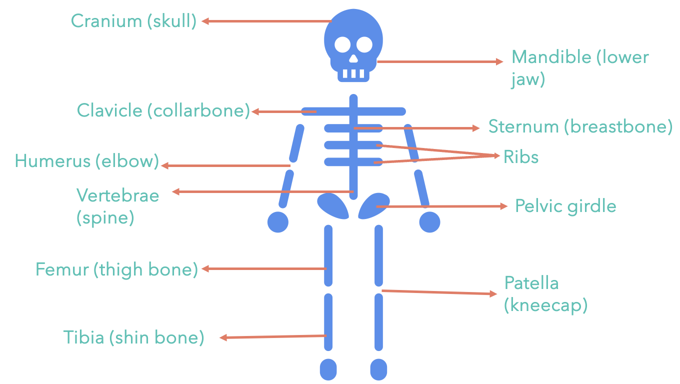
Skeletal System;/ The skeletal system has 206 separate bones. Your hand alone contains 27 bones.
Without your skeleton, you would be a jelly-like blob.
Important jobs of the bones
They form a frame which gives your body its basic shape
They protect the organs inside your body
With the help of muscles, they enable you to move.
The femur or thigh bone is the longest bone.
Stapes or stirrup bone in our ear about 3mm in length is the smallest bone.
What is in a bone?
Compact bone:
the outer layer of a bone, hard and strong.
It’s covering contains minerals (i.e. calcium and phosphorous)
Bone marrow:
Inside the bone is hollow and contains a soft tissue
Your bones are alive. They contain living cells and need a blood supply to provide oxygen and other nutrients. In order for the bone to be hard, it needs an adequate supply of two important minerals: calcium and phosphorous.
Until you reach the age of about 20, the soft cartilage that made up your skeleton when you were born is being gradually replaced.
Cartilage is very soft and rubbery, not as hard as solid bone. The trachea, nose, and ears are mostly made up of cartilage.
Ossification
The hardening of your bones as you get older is called ossification.
After ossification, the bone is made up of 70% non living and 30% living matter. As you get older, your bones may get dry and brittle. This is why older people are at a higher risk of breaking their bones.
When a bone breaks, the ends of the bone need to be put back in place so that they can grow together. If a bone is shattered into several pieces, it is sometimes possible to use pins or wires to hold the pieces in place while the bone heals.
Greenstick fracture:
occurs when the bone cracks but doesn’t break
common in children because their bones are more flexible
Osteoporosis:
a loss of bone mass that causes them to become lighter, more fragile and easily broken.
Photosynthesis and Respiration
Overview
Radiant energy (sun) → Photosynthesis (glucose) → Respiration (ATP energy) → Cell activity
Organisms that use light energy from the sun to produce food are called autotrophs.
Organisms that cannot use the sun’s energy to make food are called heterotrophs.
Photosynthesis
Photosynthesis is the process by which energy of sunlight is converted into the energy of glucose. Photosynthesis is a chemical reaction.
6CO2 + 6H2O + energy → C6H12O6 (glucose)
Q) Describe photosynthesis.
The process of changing light energy to chemical energy
Energy stored as sugar
Occurs in plants and some algae
Plants need light energy, carbon dioxide and oxygen
Takes place in chloroplasts, using chlorophyll, the green pigment in plants.
Chlorophyll is the green pigment inside chloroplasts that absorbs light for photosynthesis. As the chlorophyll in leaves decays in the autumn, the green colour fades and is replaced by the oranges and reds of carotenoids, other pigments.
What happens during photosynthesis?
Plants capture light energy and use that energy to make glucose.
Sunlight provides the energy needed by chlorophyll to change molecules of carbon dioxide and water into glucose.
Oxygen is released in this reaction
Carbon dioxide enters the leaf through stomata
CO2 combines with the stored energy in the chloroplasts through a chemical reaction to make glucose.
The sugar is moves through tubes in the leaf to the roots, stems and fruits of the plants.
Some of the sugar is used right away by the plant for energy, some is stored as starch, and some is built into plant tissue.
Cellular respiration
Cellular respiration is the release of chemical energy for use by the cells. Once the energy that was in sunlight is changed into chemical energy, an organisms has to transform the chemical energy into a form that can be used by the organism.
Cellular respiration is the process by which the energy of glucose is released into the cell to be used for life processes. Respiration occurs in all cells and can take place with or without oxygen present.
Q) Describe cellular respiration.
The breakdown of glucose molecules to release energy
Turns glucose into ATP
Takes place in all living things
Step-by-step process
Chemical formula:
C6H12O6 + O2 → 6CO2 + 6H2O + ATP