Neuromuscular PEDS
Injuries
contusions
bruise = injury to skin, soft tissue, or bone
bone (or ligament) contusion, muscle contusion = forceful or multiple hit = crushing muscle
in school age and adolescent w/ sports; toddlers when walking, or if taking anticoag or blood disorder
abuse = in odd areas → cheek, neck, <2yr w/ frequent unless disorder
collision, falls, jumping,
s/s: blue/ purple → green yellow
if severe → dec movement, pain at the injury site, swelling, ecchymosis
lab/dx:
what happen + causes = age, physical activity, location, how often sustains bruises, Rx blood thining, if lots w.o explanation; if serious → MRI
tx:
ice + rest, elevate to dec swell, no weight on affect limb and immobilize, ibuprofen for pain, PT
wearing safety gear, take steps, avoid extreme movements, remove tripping hazards, balanced diet to help heal
dislocation:
two bones come apart at joint = tear ligament
not commonly seen w/ still growing, more in older who participate in activities that put excessive force (most common the shoulder joint)
forceful movement = gynastics, basketball, football, soccer, skating, volleyball, Ehler-s Danios
s/s: swelling, bruisng around injury, pain, numb
bilateral extremities ~ be uneven, deformities present, joint movant trouble in certain way if they can measure full dislocation or subluxation
lab/dx:
aks for risks (Ehlers- Danlos syndrome)
imaging= X-R, MRI
neurovascular assessment = 5 Ps
Pain
Pallor (coloring)
Pulse
Parethesia (numbness/tingling)
Paralysis
tx:
apply ice, joint need to be immobilized, manually place bone back in socket or place sx, splint/ cast,
if not tx → sx + causes growth to be slowed = limb length discrepancies, loss of ROM
infants will cry, toddlers will point where it hurts, preschool are better bale to express but need parent supprt, school age can describe pain, OT, PT
mobility devices
give doll to show where it hurts
sprains
damage of ligament w/ stretch or twist = ankles and wrists are most common (posterior talofibular, anterior talofibular, and calcaneofibular ligaments)
due to sports overuse nto given enough time to heal
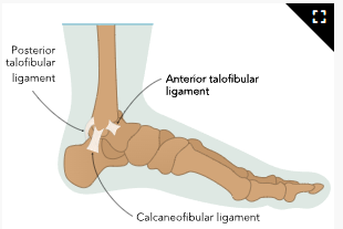
when growth plates are active, tissues grow at diff rate → low ROM
more in 10-18yr
s/s:
discoloration or ecchymosis, low ROM, pain, swelling
lab/dx: SR, MRI, CT, neurovascular
tx:
PRICE mnemonic
P: Protection
R: Rest
I: Ice
C: Compression
E: Elevation
POLICE mnemonic
P: Protection
O, L: Optimal Loading (bearing some weight)
I: Ice
C: Compression
E: Elevation
elastic bandage for supporting sprain, low discomfort, control edema
elevation and rest depend on severity, OTC, mobilizing devices, PT
Ice application:
20 minutes at a time while awake, repeating the application several times during the day for the first 72 hours after injury
After the first 72 hours, three times a day for 20 minutes each
Do not apply directly on the skin (use fabric, such as a towel or cloth, between the skin and the ice)
Only apply ice when awake and alert to prevent injury to the skin from prolonged use
stretch before a sport or running (use good form)
strains
tendon or muscle injury by excessive stretching, excess physical activity, repeated movement w/ little rest , lifting heavy object improperly= more in hamstring and lower back
in 10yr <
growth plates weaker than tendons and muscles = less occurrence in peds
s/s: redness, pain, bruise, swell, low ROM, hold area and not want to use it
lab/dx ~ sprain
tx: immobilize → crutches, wheelchair, splint, compression, PT
bending knees and not being over the back to pick up heavy object
pick up heavy object in high self = ladder or step stool
overuse
high intensity movement, during periods of rest → intense strain + inflammation
when not resting in between activity repeatedly swinging tennis or pitching baseball
>6yr
Sever disease: when growth plate in heel bone becomes infected
running
osgood-schlatter: top of the tibia (shin bone becomes infected)
flex thigh muscle → pulls knee tenon (patellar tendons) → stretch tibia tubercle
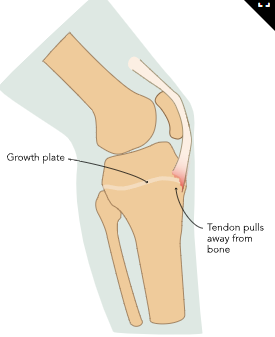
s/s will develop in few weeks
stress factures and overuse → tenderness, and not want to use extremity
lab,dx: w/ imaging
tx: orthotic heel padding, need crutches, stretching, NSAID, PT, rest, sx
encourage rest from sport 1-2 days/ week, not participate in sports all year round
Preventing Injury
Warm up before beginning an activity. The goal is to elevate the heart rate and loosen the body. Jumping jacks or running in place are examples of how to warm up.
Take time to stretch.
Use recommended equipment for the specific activity. It is there for protection.
Cool down 10 minutes before ending the physical activity by slowing the speed and intensity of the activity. This phase is complete when the skin is dry and the body feels cooled down.
Listen to body signals, and stop activity if pain is experienced.
Hydrate well to prevent dehydration.
If very physically active, schedule regular days with no physical activity to allow the body to rest.
mobilization
crutches
hand grips at the same level of hips
crutched is placed 30-5cm in front of weight-bearing foot
armpit not resting on pad, put all weight in crutch instead of injured leg
caines trouble w. balance or diff walking
warm slightly bend at the elbow, not to hold on cane on injured side, caine ahead injured limb
walkers: for more severe
most weight is in arms, keep back straight, all fur devices of legs should be placed on the ground at the same time and small steps be taken
wheelchair:
some for positioning and others for long term, lock wheels before transfer
tipping should be prevented by placing weight on back, use one direction, and avoid curbs and inclines
fractures
young children have soft bones w/ protective outer bone → severe fractures that result in long-term effects for children
complete = separate to 2 pieces of bone → sx/ incomplete
plastic deformation = bent bone but not broken → sx to straighten
buckle fractures = torus fracture under 12yr and do not require sx
excessive pressure creates bulge, more common in forearm
greenstick: slintering of bone w/o fully breaking → seen in <10yrs
simple/ closed = does not puncture skin; compound/ open
complicated: splinter into pieces and damage surrounding tissues (diff to tx)
comminuted: bone breaks in at least two places, mostly in large bone → femur
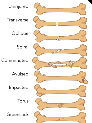
physis (between epiphysis and metaphysis) = weakest bone + growth plate
stress on bone over time; trauma; fall; automobile accident, child abuse, sport-related
idiopathic juvenile osteoporosis = weaker bones,
poor nutrition, low vit D/ ca, obesity, sports, extreme activities
screening:
assess of s/s, pain, bruising, swelling in site, unable to move injury, or bear weight, deformity → confirmed w/ XR, MRI, CT
CBC rule out infection, phosphorus, vit D/Ca
Manifestations of Pain |
Eyebrows furrowed, forehead wrinkled, tense face |
Crying (high pitch or more frequent / longer than expected), inconsolable |
Interrupted sleeping routine, unable to sleep, may take small naps |
Tension in limbs, legs drawn toward chest, hands in fists, may kick extremities |
Does not engage with others in the room, lacks interest in toys, does not eat as expected |
complications:
impaired circulation and nerve compression:
surrounding tissue damage
resultant nerve damage → numbness, pain, diff ambulating (ex sciatic nerve damage)
compartment syndrome: when fracture swells excessively → disrupts circulation = pain that continues to worsen, inability to move affected body part, tingling sensation = inc infection low circulation
physeal involvement: disrupted growth damage → growth anomalies or diff lengths
nonunion: fractured bone doesn’t heal due to lack of circulation → sx
malunion: does not heal properly → bone to be shorter/ unexpectedly shaped
risk of infection; pulmonary emboli: fat embolus
heals more rapidly → but ~ have severe growth deformities
tx:
affected area must be filled w/ blood → soft bone → hard bone
immubolizers, sx , PT, reduction (proper alignment)
traction:
pull bones, connective tissues, tendons, muscles together using pullets and weights
skeletal (Buck) traction → pins in bones w/o damaging tissue in pelvis, hip, femur, arm
casts/ splint: keep fracture stable and prevent movement
casts have inner layer for comfort and hard on the outside
splint if not as severe → wrist and easily removed
keep cast or splint dry or removed bathing
Leave the inside material of the cast intact.
Keep the cast dry. Do not submerge it in water or allow it to get wet because it can make the cast soggy, and it will no longer provide the prescribed stability.
If itching occurs, do not apply powder or lotion or insert objects inside the cast. This can break the integrity of the skin and could lead to infection.
Cool air, such as from a hair dryer on its lowest setting, may provide comfort when itching occurs.
Report a change in the shape of the cast, a break in the cast, or if the cast gets wet to the prescribing provider immediately.
under 3 require assessment of medical hx and rule out abuse
Ca = fortified or enriched foods, dairy, nuts, sardines, and green leafy vegetables
Vit D = eggs, salmon, and tuna
no processed foods or refined sugars/ no little food
physical activity for bone strength
wear safety belt, protective gear, clear clutter, no standing in high plaxes, well fitting tennis shows when running,
sx:
internal fixation: attached to bone itself; external fixation: fixates bones to be help on bracket outside of body
manually align bones
infection
osteomyelitis:
inflammation within bone and connective tissues → phagocytic cells releasing enzymes
Staphylococcus aureus
it must be damaged or have a vulnerable spot ( metaphysis → since large blood supply → once blood passes though bone = bacteria settle → necrosis)
or inadequate circulation
risk: compromised immune system + sickle cell, systemic infections, bone break, skin trauma, premature,
lab/dx: biopsy/ radiology
CBC for infection, XR, MRI
bone scan = injecting radioactive tracers, which accumulate in tissue that has changed from what is expected, and using a special camera that scans the bone(s) to create an image
s/s: cold + flu s/s= fever, nausea, lethargy, limping w/ extremity, discomfort, redness in skin, swelling above site of infection
exhaustion, discomfort, and difficulty with ADL
tx: IV abx (vancomycin), necrotic tissue → sx, NSAID
prevent skin infections = hand hygiene, open wound washed w/ soap and water → clean guaze
if 1 week to heal → HCP
movement and physical activity → strengthen bones and inc bone density loss
PT
musculoskeletal alterations
torticollis
neck looks “twisted”
congenital muscular torticollis due to short sternocleidomastoid + trapezius, and splenius or RT to cervical anomalies
if later → acquired torticollis due to dislocation of C1/C2 → cervical nerve compress nerves due to infection, trauma, reflux
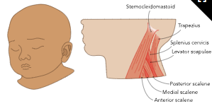
constantly turning head to one side, head appear at unexpected angle, fever, problem with balance, h/a, vision, persistent vomiting, intracranial injury, requires investigation
altered appearance
→ plagiocephaly = occur on one side of infant head, poor facial and cervical spine development
lab/dx: CBC if infection, magnetic imaging for severity of muscles involved
risk:
trauma during pregnancy or birth, rheumatoid disorder, neuro disorder, stress to muscle (sudden movement or too much physical load)
tx:
PT, tubular orthosis (collar to straighten neck), if severe → sx
if acquired = tx injury ~ 6m
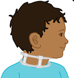
assess for hip dysplasia, infection, neuro disorders
use distraction to turn head to other side = bottle in other side
daily exercise to stretch their necks, turn head side to side, encourage affected side being used when doing quiet activity or resting
tummy time
spinal curvature
lordosis: spine tips backward and back is swayed;
cervical = neck is pushed forward
lumbar seen in L4 to S1 pushing his forward
due to muscular dystrophy, osteosarcoma, kyphosis, poor posture from pregnancy
lumbar curve if more than 40 degrees of cervical ; thoracic >50 degrees w/ protrusions in upper part of torse
tx: braces of PT → sx w/ rod in spine
kypohosis:
spine tips forward and appearance of pt hunching over = cervical and thoracic
bad posture caused by slouching = postural kyphosis
Scheuermann’s = shaped like wedge instead of rectangle
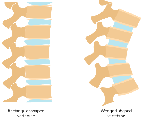
congenital = spine does not properly develop
sever causes paresthesis in legs, back tightness, pain, fatigue
tx: PT, back brace who are still growing → sx (spinal fusion)
scoliosis: curved to side (S side) → uneven shoulders
genetic = idiopathic; infant w/ scoliosis (congenital); neuromuscular dysfunction (spina bifida, cerebral palsy)
moderate > 25 degrees
uneven hips/ shoulders, asymmetrical rib cage, body leaning to one side
tx:
prevent worsening of the condition, relieve the manifestations, and improve function
spinal brace if not growing 25-45 prevents progression 16-23hrs/ day
sx for severe if >40 degrees or worsening condition = fusing spine or expandable rob
PT
→ leads to balanced issues, breathing, uneven legs ( unable to stand straight)
screening w/ physical exam
scoliosis = 10 f , 13 M
MRI, CT, inspect spine
anomalies in the vertebrae, trauma to the spine, degenerative disc disease of the vertebrae, severe slouching, and compensation in the spine.
Adam’s Forward Bend Test
The back should be exposed (either no shirt or wear a bathing suit).
Stand with feet together.
Bend forward 90 degrees at the waist with the arms loosely dangling down.
Findings that should be reported to the health care provider include unevenness in the trunk or curvature in the spine.
Mild changes in symmetry may not be obvious when inspecting the spine, but one hand may hang lower than the other.
tx:
strengthen abd muscles,
swimming and non-contact sports
Development Dysplasia of Hip
acetabulum= socket of pelvis that the ball of femur fits into
head of femoral bone does not fit into acetabulum → hip dislocation
more during infancy or early childhood = if head of femur placed weird in femur → cause hip joints to become loose → soft cartilage of acetabulum becomes damaged → shape becomes rounder
→ cause partially lose contact (subluxation), or completely separate (dislocation)
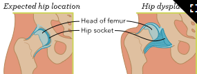
maternal hormones in female infants → loose of joints; breech during last trimester, restricted movement in womb (maldigestion), family hx of DDH or bone disorders
screen: physical exam
Barlow maneuver = adducting legs and gently pushing gown at knees to evaluate hip dislocation
Ostolani by abducting legs while lifting greater trochanter w/ 2 fingers = (+) when reduction of dislocation occurs
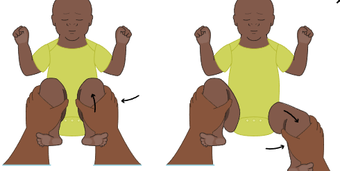
s/s:
limited ROM of hip,
toddlers w/ unexpected gait, adolescent with hip pain , diff lengthens in lower extremities
dx: US <6m / XR if >6m
tx:
Pavlik harness → 23hr/day and remove only when bathing infant;
close monitoring since can cause damage of head of femur → severe necrosis
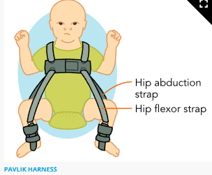
Von Rosen splint, Laudanna-developed abduction brace, Ilfeld orthosis, Frejka pillow
if untx → sx or rigid cast
<18m = closed reduction w/ external manipulation
if untx = limit ability to walk
<6m =noninvasive / >6m = invasive
maintain healthy weight
>18m= open reduction sx
orthics
for knee or hip subluxations or dislocations, bone fractures, skeletal deformities, foot pronation, or to provide support for muscle weakness or spasms
by orthotist and custom fit
Type | Application |
Foot | Special shoes (or inserts/modifications placed in shoes) that restore balance |
Ankle-foot (foot drop) | Semi-rigid braces that extend to the calf with straps |
Hip-knee-ankle-foot | Helps with upright positioning and centering of the knee joint using bands, belts, and a pelvic girdle |
Knee-ankle | Provides stability to the knee and foot for mobility and has mechanical or electrically operated hinges |
Knee | Supports and aligns the knee |
Spinal | Supports and aligns the spine and aids in sitting and standing |
Trunk-hip-knee-ankle-foot | Controls and supports trunk and spinal alignment |
Prophylactic | Rigid support for injuries |
Hand and wrist | Rigid splints to provide support |
Elbow | Supports and aligns the elbow |
Helmet | Corrects misshapen skull in infants |
help think of answers to say to peers, proper fit, schedule of braces (slowly inc time)
skin chaffing → sore, red, blister, swollen skin
Alcohol rubs, cornstarch, and specialized creams, lotions, or powder, cotton socks
or layers
check for proper fit, cracks, tears, or other indications of wear
legg-calve perthes disease
head of femur dies due to low blood flow → remodeling w. unexpected head shape
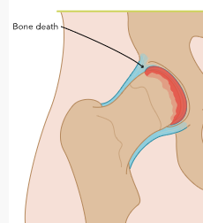
due to trauma, blood clot, family hx, male, 2nd hand smoke exposure, lbw, low SDOH
in 4-10yr
necrosis
fragmentation = bone pieces reabsorbed to body
reossification (phase ~ 1-3yrs)
remodeling: bone is flat or enlarged
s/s:
low ROM of hip, painful rotation of thigh, differing leg lengths, pain when walking, atrophy of thigh or glutes
XR height pillar stages:
a. full height, no density change → b. 50% of height → c. <50%
chronic pain, inc risk of arthritis, ~ develop sx in life
lab/dx = SR, bone scan, MRI
tx:
activity restriction <5yr, PT,, >8yr = sx
younger <5y = better outcomes
make sure limb is not bearing weight until bone has completely formed → swimming
club foot
congenital talipes equinovarus, twisted to side due to shorten tendons → front part is inward, length of total leg less than uneffected
fetal US done before 24 weeks to assess extremities (screen as early has 13 weeks), visually at birth or XR after birth
hx of clubfoot, male, cerebral palsy, birth defects, smoking during pregnancy, breech
tx:
start two weeks after birth = Ponseti method → casts + braces to correct position (HCP stretch foot before cast placement → cast change in 7 days → no more than 10 cast changes → brace = 24hr/day
will continue until 4yrs
sx if not effective or if achilles tendon require repair
unexpected neurovascular findings, infection after surgery, or damage to the cast
→ ;ater use orthdonics to keep leg straight
frequent skin checks
if unilateral = tx feet and leg will be smaller
no smoking w/ pregnnacy, or risk of muscoloskeletal alteration
weight bearing, walking, stretch
skeletal alterations
osteogenesis imperfecta
brittle bone disease = mutation of type I - IV collagen in connective tissue → fragile bones w/ unexpected shape
genetic
Types s/s:
most common + mild = half amount of collagen
curvature of spine, loss of hearing due to inner bone ear deformities, triangular head
fatal since collagen not functioning properlu
common fractures, and underdeveloped lungs
high risk of fractures, w/ smaller stature, loss of hearing, and underdeveloped muscles
fragile bones= dental problems, loss of hearing, spine curve
mutation of protein = some bones larger than expected → dislocations
~ IV = low bone mineralization → IV s/s
hip deformities, short bone = short height
growth deficiency + weak bones
leg bowing, barrel chest, sclerae discoloration (gray, purple, blue hue)
~ develop bone tumors, cardiac issue( heart valve dysfunction), loss of vision, inc resp infections, kidney stones, joint problems
lab/ dx:
skin/ bone biopsy to determine collagen, XR, gene test
tx:
mild = avoid high impact
moderate = managing fracture occurrence + orthopedic measures
severe = sx
bisphosphonate (ibandronate) = makes bones stronger
dec vit D + ca levels = supplement
PT
prevent contractures = braces + splints
regular vision and hearing checks
juvenile idiopathic arthritis
autoimmune = body attacks synovial fluid → damage cartilage + bone
Oligoarthritic (<5 joints like knees and elbows)
polyarthritis = more in hands/ feet
enthesis-RT arthritis = (where bones attach w/ other tissue) → hips and spine
systemic = whole body
psoriatic arthritis= some joints + psoriasis ( nail and skin disease → rashes, redness, depression of nail surface)
genetic, abx, c/s, breastfeed to reduce risk
s/s:
painful, swollen joints
fever, eye discomfort/ inflammation, redness in eye (enthesis ), rash/ dimpling in nails (psoriatic), diff ambulating, report stiffness
hearing loss, blurred vision developmental delay
CBC, Y-testing, inflammatory markers, US, XR, MRI
rheumatologists, corticosteroids, DMARDS, immunosuppressants (methotrexate)
OT, PT, physically active (non contact), healthy eating habits (clock method)
SLE
autoimmune attacks healthy tissue = kidney, skin, brain, joints,
stressors = sun, smoking, fatigue, stress, low vit D infections = exacerbation
check ANA screen → CBC, rheumatologist, U/A (proteinuria/ hematuria), check on body system for baseline
genetic, inc in hormones (inc in females)
s/s:
butterfly rash, joint ain, extreme fatigue, fever, hair loss, weight loss, mouth sores, swelling of hands or feet, anemia
tx:
flare ups → hydroxychloroquine
prednisone + immunosupressants
Preventing SLE exacerbation
Prioritize getting enough rest.
Keep all appointments and receive regular follow-ups with the rheumatologist.
Comply with the medication regime as prescribed.
Avoid over-exposure to sunlight and wear sunscreen.
Implement good handwashing and obtain recommended vaccines to prevent infections.
Do not smoke.
cancer
osteosarcoma = found in long bones
creates osteoid = bone cell that has not fully matured
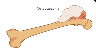
Classification:
Low-grade osteosarcoma: Slowest growing cancer
Intermediate-grade osteosarcoma: Least common, grows at a rate between low- and high-grade
High-grade osteosarcoma: Fastest growing cancer, most common in osteosarcomas
bone anomalies (exostoses), retinoblastoma, Ollier’s disease, Li-Fraumeni syndrome, polyostotic fibrous dysplasia, or Paget’s disease
exposure to ioning radiation for other cancer tx
s/s: swelling over cancer site, pain w/ activity, lump, fractures
tx: body tissue removal or amputation, but chemo/radiation 1st
ewing sarcoma:
in bone, bone marrow, surrounding tissue → aggressive (ribs/ spine)
→ immidiate tx for whole body
micro metastasis = small cells w/ round nucleus that spread to whole body → necrosis → hemorrhage
black/ asian, amle, previous radiation tx
s/s: swelling and discomfort, inc pain at night or inc activity, diff ambulating or fever
tx: chemo, sx, radiation
rhabdomyosarcoma:
skeletal muscle (in head / neck) = premature (rhabdomyoblasts → warmless white massess) → quickly multiply → aggressive
cancer stages:
good prognosis
not favorable, <5cm, not metastized
spread to lymph nodes
metastasized
Alveolar rhabdomyosarcoma (ARMS) = aggressive and in older children
genetic (rhabdomyosarcoma) w/ DNA mutations
Li-Fraumeni syndrome, neurofibromatosis, Beckwith-Wiedemann syndrome, or Costello syndrome.
s/s: depend on location, bulging eyes, pain in ear, painful elimination, vomiting
tx: bone marrow aspiration, lumbar puncture
lab/dx:
XR, CT, bone scan, MRI, PET (uses radioactive sugar to check on cancer cells),
official w/ tissue biopsy of tumor + malignant
tx:
remove tumor from localized area of body, chemo(vincristine), radiation
ADR: nausea, diarrhea, hair loss, and damage to their organs
once tissue is removed → reconstruction + joint graft replacement
exercise, no pregnancy
amputation
~ cause required assistance w/ ADL
congenital or traumatic
upper extremity sx amputation:
phalanges, hands by separating hand at the wrist; lower arm = at, below, or above the elbow.
cutting radius and ulna
if above the elbow = cut humerus
if whole arm = shoulder joint = shoulder disarticulation
lower extremity:
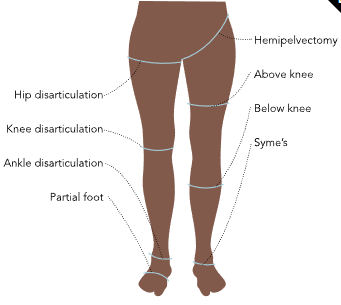
congenital: may be noticed during US
when newborn is missing limbs;
traumatic amputation:
can use tissue preservation techniques to reattachor may need additional tissue
severe burns or lack of blood flow : MVA, gunshot wounds
from interruption of blood flow → necrosis + severe infection or aggressive cancers; vascular injury → frostbite, blood clot, septic shock
osteosarcoma >10yr / <10yr = Ewing sarcoma
gangrene → black or purple → sores may occur
phantom limb pain, struggle w/ self image, grief,
lab/dx: XR, MRI, amputation, biopsy,
tx:
location and amount of tissue decided by surgeon → sx
rest of tissue formed into stump → prosthetic limb
steel implant = osseiointegration (like a living bone)
PT w/ assistive devices
if growth plate is not damaged → bone will continue to grow → ` overgrowth/spiking of bone
swelling, tender, warmth, poor socket fit → sx removal or if not bone will penetrate skin/ ulceration
comfort w/ toys showing their own amputation
make adolescent aware of door slams, reckless driving, farm equipment, fireworks, firearms,
use seatbelts, car seats
Prosthetics:
infant = prosthetic that does not does not move on its own
upper extremity = will only get operated prosthetics until 1-2yrs
lower= prosthetics w/ knee joint will be given at 2-3 yrs
achieve independence by 5 years old
adjusted qfew years to ensure comfortable fit, check for skin breakdown,
use sleeves or design of device itself to accept prosthesis.
social and physical challenges
shrinker wrap w/ stump when swelling
if sking integrity or blister = not wear prothesis
remove prosthesis 60-90mins to check for redness and open sores
take prosthesis before sleeping, care of components, liner, sheaths, or suspension sleeves
Neuromuscular Conditions
cerebral palsy (CP)
non-progressive motor dysfunction → anomalies in muscle tone, posture, motion that alters sensory perception, cognitive abilities, communication, and behavioral responses
Spastic CP presents with increased muscle tone, causing movements to be stiff and awkward.
Dyskinetic CP is characterized by impaired muscle tone and presents with slow, uncontrollable, jerky movements of the extremities.
Ataxic CP presents with poor coordination and an unsteady, wide gait.
Mixed CP is not a single type but a mixed presentation.
negative impact on fetal or neonatal brain development ~ premature, IUGR, intrauterine infection, antepartum hemorrhage, severe placental pathology, multigestation, post natal events ( cerebral malaria, seizures), genetics
hypoglycemia, meconium aspiration, lack of O2 of fetus or newborn
s/s:
in early infancy = within 3-6m = head lag, asymmetrical posture/ movements, leg stiff
hypertonia, spasticity, and dystonia, hypotonia
persistent or asymmetric hand clenching, tongue retraction, oral hypersensitivity, grimacing, and weak head control
muscle spasticity, hyperreflexia, extensor plantar responses (Babinski), clonus, weakness, atrophy, loss of dexterity, fatigability, hypotonia, dyskinesia, and ataxia.
lab/dx:
examination, other impairment : seizure, sensation, perception, cognition, communication, behavior
neuroimaging,
tx:
routine prenatal care, dec preterm birth, neuroprotective measures
PT/ OT = coordination of movements, functional task completion, strength, and conditioning
braces, orthotics, standers, seating systems, wheelchairs,
spasticity = PT/OT, orthodic devices, anticonvulsant Rx, botulism toxin → deep brain stimulation
casting + sx (ex Achilles tendon lengthening ->provide stretch + ROM),
hypotonia
muscle tone is tension when muscle is at rest → responsible for leaving upright position and maintain posture
floppy muscles
lack of resistance when holding infant or when passively stretches or engages joint during assessment and encounter no resistance
vertical suspension, horizontal suspension, and the scarf sign.
due to muscles, joints, or peripheral or central nervous systems; disorders of metabolism/ hypothyroidism; or hypoxic encephalopathy, prenatal eps, Down syndrome and Prader Willi syndrome., congenital muscular dystrophy, congenital myopathies, spinal muscular atrophy, Tay-Sachs disease, or Patau syndrome
s/s:
low APGAR
tx:
OT, ST, PT, inc nutrition, additional supprt, nasogastric or gastrotomy tubes
spinal muscular atrophy:
autosomal recessive = impacts voluntary motor movement and muscle control necessary for basic system functions, such as muscles used for breathing
SMN1 deficiency, health of motor neurons impact
screening: if carry gene but will not predict if child will have condition
Four Main Types of SMA
Type and Name(s) | Severity | Age of Presentation | Disease Presentation |
Type 0 (Type Ia, congenital) | Severe, death usually occurs by 1 month of age | At birth | Hypotonia, respiratory failure, severe weakness |
Type I (Werdnig-Hoffman disease, “non-sitters”) | Severe SMA, death by age 2 | Within the first 6 months | Limited head control, frog-like posture when supine, areflexia, hypotonia, swallowing difficulties, facial weakness with disease progression, ventilatory support required, wheelchair required |
Type II (Dubowitz disease, “sitters”) | Mild SMA | 6 to 18 months old | Hypotonia, limited areflexia, more weakness in lower extremities than upper extremities, able to sit, may need a wheelchair as disease progresses, respiratory compromise is normally the cause of death |
Type III (Kugelberg-Welander disease, “walkers”) | Mild SMA | 18 months to 3 years | Legs affected more than arms, able to walk, may need a wheelchair as disease progresses, life expectancy matches general population |
Type IV (Adult SMA) | Mildest form | 21 years or older | Mild leg weakness, life expectancy is unaffected, able to walk |
lab = physical exam, hx, noting gross motor function
tx:
neurology, pulmonology (BiPAP= weak cough, mucus plug, recurrent resp infection, inc aspiration, <5yr = chest physiotherapy + monitoring and track forced vital capacity),
gastroenterology = GI muscle weakens = constipation, gastric reflux, the risk of aspiration, and delayed gastric emptying
g-tube placed in type I
orthopedics: I/II more prone to fractures, hip subluxation, scoliosis,
speech-language pathology: I w/ diff sucking and swallowing
occupational and physical therapists, dietitians, and social workers, pallative care
Nursinersen = inc SMN protein → helps w/ type I, II, III
Risdiplam = inc SMN protein → II/ III
Onasemnogene abeparvovec =most expensive but has 100% alive pts
of under 2yrs
muscular dystrophy
generalized muscle weakness + degenerative
lack of glycoproteins (dystrophin) in muscle cell membrane = replaces muscle w/ connective tissue → pseudohypertrophy → release CK into blood as muscles are destroyed
( Duchenne (more severe= use wheelchair by 12yr) >Becker muscular dystrophy
Limb-girdle ME affects limbs and trunl
Facioscapulohumeral MD affects the face, neck, and shoulders, and onset is at 10 to 30 years old.
Oculopharyngeal MD affects the face and has an adult onset.
Myotonic MD typically affects the face and extremities, and onset is at 10 to 15 years old.
screen: check in vitro fertilization. CVS at 10-12 weeks + amniocentesis at 15-18w
X -chromosome mostly as mother being the carrier
s/s:
has developmental delays, appears clumsy, and has delayed mastery of milestones, such as walking or crawling.
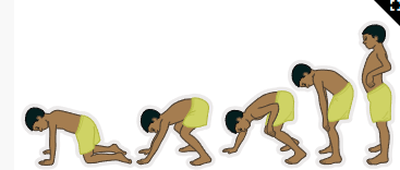
Gower’s sign = pelvic girdle becomes unstable → diff walking, waddling gait, diff standing up from seated floor position → contractures → muscle weakness, scoliosis, constant pain
DMD expectancy of <20yrs = remain ambulatory until 15yrs but BMD ~50yrs = remain ambulatory until 25yr
lab/dx:
chromosomal analysis, genetic testing, immunocytochemistry, electromyography, and a muscle biopsy, ECG
high ALT, SGPT, Aldolase, ABG (acidosis), AST, CK/CPK, CK-MB, LDH, U/A (positive for glucose), MRI, CT
tx:
delandistrogene moxeparvovec for 4-5yr
malignant hyperthermia if irregular Ca control
lose mobility, bedbound, require devices to move, skin breakdown inc → infection, pressure ulcer, pain, pressure points
glucocorticosteroid = prednisone, deflazacort → delay at 4-5yr
→ improved muscle strength, improved motor function, improved pulmonary function, decreased need for scoliosis surgery, delayed onset of cardiomyopathy, decreased inflammation, and increased total muscle mass.
Provider | Evaluation/Intervention |
Pulmonology |
|
Cardiology |
|
Orthopedics |
|
Gastroenterology |
|
Urology |
|
Neurology |
|
Endocrinology |
|