WEEK 6 Exercise Training Physiology and Biochemistry Notes
Introduction
Focus on understanding the role of hormones in exercise physiology and metabolism, which is vital for optimizing athletic performance and recovery.
Importance of considering the whole-body metabolic response during exercise, integrating organ function to achieve a balanced physiological response.
The Role of Hormones
Function of hormones:
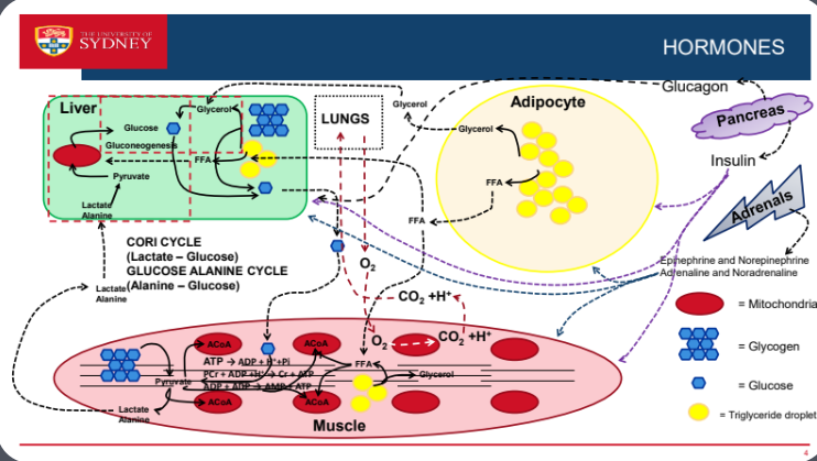
Guardians of internal regulation (homeostasis) that maintain the stability of the body's internal environment despite external changes.
Trigger adaptive responses to maintain homeostasis when disrupted, playing critical roles during periods of stress from physical activity, illness, or injury.
Serve as metabolic signals released by specific cells to target other cells, influencing metabolism, growth, and development throughout the body.
Types of Hormonal Communication
Autocrine: Hormones act on the same cell that secretes them, allowing for self-regulation of activity.
Paracrine: Hormones act on nearby cells, facilitating communication between adjacent cells and tissues, important for local responses.
Endocrine: Hormones are released into the bloodstream to affect distant cells, allowing for widespread systemic effects crucial in stress responses and recovery post-exercise. Reliant on blood flow.
Classification of Hormones by Structure
Peptide Hormones:
Chains of amino acids (e.g., Glucagon, Insulin) that are crucial in regulating metabolism and glucose homeostasis.
Typically endocrine; synthesized, stored, and released from endocrine glands, responding to various physiological signals.
Interact with cell surface receptors, triggering rapid cellular responses that modulate metabolic functions.
Catecholamine Hormones:
Derived from tyrosine (e.g., Epinephrine, Norepinephrine), which prepare the body for 'fight or flight' responses and increase heart rate, blood flow to muscles, and energy mobilization.
Function as neurotransmitters and hormones; use either cell surface or nuclear receptors, thus having versatile effects on different tissue types.
Steroid Hormones:
Made from cholesterol (e.g., Testosterone, Estradiol) that play significant roles in defining secondary sexual characteristics, muscle mass, and overall metabolic rate.
Produced in adrenal cortex, testes, and ovaries, these hormones have long-lasting effects due to their ability to enter cells and modify gene expression directly.
Eicosanoids:
Derived from polyunsaturated fatty acids (e.g., Prostaglandins), which are involved in inflammatory responses, immune function, and modulation of organ function.
Typically act in a paracrine manner and are produced as needed, allowing quick local responses to physiological changes like injury or exercise.
Hormonal Action on Receptors
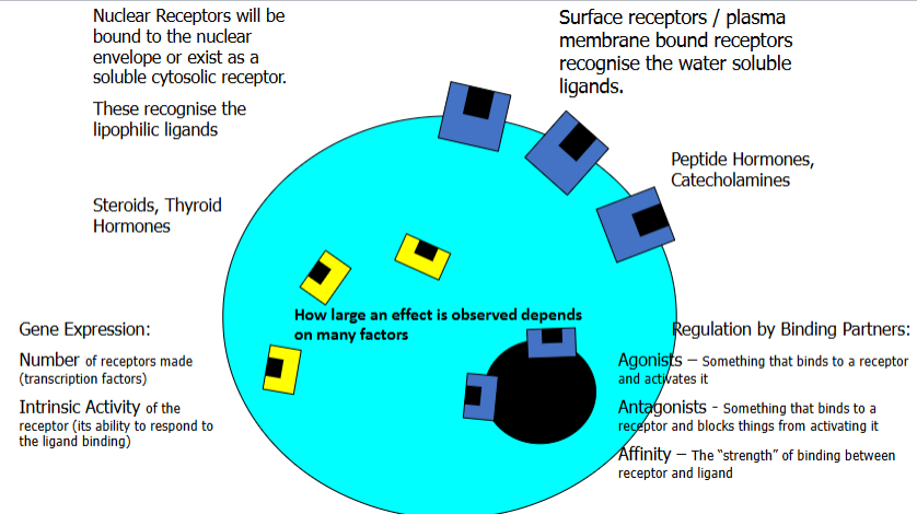
The action/ signal result of a hormone is dependent upon specific binding to a receptor. Receptors sit on the surface / nucleus surface of a cell.
The binding is non-covalent, weak but specific and will be either
Hydrogen bonding-
Ionic Bonds-
Van Der Waals forces-
Hydrophobic Interactions-
The receptor will remain active as long as the hormone is bound. The interaction is SHORT.
Types of Cell Surface Receptors
Ligand Gated Ion Channels: Mediates signals like acetylcholine, allowing immediate changes in cell membrane potential.
Single-TMS Catalytic Receptors: e.g., Insulin Tyrosine Kinases, which activate intracellular signaling cascades upon hormone binding, leading to cell growth and metabolic changes.
G-Protein Coupled Receptors (GPCRs): e.g., Epinephrine receptors, which trigger complex intracellular signaling involving secondary messengers, amplifying the physiological response.
Insulin
Binds specifically to a Tyrosine Kinase receptor, crucial for glucose uptake and metabolism.
Tyrosine Kinase Receptor is a single Transmembrane spanning (TMS) receptor
Ligand binds to the Extracellular side. This causes all the Tyrosine amino acids within the intracellular side of the protein to ‘autophosphorylate’
The process by which a kinase enzyme attaches a phosphate group to one of its own amino acid residues. In other words, the kinase catalyzes its own phosphorylation.)
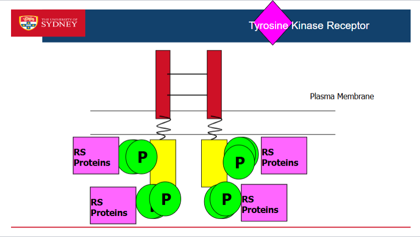
Insulin binds to the extracellular element of the receptor in the plasma membrane which autophosphrolates the tyrosine residues
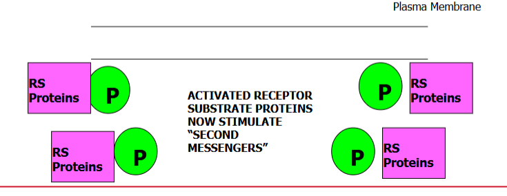
The insulin receptors create RS Protein. They will receive the phosphate created and dissociated from the receptor.
Once they leave if the hormone is still bound, the receptor can undergo additional autophosphorylation events (more RS Proteins)
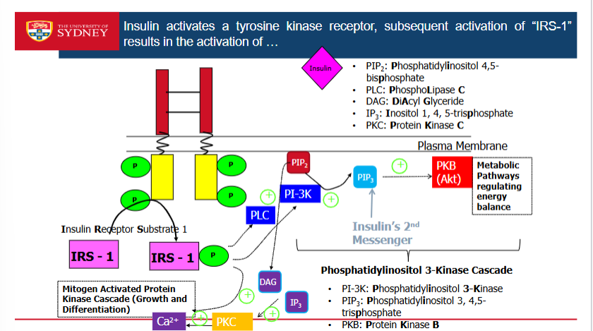
Protein PIP2 that attaches to the membrane. Wherever there is an insulin receptor, PIP2 will always be inside or near by the membrane
Insulin binds which autophosphorolates. The Insulin Receptor Substrate 1 (IRS1) comes over and phosphorylates which turns on an enzyme called PLC.
PLC breaks down PIP2 into DAG and IP3
DAG and IP3 activates Protein Kinase which then activates Calcium. Calcium activates MAP Kinase Cascade
The IRS1 activates another enzyme called PI-3K. It takes PIP2 & adds a phosphate to it to make PIP3.
PIP3 activates PKB. This activates Insulin’s 2nd Messenger. Metabolic Pathways regulating energy balance
IRS1 can activate PLC & PI3K enzymes at the same time due to there being heaps of receptor substrates.
Epinephrine
Activates G-Protein coupled receptors (Epinephrine) and Hormonal signals/ enzymes cascades
G- Coupled Protein Receptors
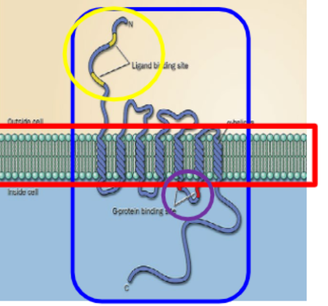
7 Transmembrane components with an inactive ‘G-Protein’
Ligand Binds to the Extracellular side
The G-Protein bound to the Intracellular side is activated
The active subunit then dissociates so to bind to and activate another protein/enzyme and the cascades begin
Called G-Protein because it is a receptor that is coupled to a protein that has GTP or GDP energy.
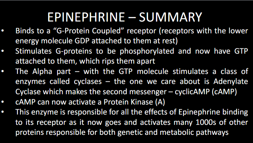
At rest the receptor has G-Protein attached to it which is a protein that has 3 sections to it as well as a low energy GDP molecule attached to it.
When Hormone binds to the receptor it changes the energy status which alters the GDP into GTP thru phosphorylation.
When it has the high energy phosphate attached to it, it breaks away leaving the alpha subunit to activate something else
The alpha subunit activates the enzyme called ‘adenylate Cyclase’
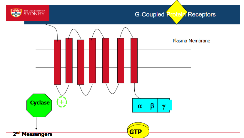
Adenylate Cyclase takes ATP to make cAMP- can actiavte metabolic and genetic changes
cAMP is an inert signal, however CAN actiavte Protein Kinase A which can then activate all the metabolic effects such as glycolsis, glycogenolysis and Gene expression of PKA
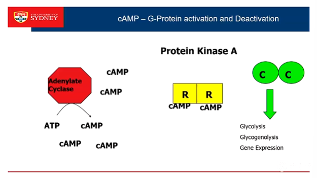
The only way to turn off the signal for epinephrine is to remove the cAMP. PDE breaks down the cAMP into AMP, effectively terminating the signaling pathway and allowing the cell to reset its response to epinephrine stimulation.